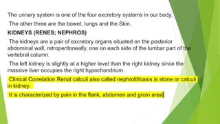
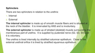
SlideShare a Scribd company logo
1 of 93
URINARY
SYSTEM
MRS RAMESHWORI TH
ASSOCIATE PROFESSOR
PARTS OF URINARY SYSTEM
The urinary system comprises-
two kidneys, two ureters, urinary bladder and urethra.
The kidneys remove waste products of metabolism, excess water and salts
from blood and maintain the pH.
The ureters convey urine from the kidneys to the urinary bladder.
The urinary bladder is the muscular reservoir of urine and the urethra is the
channel to the exterior.
FUNCTIONS OF URINARY SYSTEM
The kidneys maintain a stable internal environment by regulating
the volume and composition of body fluids as well as by excreting
the waste products and excess water.
For maintaining homeostasis, kidneys do multiple functions as
follows:
 Excretion of metabolic waste products and chemicals,
like urea, uric acid, creatinine and many drugs.
 Regulation of body fluid volume and osmolality by
excreting either dilute or concentrated urine.
 Regulation of concentration of electrolytes and various
ions.
 Regulation of acid-base balance by excreting either
excess acid or base.
 Regulation of arterial blood pressure by adjusting Na* and
water excretion.
 Secretion and production of some hormones, like
erythropoietin, 1,25-dihydroxycholecalciferol, renin,
prostaglandins, etc.
 Metabolism of various hormones, like insulin, glucagon,
parathyroid hormone, etc.
The urinary system is one of the four excretory systems in our body.
The other three are the bowel, Iungs and the Skin.
KIDNEYS (RENES; NEPHROS)
The kidneys are a pair of excretory organs situated on the posterior
abdominal wall, retroperitoneally, one on each side of the lumbar part of the
vertebral column.
The left kidney is slightly at a higher level than the right kidney since the
massive liver occupies the right hypochondrium.
Clinical Correlation Renal calculi also called nephrolithiasis is stone or calculi
in kidney.
It is characterized by pain in the flank, abdomen and groin area
Shape and Size
Each kidney is bean-shaped and measures 7.5-10 cm in length, 5 cm in width and is 2.5 cm
thick. Each kidney weighs 150 g.
It has two poles-upper and lower; two borders-medial and lateral; and two surfaces-anterior and
posterior.
1. The upper pole is broad and is closely related to the suprarenal (adrenal) gland. The lower
pole is pointed. The upper poles are closer to the median plane than the lower poles.
2. The lateral border is convex.
3. The medial border is concave.
The middle part of the medial border is depressed and is known as the hilum (hilus).
.
Structures Present in the Hilum of Kidney
The following structures are seen in the hilum of kidney (from anterior to
posterior):
• The renal vein
• The renal artery
• The renal pelvis-the upper extended portion of ureter.
In addition to these three structures, the hilum also transmits nerves and
lymphatics
Coverings
The kidney has three coverings:
 Inner most fibrous capsule or true capsule.
• Middle fatty capsule or perinephric fat-it is a collection of fatty tissue.
It acts as a shock absorber and also helps to maintain the kidney in its position.
 The false capsule is made of renal fascia. It has two layers-anterior and
posterior. Superiorly, the two layers enclose the suprarenal gland and then merge
with diaphragmatic fascia. (That is why the kidneys move with respiration.)
 Pararenal fat-forms a cushion for the kidney.
Organs Associated with Kidney
As the kidneys lie on either side of
the vertebral column, each is
associated with a different group of
structures.
Right Kidney
Anteriorly :the duodenum(second
part),the hepatic flexure of the colon(
colic area in the fig) and the right lobe
of the liver.
.
Posteriorly:
Diaphragm,muscles of the posterior abdominal wall (psoas
major, quadratus lumborum and transverse abdomen is), and
subcostal vessels gr nerves.
Superiorly:
The right adrenal gland
Left Kidney
• Anteriorly: The spleen and splenic
vessels, jejunum and splenic flexure of
the colon (colic area in the
figure). pancreas and stomach.
• Posteriorly: The diaphragm and
muscles of the posterior abdominal wall
(psoas major, quadratus lumborum and
transversus abdominis), and subcostal
vessels and nerves.
Superiorly: The left adrenal gland
Gross and Microscopic Structure of Kidney
GROSS STRUCTURE
The naked-eye examination of a coronal section
shows the following features:
Characteristic bean-shape
 It has a convex lateral margin and a concave
medial hilum (hilus).
 The hilum leads into a space called renal
sinus. This sinus is occupied by the upper
expanded part of the ureter, which is called the
renal pelvis (pelvis=basin).
 Within the sinus, the renal pelvis divides into two or
three major calyces (calyx=cup).
 Each major calyx divides into minor calyces.
The kidney tissue, covered by a
fibrous capsule or true capsule,
consists of an outer cortex and inner
medulla.
 The outer cortex, which lies
immediately below the capsule
and outside the pyramid, is light in
color.
 The inner medulla is made up of
triangular are as of renal tissue
called renal pyramids and appears
dark in color.
 The apex of the papilla which fits
into the minor calyx.
 The bases of the pyramids are
capped by renal cortex called
cortical arches.
 The parts of the renal cortex
projecting between the pyramids
are called the renal columns
MICROSCOPIC STRUCTURE
The kidney may be regarded as a collection
of millions of uriniferous tubules.
Each uriniferous tubule consists of an
excretory part called the nephron and of a
collecting tubule.
There are 1-2 million nephrons in a kidney.
Parts of a Nephron
Nephron, the functional unit of kidney, has the following parts:
A renal corpuscle or Malpighian corpuscle is a rounded structure
consisting of a tuft of capillaries called glomerulus and a cup-like double-
layered covering called the Bowman's capsule or glomerular capsule.
The two layers of glomerular capsule are separated by the urinary space.
The outer layer is lined by squamous cells.
With the light microscope, the inner wall also appears to be lined by
squamous cells, but the electron microscope (EM) shows that these cells,
called podocytes, have a highly specialized structure
The renal tubule (tubular part) is a long
complicated tubule. It has different parts.
These are:
 Proximal convoluted tubule (PCT): The PCT
is lined with cuboidal or low columnar
epithelium. The cells are provided with
microvilli and numerous mitochondria.
 Loop of Henle: It has a descending limb, a
loop and an ascending limb. The loop of
Henle is lined with low cuboidal or squamous
epithelium.
 Distal convoluted tubule (DCT): The DCT is
lined with cuboidal cells. These cells do not
have microvilli.
.
The DCT ends by joining a collecting tubule.
 The collecting tubules are lined with cuboidal or low
columnar cells. The collecting tubules draining different
nephrons join to form larger tubules called papillary ducts
of Bellini, which open into a minor calyx at the apex of a
renal papilla.
 A minor calyx receives urine from papilla and delivers it
to major calyx.
 From the major calyces, urine drains into the renal pelvis
and then out through the ureter to the urinary bladder
 The renal artery after entering the kidney divides
repeatedly
Parts of a nephron
Nephron
Renal corpuscle Tubular part
Glomerulus Bowman's capsule
Proximal convoluted tubule Loop of Henle Distal convoluted tubule
Key Points
• The renal corpuscles and major parts of PCT and DCT are located in the cortex of the kidney.
• The loops of Henle and the collecting ducts lie in the medulla
Path of urine
Nephron
Collecting duct
Papillary duct of Bellini
Minor calyx
Major calyx
Renal pelvis
Ureter
Urinary bladder
Functionally there are 2 sets of arterioles and capillaries.
 • The first capillary system present in the glomeruli is
concerned exclusively with the removal of waste products
from blood.
 It does not supply oxygen to the renal tissues.
 The second set of capillaries present around the tubules is
concerned with the exchange of gases
FUNCTIONS OF DIFFERENT PARTS OF A NEPHRON
PART FUNCTION
GLOMERULAR CAPSULE FILTRATION OF MOLECULES AND WATER FROM BLOOD
PROXIMAL CONVULATED
TUBULE
SELECTIVE REABSORPTION OF MANY SUBSTANCES FROM GLOMERULAR
FILTRATE,EG: GLUCOSE,AMINO ACIDS,IONS LIKE SODIUM AND CHORIDE,ETC
LOOP OF HENLE CREATES A HYPERTONIC ENVIRONMENT IN THE MEDULLA TO FACILITATE WATER
REABSORPTION BY COLLECTING DUCTS
DISTAL CONVULATED TUBULE SELECTIVE REABSORPTION OF IONS(MAINLY SODIUM BICARBONATE) AND
SECRETION OF HYDROGEN IONS
COLLECTING DUCTS FINAL CONCENTRATION OF URINE
Blood Supply
About 25% of the total cardiac output flows through
kidneys, ie., 1200 mL/minute at resting condition.
However, kidney is the organ in our body
possessing highest blood supply, i.e., about 400
mL/min/100 g.
The blood supply to the kidney is through the renal
artery.
medullary nephrons are called vasa recta
The renal artery (branch of abdominal aorta) branches progressively to form the
interlobar arteries, the arcuate arteries, the cortical arteries or the interlobular
arteries and the afferent arterioles.
Each afferent arteriole breaks up into the glomerular capillary network which joins
together to form the efferent arteriole which again divides into a second capillary
network.
The second capillary networks around the cortical nephrons are called peritubular
capillaries and those surrounding juxta
Flowchart : Path of blood in kidney.
 Renal artery
 Interlobar arteries
 Arcuate arteries
 Interlobular arteries
 Afferent arterioles
 Glomerular capillaries
 Efferent arterioles
 Peritubular capillaries/Vasa recta
 Interlobular veins
 Arcuate veins
 Interlobar veins
 Renal vein
Both peritubular capillaries and vasa recta drain into the vessels of venous
system running parallel to the arterial vessels and branch progressively to form
the interlobular or cortical vein, the arcuate vein, the interlobar vein and finally
the renal vein. The renal circulation possesses the feature of double capillary
network, which is also known as portal circulation.
Nerve Supply
 Kidneys are innervated by autonomic nervous system.
 Sympathetic fibers are derived from T10-L1 segments.
 Parasympathetic fibers are derived from the vagus nerves.
Lymphatics from the kidneys drain into the lateral aortic nodes.
Juxtaglomerular Apparatus
(JGA)
• Juxtaglomerular apparatus
is a specialized organ
situated near the glomerulus
of each nephron
(juxta=near).
The juxtaglomerular
apparatus is formed by three
different structures:
1. Juxtaglomerular cells
2. Macula densa
3. Lacis cells
Parts
Juxtaglomerular Cells
 A part of the distal convoluted tubule (at the junction of its straight and
convoluted parts) lies close to the
vascular pole of the renal corpuscle, between the afferent and efferent arterioles
 In this region, the muscle cells in the wall of the afferent arteriole are modified.
 They are large and rounded (epithelioid) and have spherical nuclei.
 Their cytoplasm contains granules that can be stained with
special methods.
 These are juxtaglomerular cells. They are innervated by
unmyelinated adrenergic nerve fibers.
 Juxtamedullary cells are regarded, by some, as highly
modified myoepithelial cells as they contain contractile
filaments in the cytoplasm.
The granules of the juxtaglomerular
cells are seen by EM to be
membrane-bound secretory
granules.
They contain an enzyme called
renin.
The juxtaglomerular cells also
probably act as baroreceptors
reacting to a fall in blood pressure by
release of renin.
Secretion of renin is also stimulated
by low sodium blood levels and by
sympathetic stimulation.
Note: In addition to renin, the kidney
produces the hormone erythropoietin
(which stimulates erythrocyte
production). Some workers have
claimed that erythropoietin is produced
by juxtaglomerular cells, but the site of
production of the hormone is uncertain
Macula Densa
 The wall of the distal convoluted tubule is also modified
at the site of contact with the arteriole.
 Here the cells lining it are densely packed together,
and are columnar (rather than cuboidal as in the rest of
the tubule). •
 These cells form the macula densa. The cells of the
macula densa lie in close contact with the
juxtaglomerular cells.
 In addition to the renin-producing cells, and the
macula densa, the juxtaglomerular apparatus has
a third component: these are lacis cells.
 These cells are so called as they bear processes
that form a lace-like network
 They are located in the interval between the
macula densa and the afferent and efferent
arterioles.
 The function of lacis cells is unknown
Functions
 The primary function of juxtaglomerular apparatus is the secretion of
hormonal substances.
 It also regulates the glomerular blood flow and glomerular filtration
rate.
Secretion of Renin
The juxtaglomerular cells secrete renin.
Renin is a peptide with 340 amino acids. Along with angiotensins, renin
forms the renin-angiotensin system, which is a hormone system that
plays an important role in the maintenance of blood pressure.
Renin-Angiotensin System
When renin is released into the blood, it acts on angiotensinogen and converts it into angiotensin
I. Angiotensin I is converted into angiotensin II by the activity of angiotensin converting enzyme
(ACE) secreted from lungs. Most of the conversion of angiotensin I into angiotensin II takes place
in lungs.
Angiotensin II has a short half-life of about 1-2 minutes. Then it is degraded into angiotensin III
by angiotensinases, which are present in RBCS and vascular beds in many tissues. Finally,
angiotensin III is converted into angiotensin IV
Actions of Angiotensin
Angiotensin I
It is physiologically inactive and serves only as the precursor of angiotensin II.
Angiotensin II
Angiotensin II is the most active form. Its actions are:
It increases arterial blood pressure by causing vasoconstriction and inhibiting baroreceptor reflex.
It stimulates zona glomerulosa of adrenal cortex to secrete aldosterone.
It regulates glomerular filtration. It increases sodium reabsorption from renal tubules.
It increases water intake by stimulating the thirst center.
It increases secretion of antidiuretic hormone (ADH) from hypothalamus.
Secretion of Other Substances
The extraglomerular mesangial cells of juxtaglomerular apparatus secrete prostaglandin.
Flowchart : Conversion of angiotensinogen into angiotensin II.
Renin ACE
Angiotensinogen Angiotensin I Angiotensin II (ACE: angiotensin converting enzyme)
Angiotensin IV Angiotensin III
Regulation of Glomerular Blood Flow and
Glomerular Filtration Rate
Macula densa of juxtaglomerular apparatus plays an important role
in the feedback mechanism called tubuloglomerular feedback
mechanism, which regulates the renal blood low and glomerular
filtration rate.
Mode of Action of Juxtaglomerular Apparatus
The juxtaglomerular apparatus is a mechanism that
controls the degree of reabsorption of ions by the renal
tubule.
It appears that cells of the macula densa monitor the
ionic constitution of the fluid passing across them
(within the tubule).
The cells of the macula densa appear to influence the
release of renin by the juxtaglomerular cells.
Functions of Kidney
The functions of kidney are the same as that of urinary system and also summarized in
Flowchart
PHYSIOLOGY OF URINE
FORMATION
 Key Points Formation of urine involves
three basic processes:
 1. Glomerular filtration
 2. Tubular reabsorption
 3. Tubular secretion
 Glomerulusis responsible for filtration and tubular parts of nephron are
responsible for reabsorption and secretion.
About 1800 L/day (1.2 L/min) of blood flows through both the kidneys. Of this,
about 180 L/day is filtered through the glomeruli.
But only less than 1% of the filtered water and variable amounts of many of the
solutes are excreted in the urine.
This is made possible by the processes of tubular reabsorption and secretion.
Thus, the renal tubules precisely control the volume, composition and pH of the
body fluids.
GLOMERULAR FILTRATION
It means the ultrafiltration of plasma by the
glomerulus. The first step in the formation of urine
is filtration of plasma through the glomerular
capillaries.
The fluid that enters the capsular space is called
the glomerular fltrate (ultraflltrate).
The composition of filtrate is similar to plasma
except that it contains no proteins.
The fraction of blood plasma in the afferent
arterioles, which becomes glomerular filtrate, is the
flltration fraction.
The filtration fraction is 0.16-0.20 (16-20%).
Filtration occurs across the glomerular filtration
membrane or barrier
. Glomerular Filtration Barrier
The capillary endothelium, basement membrane and
the foot processes of podocytes form the glomerular
flltration membrane or fltration barrier.
The endothelium is fenestrated with many pores,
through which small solutes and water can easily
pass. Also the pores are lined by sialoproteins, which
provide a negative charge to them.
The basement membrane or basal lamina is also porous.
It is a layer of a cellular material formed by type-IV collagen,
proteoglycan and other proteins found between the endothelium
and podocytes.
The podocytes of the visceral layer of Bowman's capsule have
long foot-like processes that encircle the outer surface of the
capillaries.
In between these foot processes, there are filtration sites that are
lined by negatively charged glycoproteins.
Because of the negative charges lining the endothelium,
basement membrane and filtration slits, the plasma proteins are
repelled and their filtration is prevented.
 All Solutes Up to 4 Nm Size Can Freely Pass Through The filtering
Membrane.
 Solutes with Size More Than 8 NM Are Not Filtered. Among The
Charged Ions, Cations Are More Easily filtered Than Neutral Ions,
And Then Anions. The Cellular Elements in the Blood Are Not
Filtered.
 The Total Area of the Filtering Membrane of Both Kidneys is 0.8
Square Meter.
Glomerular Filtration Rate
Definition :
Glomerular Filtration Rate (GFR) is defined as the amount of filtrate formed in all
the nephrons of both kidneys in one minute. The Total Quantity of Ultrafiltrate
formed In all the nephrons of both Kidneys per Minute Is Termed as Glomerular
filtration rate or GFR.
It is the sum of the filtration rate of individual nephrons and hence gives an index
of Kidney Function.
Normal GFR IS About 125 ml / min or 180 l / day.
The Number of Functioning Nephrons Decreases as The Age Advances and,
Hence, The GFR Deceases in Old Age
Measurement of GFR
 The IDEAL Substance to Measure GFR is Inulin, Which is Fructose Polymer.
Inulin Is Preferred Because of The Following Reasons
 It is freely Filtered by The Glomerulus
 * It is not reabsorbed Or secreted by Renal Tubules
 * It is not Synthesized, Metabolized Or Stored in the Kidney
 * It is Nontoxic Substance, Soluble in Plasma
 Its concentration in Plasma and Urine Can Be Easily Measured.
Net Filtration Pressure
Filtration across the glomerular membrane depends on three main pressures-glomerular capillary
hydrostatic pressure, colloidal osmotic pressure and hydrostatic pressure in Bowman's capsule .
 The glomerular hydrostatic capillary pressure is the pressure exerted by blood in glomerular
capillaries. It is = 55-60 mm Hg and the pressure favors GFR.
 Colloidal osmotic pressure is the pressure exerted by plasma proteins in the glomeruli. It is =
25-30 mmHg and opposes GFR.
 Hydrostatic pressure exerted by filtrate in Bowman's capsule. It is - 15 mmHg and opposes
GFR.
Net filtration pressure =Glomerular capillary pressure - (Hydrostatic pressure +
Colloidal osmotic pressure)
= 60-(15+25) = 10-20 mmHg
Factors affecting GFR
 Net filtration pressure: NFP from 10 to 20
mm is ideal for glomerular filtration.
 Renal blood flow: GFR is directly
proportional to renal blood flow.
 Permeability of glomerular membrane:
Substances with diameter less than 4 nm
are freely filtered. Substances having
diameter more than 8 nm will not be
filtered.
Substances having diameter between 4 and
8 nm are filtered depending on their charge.
 Surface area of ​​filtering membrane: When
the area of ​​the filtering membrane is
decreased, less fluid is filtered.
TUBULAR REABSORPTION
Definition
As filtered fluid flows through the renal tubules, the tubules reabsorb 99%
of water and solutes. This is called tubular reabsorption
About 180 liters of glomerular filtrate is formed per day.
Only 1% of this volume is lost as urine.
Large quantity of water (more than 99%), electrolytes and other
substances are reabsorbed by the tubular epithelial cells. The absorbed
substances move into the interstitial fluid of renal medulla. And, from here,
the sub- stances move into the blood in peritubular capillaries.
 Tubular reabsorption is known as selective reabsorption
because the tubular cells reabsorb only the substances
necessary for the body.
 Substances reabsorbed by the renal tubules include water,
glucose, amino acids and electrolytes. Reabsorption of
substances occurs by active transport, passive transport
and by osmosis.
Hormones Influencing Selective Reabsorption summary of which is given in Table 2.1
URINE
Properties of Urine
 Volume: 1000 to 1500ml per day
 Reaction: Slightly acidic with pH of 4.5 to 6
 Specific gravity: 1.010 to 1.025
 Color: Normally, urine is straw-colored. Urine is clear and amber-colored due
to the presence of urobilin, a bile pigment altered in the intestine, reabsorbed
and then excreted by the kidneys.
 Odor: Fresh urine has light aromatic odor. If stored for some time, the odor
becomes stronger due to bacterial decomposition.
 Composition: For composition of urine
Role of ADH in the Concentration of
Urine
 Normally, the distal convoluted tubule
and collecting duct are not permeable
to water.
 Antidiuretic hormone (ADH) from
posterior pituitary makes the tubular
cells of collecting duct permeable to
water.
 The stimulus for its secretion is the
decreased body fluid volume and/or
increased sodium concentration
(hyperosmolarity).
 ADH increases the water reabsorption from distal convoluted tubule
and collecting duct resulting in concentration of urine.
 But when the volume of body fluid increases or the osmolarity of body
fluid decreases, ADH secretion stops.
 So, water reabsorption from renal tubules does not take place, leading
to excretion of large amount of water in urine making the urine dilute.
 It brings back the normalcy of water content and osmolarity of body
fluids.
WATER BALANCE
 The compositions of the ECF and ICF differ from each other and are
maintained in a steady-state condition by a variety of regulatory processes
called homeostatic mechanisms.
 The composition of the ECF is maintained by the cardiovascular system,
respiratory, renal, gastrointestinal, endocrine and nervous systems acting in a
coordinated fashion.
 The composition of the ICF is maintained by the cell membrane, which
mediates the transport of material between the ICF and ECF. · We are
discussing the major homeostatic mechanisms that operate, primarily
through the kidneys and the lungs, to maintain the tonicity and the
volume of body fluids.
 Maintenance of tonicity: Normal plasma osmolality is: 280-295
mosm/L.
 Maintenance of tonicity is primarily the function of:
ADH (vasopressin), and Thirst mechanism.
 The intensity of thirst and ADH secretion is directly proportional to the
plasma osmolality.
Body Fluid Volume Volume of the ECF is determined by two mechanisms:
 1. By the plasma osmolality primarily, and
 2. By control of water excretion through:
ADH;
Angiotensin II; and –
Atrial Natriuretic Peptide (ANP)
.
Control of Water Excretion
Role of ADH
In general, increase in ECF volume inhibits ADH secretion, whereas decrease in
ECF volume stimulates it. However, major stimuli for ADH secretions are: plasma
hyper-osmolality and hypovolemia. Therefore:
a. 1% to 2% increase in plasma osmolality stimulates osmoreceptors (located in
the anterior hypothalamus); and
b. 10% decrease in effective circulating blood volume + decreases firing from
Baroreceptors (venous and arterial).
(a) and (b) → ↑ ADH secretion → directly ↑ permeability of DCT and collecting
tubules to ↑ water reabsorption
Key Point Since the plasma (Na'] accounts for 95% of the effective osmotic
pressure, therefore, in general, the plasma [Na] is the primary determinant of
ADH secretion
Role of Atrial Natriuretic Peptide (ANP)
ANP refers to a group of polypeptides produced by the atrial muscle cells that increases the
urinary excretion of sodium.
ANP is secreted when NaCl intake is increased and/or increased in ECF volume.
↑ ECF volume → stimulate atrial stretch receptors in the right atrium
↑ Secretion of ANP > (a) Natriuresis (increased sodium excretion), and
(b) Diuresis (increased water excretion).
Note: ANP inhibits renin secretion. It also antagonizes the action of many vasoconstrictor
agents and, thus, decreases arterial BP.
URETERS
The ureters are tubular structures which serve to conduct urine from
the kidneys to the urinary bladder. They are approximately 25 cm in
length with a diameter of 0.6 cm.
Extent
Each ureter starts from within the renal sinus as a funnel-shaped
expanded part called the pelvis of ureter.
The ureter proper runs downwards and medially on the psoas major,
crosses the pelvic brim to enter the pelvic cavity; where it ends by
opening into the lateral angles of the bladder.
Parts
For the purposes of description, the ureter is divided into two
parts:
1. Abdominal part-from the site of origin to pelvic brim
2. Pelvic part-from pelvic brim to the entry into urinary bladder
Constrictions
There are three natural constrictions in the
ureter:
1. At the pelviureteric junction
2. At the pelvic brim
3. Just before it enters the bladder
The renal stones tend to get arrested at
these sites.
Blood Supply
Ureter is supplied by branches of renal artery, abdominal
aorta, gonadal artery, common iliac artery, internal iliac artery
and inferior vesical artery. These branches form a continuous
arterial chain. The veins correspond to the arteries.
Nerve Supply
The ureter is supplied by sympathetic from T10-L1 segments
and parasympathetic from S2-S4 nerves.
Applied Anatomy
Ureteric calculi: Calculi may be located in the calyces of the
kidneys, ureters or urinary bladder.
* A kidney stone may pass from the kidney to the ureter, causing
partial or complete obstruction.
* It causes severe rhythmic pain called ureteric colic.
* The ureteric colic is a sharp, stabbing pain, which passes
inferolaterally from loin to groin.
* Ureter can be injured or accidentally tied during surgeries of
ovary or during hysterectomy (removal of uterus)
Congenital anomalies:Bifid uterus or double ureters may be seen
URINARY BLADDER
The urinary bladder is a hollow,
muscular organ, which functions
as the reservoir for the urine
received from the kidneys and to
discharge it out periodically.
Shape
The empty bladder resembles a
four-sided pyramid.
It has:
Four angles-apex, neck and two
lateral angles
.
Four surfaces
Base (posterior surface)
Two inferolateral surfaces
Superior surface
When distended, it is ovoid in shape.
Position
Empty bladder in the adult is situated within the true pelvis.
When distended, it rises up into the abdominal cavity and
becomes an abdominopelvic organ.
In the newborn, it is abdominal in position.
Capacity
The normal capacity is 200-300 cc. Relations The organs related to urinary bladder in males and females are
shown in Figure 12.20
Interior of Bladder
 The mucous membrane is straw-
colored and is thrown into folds. When
bladder is distended, these folds
disappear. •
 The posterior wall shows a smooth
triangular area called trigone.
 There are no mucous folds in this
region.
 The mucosa is pink in color. It is richly innervated
and highly sensitive.
 At the upper lateral angles of the trigone are the
ureteric openings.
 At its inferior angle is the internal urethral orifice.
Histology of Urinary Bladder
 • Outer fibroelastic coat
 • The middle muscle coat made of smooth muscle fibers called detrusor
muscle
 .* Inner mucous coat is lined by transitional epithelium.It rests on lamina
propria, made chiefly of collagen fibers
Blood Supply
 Branches of internal iliac artery The corresponding veins form a plexus and
drain into the internal iliac vein.
Nerve Supply
 The vesical plexus, composed of sympathetic and parasympathetic fibers,
innervate the bladder.
 Parasympathetic fibers are derived from S2, S3 and S4 segments of spinal
cord. Sympathetic fibers are derived from L1 segment.
URETHRA
The urethra is a tubular passage extending from the neck of the bladder to the external urethral meatus
or orifice.
Female Urethra
 Female urethra is 3.75-4 cm long. It extends downward and forward, closely related to the anterior
wall of the vagina.
 It is surrounded by the sphincter urethrae muscle.
 It ends at the external urethral orifice in the vestibule.
 The mucosa is folded. There are a number of paraurethral glands in the submucosa which open by
small ducts on the mucous membrane (These glands are said to be homologous to the prostate
gland of a male).
Male Urethra
 The male urethra is 18-20 cm long,
In the flaccid state of penis, the
urethra is S-shaped. When penis is
erect, it becomes J-shaped. The
male urethra forms a part of the
urinary system as well
as. reproductive system.
The male urethra is divided into
three parts:
1. Prostatic part
2. Membranous part
3. Spongy or penile part
Prostatic part is 3 cm long and passes through the prostate gland.
It is the widest and most dilatable part of the male urethra. It receives the
openings of ejaculatory ducts on a raised area called verumontanum. It also
receives the openings of the glands of the prostate.
Membranous part: About 1 cm in length, it is the narrowest and least
distensible part. It passes through the urogenital diaphragm and is
surrounded by the sphincter urethrae muscle.
Spongy or penile part: About 15-16 cm in length, it passes through the bulb
and corpus spongiosum of penis. It is narrow, with a diameter of 6 mm. It
shows two dilatations.
There are a number of openings of urethral and bulbourethral glands into
the urethra.
Applied Anatomy
Catheterization of bladder: In some cases, the patient is
unable to pass urine (retention of urine). In such cases, a
rubber or metal tube (catheter) is introduced into the bladder
through the urethra. While passing a metallic catheter in the
males, the normal curvature shave to be
considered. Forceful insertion may rupture the
urethra. Catheterization of female urethra is easy because it
is short and wide.
Clinical Correlation
Congenital anomalies:
• Hypospadias-the urethra opens on the ventral surface of penis (normally
opens at the tip)
• Epispadias-urethra opens on the dorsal side of penis Infection of urethra is
called urethritis Rupture of urethra can occur following a fracture of the
pelvis. Usually, the membranous part is involved.
Sphincters
There are two sphincters in relation to the urethra:
1. Internal
2. External
The internal sphincter is made up of smooth muscle fibers and is situated at
the neck of the bladder. It is innervated by ANS and is involuntary.
The external sphincter is made up of striated (skeletal) muscle surrounding the
membranous part of urethra. It is supplied by pudendal nerve (S2, S3, S4) and
it is voluntary.
The urethra is lined internally by stratified columnar epithelium. Close to the
external urethral orifice it is lined by stratified squamous epithelium.
MICTURITION
It is the process by which the urinary bladder empties when it
becomes filled with urine.
The urinary bladder fills progressively until the pressure inside it
(called intravesical pressure) rises above a particular threshold
level.
Then it initiates the micturition reflex as follows:
 Several stretch receptors are present in the bladder wall,
which get stimulated when it is filled with urine.
 They send signals to the "micturition center" in the spinal
cord via the pelvic nerves and micturition contractions are
initiated in the bladder.
 Initial contractions will further stimulate the stretch
receptors and so micturition contractions are said to be self-
regenerative. This lasts for about a few seconds to one
minute.
 As the bladder becomes more and more filled, micturition
reflexes occur more frequently and more powerfully, and
urge to urination (micturition) occurs.
The whole process of
micturition can be summarized
here:
The micturition reflex is an
autonomic spinal cord reflex, but it
can be suppressed or facilitated
voluntarily by several centers in
the brain, including cerebral
cortex, posterior hypothalamus
and midbrain.
They keep the micturition reflex
partially inhibited except when it is
desired.
Micturition can also be initiated
voluntarily by contraction of
abdominal muscles.
After micturition, female urethra
empties by gravity and male
urethra by contraction of

More Related Content

Similar to URINARY SYSTEM.pptx

Similar to URINARY SYSTEM.pptx (20)

Anatomy of Urinary system
Anatomy of Urinary systemAnatomy of Urinary system
Anatomy of Urinary system
 
Urinary System.pdf
Urinary System.pdfUrinary System.pdf
Urinary System.pdf
 
urinary system (1).pptx
urinary system (1).pptxurinary system (1).pptx
urinary system (1).pptx
 
Urinary system
Urinary systemUrinary system
Urinary system
 
The urinary system
The urinary systemThe urinary system
The urinary system
 
Urinary system
Urinary systemUrinary system
Urinary system
 
urinary system
urinary systemurinary system
urinary system
 
The urinary system
The urinary systemThe urinary system
The urinary system
 
Urinary Systemjjejejwisdkejejekidjjsjejsisus
Urinary SystemjjejejwisdkejejekidjjsjejsisusUrinary Systemjjejejwisdkejejekidjjsjejsisus
Urinary Systemjjejejwisdkejejekidjjsjejsisus
 
Excretory system
Excretory systemExcretory system
Excretory system
 
Anaphysio ch 12 urinary
Anaphysio ch 12   urinaryAnaphysio ch 12   urinary
Anaphysio ch 12 urinary
 
Uninary System
Uninary SystemUninary System
Uninary System
 
Anatomy of urinary system
Anatomy of urinary systemAnatomy of urinary system
Anatomy of urinary system
 
Washing machine of body
Washing machine of bodyWashing machine of body
Washing machine of body
 
URINARY SYSTEM
URINARY  SYSTEMURINARY  SYSTEM
URINARY SYSTEM
 
Urinary and Excretory system
Urinary and Excretory system Urinary and Excretory system
Urinary and Excretory system
 
“Anatomy Renal System” Unidad Ii
“Anatomy Renal System”  Unidad Ii“Anatomy Renal System”  Unidad Ii
“Anatomy Renal System” Unidad Ii
 
Anatomy of kidney By M.Adil
Anatomy of kidney By M.AdilAnatomy of kidney By M.Adil
Anatomy of kidney By M.Adil
 
Anatomy of kidneys
Anatomy of  kidneysAnatomy of  kidneys
Anatomy of kidneys
 
Urinary system
Urinary systemUrinary system
Urinary system
 

More from RameeThj

ASSESSMENT AND MANAGEMENT OF WOMEN DURING POSTNATAL PERIOD.pptx
ASSESSMENT AND MANAGEMENT OF WOMEN DURING POSTNATAL PERIOD.pptxASSESSMENT AND MANAGEMENT OF WOMEN DURING POSTNATAL PERIOD.pptx
ASSESSMENT AND MANAGEMENT OF WOMEN DURING POSTNATAL PERIOD.pptxRameeThj
 
ANTENATAL CARE.pptx
ANTENATAL CARE.pptxANTENATAL CARE.pptx
ANTENATAL CARE.pptxRameeThj
 
Brow presentation.pptx
Brow presentation.pptxBrow presentation.pptx
Brow presentation.pptxRameeThj
 
Shoulder dystocia.pdf
Shoulder dystocia.pdfShoulder dystocia.pdf
Shoulder dystocia.pdfRameeThj
 
Genetic testing in the neonates and children.pptx
Genetic testing in the neonates and children.pptxGenetic testing in the neonates and children.pptx
Genetic testing in the neonates and children.pptxRameeThj
 
Eclampsia.pptx
Eclampsia.pptxEclampsia.pptx
Eclampsia.pptxRameeThj
 
EPISIOTOMY.pptx
EPISIOTOMY.pptxEPISIOTOMY.pptx
EPISIOTOMY.pptxRameeThj
 
1_ ResU-V, Sampling methods.pptx
1_ ResU-V, Sampling methods.pptx1_ ResU-V, Sampling methods.pptx
1_ ResU-V, Sampling methods.pptxRameeThj
 
1759-MICRO BLUE PRINT.docx
1759-MICRO  BLUE PRINT.docx1759-MICRO  BLUE PRINT.docx
1759-MICRO BLUE PRINT.docxRameeThj
 
1001224_perioperative nursing care 1.ppt
1001224_perioperative nursing care 1.ppt1001224_perioperative nursing care 1.ppt
1001224_perioperative nursing care 1.pptRameeThj
 
Introduction to Midwifery.pptx
Introduction to Midwifery.pptxIntroduction to Midwifery.pptx
Introduction to Midwifery.pptxRameeThj
 
WOUND.pptx
WOUND.pptxWOUND.pptx
WOUND.pptxRameeThj
 
ASSESSMENT.pptx
ASSESSMENT.pptxASSESSMENT.pptx
ASSESSMENT.pptxRameeThj
 
High risk pregnancy.pptx
High risk pregnancy.pptxHigh risk pregnancy.pptx
High risk pregnancy.pptxRameeThj
 
ANTENATAL EXERCISES.pptx
ANTENATAL EXERCISES.pptxANTENATAL EXERCISES.pptx
ANTENATAL EXERCISES.pptxRameeThj
 
DIAGNOSIS MODALITIES OF PREGNANCY.pptx
DIAGNOSIS MODALITIES OF PREGNANCY.pptxDIAGNOSIS MODALITIES OF PREGNANCY.pptx
DIAGNOSIS MODALITIES OF PREGNANCY.pptxRameeThj
 
Public relations.pptx
Public relations.pptxPublic relations.pptx
Public relations.pptxRameeThj
 
PRE AND POST –OPERATIVE NURSING CARE.pptx
PRE AND POST –OPERATIVE NURSING CARE.pptxPRE AND POST –OPERATIVE NURSING CARE.pptx
PRE AND POST –OPERATIVE NURSING CARE.pptxRameeThj
 
Review of literature [Autosaved].pptx
Review of literature [Autosaved].pptxReview of literature [Autosaved].pptx
Review of literature [Autosaved].pptxRameeThj
 
Discomforts of pregnancy.pptx
Discomforts of pregnancy.pptxDiscomforts of pregnancy.pptx
Discomforts of pregnancy.pptxRameeThj
 

More from RameeThj (20)

ASSESSMENT AND MANAGEMENT OF WOMEN DURING POSTNATAL PERIOD.pptx
ASSESSMENT AND MANAGEMENT OF WOMEN DURING POSTNATAL PERIOD.pptxASSESSMENT AND MANAGEMENT OF WOMEN DURING POSTNATAL PERIOD.pptx
ASSESSMENT AND MANAGEMENT OF WOMEN DURING POSTNATAL PERIOD.pptx
 
ANTENATAL CARE.pptx
ANTENATAL CARE.pptxANTENATAL CARE.pptx
ANTENATAL CARE.pptx
 
Brow presentation.pptx
Brow presentation.pptxBrow presentation.pptx
Brow presentation.pptx
 
Shoulder dystocia.pdf
Shoulder dystocia.pdfShoulder dystocia.pdf
Shoulder dystocia.pdf
 
Genetic testing in the neonates and children.pptx
Genetic testing in the neonates and children.pptxGenetic testing in the neonates and children.pptx
Genetic testing in the neonates and children.pptx
 
Eclampsia.pptx
Eclampsia.pptxEclampsia.pptx
Eclampsia.pptx
 
EPISIOTOMY.pptx
EPISIOTOMY.pptxEPISIOTOMY.pptx
EPISIOTOMY.pptx
 
1_ ResU-V, Sampling methods.pptx
1_ ResU-V, Sampling methods.pptx1_ ResU-V, Sampling methods.pptx
1_ ResU-V, Sampling methods.pptx
 
1759-MICRO BLUE PRINT.docx
1759-MICRO  BLUE PRINT.docx1759-MICRO  BLUE PRINT.docx
1759-MICRO BLUE PRINT.docx
 
1001224_perioperative nursing care 1.ppt
1001224_perioperative nursing care 1.ppt1001224_perioperative nursing care 1.ppt
1001224_perioperative nursing care 1.ppt
 
Introduction to Midwifery.pptx
Introduction to Midwifery.pptxIntroduction to Midwifery.pptx
Introduction to Midwifery.pptx
 
WOUND.pptx
WOUND.pptxWOUND.pptx
WOUND.pptx
 
ASSESSMENT.pptx
ASSESSMENT.pptxASSESSMENT.pptx
ASSESSMENT.pptx
 
High risk pregnancy.pptx
High risk pregnancy.pptxHigh risk pregnancy.pptx
High risk pregnancy.pptx
 
ANTENATAL EXERCISES.pptx
ANTENATAL EXERCISES.pptxANTENATAL EXERCISES.pptx
ANTENATAL EXERCISES.pptx
 
DIAGNOSIS MODALITIES OF PREGNANCY.pptx
DIAGNOSIS MODALITIES OF PREGNANCY.pptxDIAGNOSIS MODALITIES OF PREGNANCY.pptx
DIAGNOSIS MODALITIES OF PREGNANCY.pptx
 
Public relations.pptx
Public relations.pptxPublic relations.pptx
Public relations.pptx
 
PRE AND POST –OPERATIVE NURSING CARE.pptx
PRE AND POST –OPERATIVE NURSING CARE.pptxPRE AND POST –OPERATIVE NURSING CARE.pptx
PRE AND POST –OPERATIVE NURSING CARE.pptx
 
Review of literature [Autosaved].pptx
Review of literature [Autosaved].pptxReview of literature [Autosaved].pptx
Review of literature [Autosaved].pptx
 
Discomforts of pregnancy.pptx
Discomforts of pregnancy.pptxDiscomforts of pregnancy.pptx
Discomforts of pregnancy.pptx
 

Recently uploaded

💚😋Chandigarh Escort Service Call Girls, ₹5000 To 25K With AC💚😋
💚😋Chandigarh Escort Service Call Girls, ₹5000 To 25K With AC💚😋💚😋Chandigarh Escort Service Call Girls, ₹5000 To 25K With AC💚😋
💚😋Chandigarh Escort Service Call Girls, ₹5000 To 25K With AC💚😋Sheetaleventcompany
 
VIP Kolkata Call Girl New Town 👉 8250192130 Available With Room
VIP Kolkata Call Girl New Town 👉 8250192130  Available With RoomVIP Kolkata Call Girl New Town 👉 8250192130  Available With Room
VIP Kolkata Call Girl New Town 👉 8250192130 Available With Roomdivyansh0kumar0
 
Dehradun Call Girls Service ❤️🍑 8854095900 👄🫦Independent Escort Service Dehradun
Dehradun Call Girls Service ❤️🍑 8854095900 👄🫦Independent Escort Service DehradunDehradun Call Girls Service ❤️🍑 8854095900 👄🫦Independent Escort Service Dehradun
Dehradun Call Girls Service ❤️🍑 8854095900 👄🫦Independent Escort Service DehradunNiamh verma
 
Nanded Call Girls 👙 6297143586 👙 Genuine WhatsApp Number for Real Meet
Nanded Call Girls 👙 6297143586 👙 Genuine WhatsApp Number for Real MeetNanded Call Girls 👙 6297143586 👙 Genuine WhatsApp Number for Real Meet
Nanded Call Girls 👙 6297143586 👙 Genuine WhatsApp Number for Real MeetCall Girls Service
 
VIP Call Girls Noida Sia 9711199171 High Class Call Girl Near Me
VIP Call Girls Noida Sia 9711199171 High Class Call Girl Near MeVIP Call Girls Noida Sia 9711199171 High Class Call Girl Near Me
VIP Call Girls Noida Sia 9711199171 High Class Call Girl Near Memriyagarg453
 
💚😋Mumbai Escort Service Call Girls, ₹5000 To 25K With AC💚😋
💚😋Mumbai Escort Service Call Girls, ₹5000 To 25K With AC💚😋💚😋Mumbai Escort Service Call Girls, ₹5000 To 25K With AC💚😋
💚😋Mumbai Escort Service Call Girls, ₹5000 To 25K With AC💚😋Sheetaleventcompany
 
❤️♀️@ Jaipur Call Girls ❤️♀️@ Meghna Jaipur Call Girls Number CRTHNR Call G...
❤️♀️@ Jaipur Call Girls ❤️♀️@ Meghna Jaipur Call Girls Number CRTHNR   Call G...❤️♀️@ Jaipur Call Girls ❤️♀️@ Meghna Jaipur Call Girls Number CRTHNR   Call G...
❤️♀️@ Jaipur Call Girls ❤️♀️@ Meghna Jaipur Call Girls Number CRTHNR Call G...Gfnyt.com
 
Chandigarh Call Girls 👙 7001035870 👙 Genuine WhatsApp Number for Real Meet
Chandigarh Call Girls 👙 7001035870 👙 Genuine WhatsApp Number for Real MeetChandigarh Call Girls 👙 7001035870 👙 Genuine WhatsApp Number for Real Meet
Chandigarh Call Girls 👙 7001035870 👙 Genuine WhatsApp Number for Real Meetpriyashah722354
 
pOOJA sexy Call Girls In Sector 49,9999965857 Young Female Escorts Service In...
pOOJA sexy Call Girls In Sector 49,9999965857 Young Female Escorts Service In...pOOJA sexy Call Girls In Sector 49,9999965857 Young Female Escorts Service In...
pOOJA sexy Call Girls In Sector 49,9999965857 Young Female Escorts Service In...Call Girls Noida
 
💚😋Kolkata Escort Service Call Girls, ₹5000 To 25K With AC💚😋
💚😋Kolkata Escort Service Call Girls, ₹5000 To 25K With AC💚😋💚😋Kolkata Escort Service Call Girls, ₹5000 To 25K With AC💚😋
💚😋Kolkata Escort Service Call Girls, ₹5000 To 25K With AC💚😋Sheetaleventcompany
 
Hot Call Girl In Ludhiana 👅🥵 9053'900678 Call Girls Service In Ludhiana
Hot  Call Girl In Ludhiana 👅🥵 9053'900678 Call Girls Service In LudhianaHot  Call Girl In Ludhiana 👅🥵 9053'900678 Call Girls Service In Ludhiana
Hot Call Girl In Ludhiana 👅🥵 9053'900678 Call Girls Service In LudhianaRussian Call Girls in Ludhiana
 
Call Girls Service Chandigarh Gori WhatsApp ❤7710465962 VIP Call Girls Chandi...
Call Girls Service Chandigarh Gori WhatsApp ❤7710465962 VIP Call Girls Chandi...Call Girls Service Chandigarh Gori WhatsApp ❤7710465962 VIP Call Girls Chandi...
Call Girls Service Chandigarh Gori WhatsApp ❤7710465962 VIP Call Girls Chandi...Niamh verma
 
Call Girls Amritsar 💯Call Us 🔝 8725944379 🔝 💃 Independent Escort Service Amri...
Call Girls Amritsar 💯Call Us 🔝 8725944379 🔝 💃 Independent Escort Service Amri...Call Girls Amritsar 💯Call Us 🔝 8725944379 🔝 💃 Independent Escort Service Amri...
Call Girls Amritsar 💯Call Us 🔝 8725944379 🔝 💃 Independent Escort Service Amri...Niamh verma
 
Nepali Escort Girl * 9999965857 Naughty Call Girls Service in Faridabad
Nepali Escort Girl * 9999965857 Naughty Call Girls Service in FaridabadNepali Escort Girl * 9999965857 Naughty Call Girls Service in Faridabad
Nepali Escort Girl * 9999965857 Naughty Call Girls Service in Faridabadgragteena
 
VIP Call Girls Sector 67 Gurgaon Just Call Me 9711199012
VIP Call Girls Sector 67 Gurgaon Just Call Me 9711199012VIP Call Girls Sector 67 Gurgaon Just Call Me 9711199012
VIP Call Girls Sector 67 Gurgaon Just Call Me 9711199012Call Girls Service Gurgaon
 
Vip Kolkata Call Girls Cossipore 👉 8250192130 ❣️💯 Available With Room 24×7
Vip Kolkata Call Girls Cossipore 👉 8250192130 ❣️💯 Available With Room 24×7Vip Kolkata Call Girls Cossipore 👉 8250192130 ❣️💯 Available With Room 24×7
Vip Kolkata Call Girls Cossipore 👉 8250192130 ❣️💯 Available With Room 24×7Miss joya
 
Hot Call Girl In Chandigarh 👅🥵 9053'900678 Call Girls Service In Chandigarh
Hot  Call Girl In Chandigarh 👅🥵 9053'900678 Call Girls Service In ChandigarhHot  Call Girl In Chandigarh 👅🥵 9053'900678 Call Girls Service In Chandigarh
Hot Call Girl In Chandigarh 👅🥵 9053'900678 Call Girls Service In ChandigarhVip call girls In Chandigarh
 
❤️Call girls in Jalandhar ☎️9876848877☎️ Call Girl service in Jalandhar☎️ Jal...
❤️Call girls in Jalandhar ☎️9876848877☎️ Call Girl service in Jalandhar☎️ Jal...❤️Call girls in Jalandhar ☎️9876848877☎️ Call Girl service in Jalandhar☎️ Jal...
❤️Call girls in Jalandhar ☎️9876848877☎️ Call Girl service in Jalandhar☎️ Jal...chandigarhentertainm
 
(Sonam Bajaj) Call Girl in Jaipur- 09257276172 Escorts Service 50% Off with C...
(Sonam Bajaj) Call Girl in Jaipur- 09257276172 Escorts Service 50% Off with C...(Sonam Bajaj) Call Girl in Jaipur- 09257276172 Escorts Service 50% Off with C...
(Sonam Bajaj) Call Girl in Jaipur- 09257276172 Escorts Service 50% Off with C...indiancallgirl4rent
 
Enjoyment ★ 8854095900 Indian Call Girls In Dehradun 🍆🍌 By Dehradun Call Girl ★
Enjoyment ★ 8854095900 Indian Call Girls In Dehradun 🍆🍌 By Dehradun Call Girl ★Enjoyment ★ 8854095900 Indian Call Girls In Dehradun 🍆🍌 By Dehradun Call Girl ★
Enjoyment ★ 8854095900 Indian Call Girls In Dehradun 🍆🍌 By Dehradun Call Girl ★indiancallgirl4rent
 

Recently uploaded (20)

💚😋Chandigarh Escort Service Call Girls, ₹5000 To 25K With AC💚😋
💚😋Chandigarh Escort Service Call Girls, ₹5000 To 25K With AC💚😋💚😋Chandigarh Escort Service Call Girls, ₹5000 To 25K With AC💚😋
💚😋Chandigarh Escort Service Call Girls, ₹5000 To 25K With AC💚😋
 
VIP Kolkata Call Girl New Town 👉 8250192130 Available With Room
VIP Kolkata Call Girl New Town 👉 8250192130  Available With RoomVIP Kolkata Call Girl New Town 👉 8250192130  Available With Room
VIP Kolkata Call Girl New Town 👉 8250192130 Available With Room
 
Dehradun Call Girls Service ❤️🍑 8854095900 👄🫦Independent Escort Service Dehradun
Dehradun Call Girls Service ❤️🍑 8854095900 👄🫦Independent Escort Service DehradunDehradun Call Girls Service ❤️🍑 8854095900 👄🫦Independent Escort Service Dehradun
Dehradun Call Girls Service ❤️🍑 8854095900 👄🫦Independent Escort Service Dehradun
 
Nanded Call Girls 👙 6297143586 👙 Genuine WhatsApp Number for Real Meet
Nanded Call Girls 👙 6297143586 👙 Genuine WhatsApp Number for Real MeetNanded Call Girls 👙 6297143586 👙 Genuine WhatsApp Number for Real Meet
Nanded Call Girls 👙 6297143586 👙 Genuine WhatsApp Number for Real Meet
 
VIP Call Girls Noida Sia 9711199171 High Class Call Girl Near Me
VIP Call Girls Noida Sia 9711199171 High Class Call Girl Near MeVIP Call Girls Noida Sia 9711199171 High Class Call Girl Near Me
VIP Call Girls Noida Sia 9711199171 High Class Call Girl Near Me
 
💚😋Mumbai Escort Service Call Girls, ₹5000 To 25K With AC💚😋
💚😋Mumbai Escort Service Call Girls, ₹5000 To 25K With AC💚😋💚😋Mumbai Escort Service Call Girls, ₹5000 To 25K With AC💚😋
💚😋Mumbai Escort Service Call Girls, ₹5000 To 25K With AC💚😋
 
❤️♀️@ Jaipur Call Girls ❤️♀️@ Meghna Jaipur Call Girls Number CRTHNR Call G...
❤️♀️@ Jaipur Call Girls ❤️♀️@ Meghna Jaipur Call Girls Number CRTHNR   Call G...❤️♀️@ Jaipur Call Girls ❤️♀️@ Meghna Jaipur Call Girls Number CRTHNR   Call G...
❤️♀️@ Jaipur Call Girls ❤️♀️@ Meghna Jaipur Call Girls Number CRTHNR Call G...
 
Chandigarh Call Girls 👙 7001035870 👙 Genuine WhatsApp Number for Real Meet
Chandigarh Call Girls 👙 7001035870 👙 Genuine WhatsApp Number for Real MeetChandigarh Call Girls 👙 7001035870 👙 Genuine WhatsApp Number for Real Meet
Chandigarh Call Girls 👙 7001035870 👙 Genuine WhatsApp Number for Real Meet
 
pOOJA sexy Call Girls In Sector 49,9999965857 Young Female Escorts Service In...
pOOJA sexy Call Girls In Sector 49,9999965857 Young Female Escorts Service In...pOOJA sexy Call Girls In Sector 49,9999965857 Young Female Escorts Service In...
pOOJA sexy Call Girls In Sector 49,9999965857 Young Female Escorts Service In...
 
💚😋Kolkata Escort Service Call Girls, ₹5000 To 25K With AC💚😋
💚😋Kolkata Escort Service Call Girls, ₹5000 To 25K With AC💚😋💚😋Kolkata Escort Service Call Girls, ₹5000 To 25K With AC💚😋
💚😋Kolkata Escort Service Call Girls, ₹5000 To 25K With AC💚😋
 
Hot Call Girl In Ludhiana 👅🥵 9053'900678 Call Girls Service In Ludhiana
Hot  Call Girl In Ludhiana 👅🥵 9053'900678 Call Girls Service In LudhianaHot  Call Girl In Ludhiana 👅🥵 9053'900678 Call Girls Service In Ludhiana
Hot Call Girl In Ludhiana 👅🥵 9053'900678 Call Girls Service In Ludhiana
 
Call Girls Service Chandigarh Gori WhatsApp ❤7710465962 VIP Call Girls Chandi...
Call Girls Service Chandigarh Gori WhatsApp ❤7710465962 VIP Call Girls Chandi...Call Girls Service Chandigarh Gori WhatsApp ❤7710465962 VIP Call Girls Chandi...
Call Girls Service Chandigarh Gori WhatsApp ❤7710465962 VIP Call Girls Chandi...
 
Call Girls Amritsar 💯Call Us 🔝 8725944379 🔝 💃 Independent Escort Service Amri...
Call Girls Amritsar 💯Call Us 🔝 8725944379 🔝 💃 Independent Escort Service Amri...Call Girls Amritsar 💯Call Us 🔝 8725944379 🔝 💃 Independent Escort Service Amri...
Call Girls Amritsar 💯Call Us 🔝 8725944379 🔝 💃 Independent Escort Service Amri...
 
Nepali Escort Girl * 9999965857 Naughty Call Girls Service in Faridabad
Nepali Escort Girl * 9999965857 Naughty Call Girls Service in FaridabadNepali Escort Girl * 9999965857 Naughty Call Girls Service in Faridabad
Nepali Escort Girl * 9999965857 Naughty Call Girls Service in Faridabad
 
VIP Call Girls Sector 67 Gurgaon Just Call Me 9711199012
VIP Call Girls Sector 67 Gurgaon Just Call Me 9711199012VIP Call Girls Sector 67 Gurgaon Just Call Me 9711199012
VIP Call Girls Sector 67 Gurgaon Just Call Me 9711199012
 
Vip Kolkata Call Girls Cossipore 👉 8250192130 ❣️💯 Available With Room 24×7
Vip Kolkata Call Girls Cossipore 👉 8250192130 ❣️💯 Available With Room 24×7Vip Kolkata Call Girls Cossipore 👉 8250192130 ❣️💯 Available With Room 24×7
Vip Kolkata Call Girls Cossipore 👉 8250192130 ❣️💯 Available With Room 24×7
 
Hot Call Girl In Chandigarh 👅🥵 9053'900678 Call Girls Service In Chandigarh
Hot  Call Girl In Chandigarh 👅🥵 9053'900678 Call Girls Service In ChandigarhHot  Call Girl In Chandigarh 👅🥵 9053'900678 Call Girls Service In Chandigarh
Hot Call Girl In Chandigarh 👅🥵 9053'900678 Call Girls Service In Chandigarh
 
❤️Call girls in Jalandhar ☎️9876848877☎️ Call Girl service in Jalandhar☎️ Jal...
❤️Call girls in Jalandhar ☎️9876848877☎️ Call Girl service in Jalandhar☎️ Jal...❤️Call girls in Jalandhar ☎️9876848877☎️ Call Girl service in Jalandhar☎️ Jal...
❤️Call girls in Jalandhar ☎️9876848877☎️ Call Girl service in Jalandhar☎️ Jal...
 
(Sonam Bajaj) Call Girl in Jaipur- 09257276172 Escorts Service 50% Off with C...
(Sonam Bajaj) Call Girl in Jaipur- 09257276172 Escorts Service 50% Off with C...(Sonam Bajaj) Call Girl in Jaipur- 09257276172 Escorts Service 50% Off with C...
(Sonam Bajaj) Call Girl in Jaipur- 09257276172 Escorts Service 50% Off with C...
 
Enjoyment ★ 8854095900 Indian Call Girls In Dehradun 🍆🍌 By Dehradun Call Girl ★
Enjoyment ★ 8854095900 Indian Call Girls In Dehradun 🍆🍌 By Dehradun Call Girl ★Enjoyment ★ 8854095900 Indian Call Girls In Dehradun 🍆🍌 By Dehradun Call Girl ★
Enjoyment ★ 8854095900 Indian Call Girls In Dehradun 🍆🍌 By Dehradun Call Girl ★
 

URINARY SYSTEM.pptx

  • 2.
  • 3. PARTS OF URINARY SYSTEM The urinary system comprises- two kidneys, two ureters, urinary bladder and urethra. The kidneys remove waste products of metabolism, excess water and salts from blood and maintain the pH. The ureters convey urine from the kidneys to the urinary bladder. The urinary bladder is the muscular reservoir of urine and the urethra is the channel to the exterior.
  • 4. FUNCTIONS OF URINARY SYSTEM The kidneys maintain a stable internal environment by regulating the volume and composition of body fluids as well as by excreting the waste products and excess water. For maintaining homeostasis, kidneys do multiple functions as follows:  Excretion of metabolic waste products and chemicals, like urea, uric acid, creatinine and many drugs.  Regulation of body fluid volume and osmolality by excreting either dilute or concentrated urine.  Regulation of concentration of electrolytes and various ions.
  • 5.  Regulation of acid-base balance by excreting either excess acid or base.  Regulation of arterial blood pressure by adjusting Na* and water excretion.  Secretion and production of some hormones, like erythropoietin, 1,25-dihydroxycholecalciferol, renin, prostaglandins, etc.  Metabolism of various hormones, like insulin, glucagon, parathyroid hormone, etc.
  • 6. The urinary system is one of the four excretory systems in our body. The other three are the bowel, Iungs and the Skin. KIDNEYS (RENES; NEPHROS) The kidneys are a pair of excretory organs situated on the posterior abdominal wall, retroperitoneally, one on each side of the lumbar part of the vertebral column. The left kidney is slightly at a higher level than the right kidney since the massive liver occupies the right hypochondrium. Clinical Correlation Renal calculi also called nephrolithiasis is stone or calculi in kidney. It is characterized by pain in the flank, abdomen and groin area
  • 7. Shape and Size Each kidney is bean-shaped and measures 7.5-10 cm in length, 5 cm in width and is 2.5 cm thick. Each kidney weighs 150 g. It has two poles-upper and lower; two borders-medial and lateral; and two surfaces-anterior and posterior. 1. The upper pole is broad and is closely related to the suprarenal (adrenal) gland. The lower pole is pointed. The upper poles are closer to the median plane than the lower poles. 2. The lateral border is convex. 3. The medial border is concave. The middle part of the medial border is depressed and is known as the hilum (hilus). .
  • 8. Structures Present in the Hilum of Kidney The following structures are seen in the hilum of kidney (from anterior to posterior): • The renal vein • The renal artery • The renal pelvis-the upper extended portion of ureter. In addition to these three structures, the hilum also transmits nerves and lymphatics
  • 9. Coverings The kidney has three coverings:  Inner most fibrous capsule or true capsule. • Middle fatty capsule or perinephric fat-it is a collection of fatty tissue. It acts as a shock absorber and also helps to maintain the kidney in its position.  The false capsule is made of renal fascia. It has two layers-anterior and posterior. Superiorly, the two layers enclose the suprarenal gland and then merge with diaphragmatic fascia. (That is why the kidneys move with respiration.)  Pararenal fat-forms a cushion for the kidney.
  • 10.
  • 11. Organs Associated with Kidney As the kidneys lie on either side of the vertebral column, each is associated with a different group of structures. Right Kidney Anteriorly :the duodenum(second part),the hepatic flexure of the colon( colic area in the fig) and the right lobe of the liver. .
  • 12. Posteriorly: Diaphragm,muscles of the posterior abdominal wall (psoas major, quadratus lumborum and transverse abdomen is), and subcostal vessels gr nerves. Superiorly: The right adrenal gland
  • 13. Left Kidney • Anteriorly: The spleen and splenic vessels, jejunum and splenic flexure of the colon (colic area in the figure). pancreas and stomach. • Posteriorly: The diaphragm and muscles of the posterior abdominal wall (psoas major, quadratus lumborum and transversus abdominis), and subcostal vessels and nerves. Superiorly: The left adrenal gland
  • 14. Gross and Microscopic Structure of Kidney GROSS STRUCTURE The naked-eye examination of a coronal section shows the following features: Characteristic bean-shape  It has a convex lateral margin and a concave medial hilum (hilus).  The hilum leads into a space called renal sinus. This sinus is occupied by the upper expanded part of the ureter, which is called the renal pelvis (pelvis=basin).  Within the sinus, the renal pelvis divides into two or three major calyces (calyx=cup).  Each major calyx divides into minor calyces.
  • 15. The kidney tissue, covered by a fibrous capsule or true capsule, consists of an outer cortex and inner medulla.  The outer cortex, which lies immediately below the capsule and outside the pyramid, is light in color.
  • 16.  The inner medulla is made up of triangular are as of renal tissue called renal pyramids and appears dark in color.  The apex of the papilla which fits into the minor calyx.  The bases of the pyramids are capped by renal cortex called cortical arches.  The parts of the renal cortex projecting between the pyramids are called the renal columns
  • 17.
  • 18. MICROSCOPIC STRUCTURE The kidney may be regarded as a collection of millions of uriniferous tubules. Each uriniferous tubule consists of an excretory part called the nephron and of a collecting tubule. There are 1-2 million nephrons in a kidney.
  • 19. Parts of a Nephron Nephron, the functional unit of kidney, has the following parts: A renal corpuscle or Malpighian corpuscle is a rounded structure consisting of a tuft of capillaries called glomerulus and a cup-like double- layered covering called the Bowman's capsule or glomerular capsule. The two layers of glomerular capsule are separated by the urinary space. The outer layer is lined by squamous cells. With the light microscope, the inner wall also appears to be lined by squamous cells, but the electron microscope (EM) shows that these cells, called podocytes, have a highly specialized structure
  • 20. The renal tubule (tubular part) is a long complicated tubule. It has different parts. These are:  Proximal convoluted tubule (PCT): The PCT is lined with cuboidal or low columnar epithelium. The cells are provided with microvilli and numerous mitochondria.  Loop of Henle: It has a descending limb, a loop and an ascending limb. The loop of Henle is lined with low cuboidal or squamous epithelium.  Distal convoluted tubule (DCT): The DCT is lined with cuboidal cells. These cells do not have microvilli. .
  • 21. The DCT ends by joining a collecting tubule.  The collecting tubules are lined with cuboidal or low columnar cells. The collecting tubules draining different nephrons join to form larger tubules called papillary ducts of Bellini, which open into a minor calyx at the apex of a renal papilla.  A minor calyx receives urine from papilla and delivers it to major calyx.  From the major calyces, urine drains into the renal pelvis and then out through the ureter to the urinary bladder  The renal artery after entering the kidney divides repeatedly
  • 22. Parts of a nephron Nephron Renal corpuscle Tubular part Glomerulus Bowman's capsule Proximal convoluted tubule Loop of Henle Distal convoluted tubule Key Points • The renal corpuscles and major parts of PCT and DCT are located in the cortex of the kidney. • The loops of Henle and the collecting ducts lie in the medulla
  • 23.
  • 24. Path of urine Nephron Collecting duct Papillary duct of Bellini Minor calyx Major calyx Renal pelvis Ureter Urinary bladder
  • 25. Functionally there are 2 sets of arterioles and capillaries.  • The first capillary system present in the glomeruli is concerned exclusively with the removal of waste products from blood.  It does not supply oxygen to the renal tissues.  The second set of capillaries present around the tubules is concerned with the exchange of gases
  • 26. FUNCTIONS OF DIFFERENT PARTS OF A NEPHRON PART FUNCTION GLOMERULAR CAPSULE FILTRATION OF MOLECULES AND WATER FROM BLOOD PROXIMAL CONVULATED TUBULE SELECTIVE REABSORPTION OF MANY SUBSTANCES FROM GLOMERULAR FILTRATE,EG: GLUCOSE,AMINO ACIDS,IONS LIKE SODIUM AND CHORIDE,ETC LOOP OF HENLE CREATES A HYPERTONIC ENVIRONMENT IN THE MEDULLA TO FACILITATE WATER REABSORPTION BY COLLECTING DUCTS DISTAL CONVULATED TUBULE SELECTIVE REABSORPTION OF IONS(MAINLY SODIUM BICARBONATE) AND SECRETION OF HYDROGEN IONS COLLECTING DUCTS FINAL CONCENTRATION OF URINE
  • 27. Blood Supply About 25% of the total cardiac output flows through kidneys, ie., 1200 mL/minute at resting condition. However, kidney is the organ in our body possessing highest blood supply, i.e., about 400 mL/min/100 g. The blood supply to the kidney is through the renal artery. medullary nephrons are called vasa recta
  • 28. The renal artery (branch of abdominal aorta) branches progressively to form the interlobar arteries, the arcuate arteries, the cortical arteries or the interlobular arteries and the afferent arterioles. Each afferent arteriole breaks up into the glomerular capillary network which joins together to form the efferent arteriole which again divides into a second capillary network. The second capillary networks around the cortical nephrons are called peritubular capillaries and those surrounding juxta
  • 29.
  • 30.
  • 31. Flowchart : Path of blood in kidney.  Renal artery  Interlobar arteries  Arcuate arteries  Interlobular arteries  Afferent arterioles  Glomerular capillaries  Efferent arterioles  Peritubular capillaries/Vasa recta  Interlobular veins  Arcuate veins  Interlobar veins  Renal vein
  • 32. Both peritubular capillaries and vasa recta drain into the vessels of venous system running parallel to the arterial vessels and branch progressively to form the interlobular or cortical vein, the arcuate vein, the interlobar vein and finally the renal vein. The renal circulation possesses the feature of double capillary network, which is also known as portal circulation. Nerve Supply  Kidneys are innervated by autonomic nervous system.  Sympathetic fibers are derived from T10-L1 segments.  Parasympathetic fibers are derived from the vagus nerves. Lymphatics from the kidneys drain into the lateral aortic nodes.
  • 33. Juxtaglomerular Apparatus (JGA) • Juxtaglomerular apparatus is a specialized organ situated near the glomerulus of each nephron (juxta=near). The juxtaglomerular apparatus is formed by three different structures: 1. Juxtaglomerular cells 2. Macula densa 3. Lacis cells
  • 34. Parts Juxtaglomerular Cells  A part of the distal convoluted tubule (at the junction of its straight and convoluted parts) lies close to the vascular pole of the renal corpuscle, between the afferent and efferent arterioles  In this region, the muscle cells in the wall of the afferent arteriole are modified.  They are large and rounded (epithelioid) and have spherical nuclei.
  • 35.  Their cytoplasm contains granules that can be stained with special methods.  These are juxtaglomerular cells. They are innervated by unmyelinated adrenergic nerve fibers.  Juxtamedullary cells are regarded, by some, as highly modified myoepithelial cells as they contain contractile filaments in the cytoplasm.
  • 36. The granules of the juxtaglomerular cells are seen by EM to be membrane-bound secretory granules. They contain an enzyme called renin. The juxtaglomerular cells also probably act as baroreceptors reacting to a fall in blood pressure by release of renin. Secretion of renin is also stimulated by low sodium blood levels and by sympathetic stimulation.
  • 37. Note: In addition to renin, the kidney produces the hormone erythropoietin (which stimulates erythrocyte production). Some workers have claimed that erythropoietin is produced by juxtaglomerular cells, but the site of production of the hormone is uncertain
  • 38.
  • 39. Macula Densa  The wall of the distal convoluted tubule is also modified at the site of contact with the arteriole.  Here the cells lining it are densely packed together, and are columnar (rather than cuboidal as in the rest of the tubule). •  These cells form the macula densa. The cells of the macula densa lie in close contact with the juxtaglomerular cells.
  • 40.  In addition to the renin-producing cells, and the macula densa, the juxtaglomerular apparatus has a third component: these are lacis cells.  These cells are so called as they bear processes that form a lace-like network  They are located in the interval between the macula densa and the afferent and efferent arterioles.  The function of lacis cells is unknown
  • 41. Functions  The primary function of juxtaglomerular apparatus is the secretion of hormonal substances.  It also regulates the glomerular blood flow and glomerular filtration rate. Secretion of Renin The juxtaglomerular cells secrete renin. Renin is a peptide with 340 amino acids. Along with angiotensins, renin forms the renin-angiotensin system, which is a hormone system that plays an important role in the maintenance of blood pressure.
  • 42. Renin-Angiotensin System When renin is released into the blood, it acts on angiotensinogen and converts it into angiotensin I. Angiotensin I is converted into angiotensin II by the activity of angiotensin converting enzyme (ACE) secreted from lungs. Most of the conversion of angiotensin I into angiotensin II takes place in lungs. Angiotensin II has a short half-life of about 1-2 minutes. Then it is degraded into angiotensin III by angiotensinases, which are present in RBCS and vascular beds in many tissues. Finally, angiotensin III is converted into angiotensin IV
  • 43. Actions of Angiotensin Angiotensin I It is physiologically inactive and serves only as the precursor of angiotensin II. Angiotensin II Angiotensin II is the most active form. Its actions are: It increases arterial blood pressure by causing vasoconstriction and inhibiting baroreceptor reflex. It stimulates zona glomerulosa of adrenal cortex to secrete aldosterone. It regulates glomerular filtration. It increases sodium reabsorption from renal tubules. It increases water intake by stimulating the thirst center. It increases secretion of antidiuretic hormone (ADH) from hypothalamus. Secretion of Other Substances The extraglomerular mesangial cells of juxtaglomerular apparatus secrete prostaglandin. Flowchart : Conversion of angiotensinogen into angiotensin II. Renin ACE Angiotensinogen Angiotensin I Angiotensin II (ACE: angiotensin converting enzyme) Angiotensin IV Angiotensin III
  • 44. Regulation of Glomerular Blood Flow and Glomerular Filtration Rate Macula densa of juxtaglomerular apparatus plays an important role in the feedback mechanism called tubuloglomerular feedback mechanism, which regulates the renal blood low and glomerular filtration rate.
  • 45. Mode of Action of Juxtaglomerular Apparatus The juxtaglomerular apparatus is a mechanism that controls the degree of reabsorption of ions by the renal tubule. It appears that cells of the macula densa monitor the ionic constitution of the fluid passing across them (within the tubule). The cells of the macula densa appear to influence the release of renin by the juxtaglomerular cells.
  • 46. Functions of Kidney The functions of kidney are the same as that of urinary system and also summarized in Flowchart
  • 47. PHYSIOLOGY OF URINE FORMATION  Key Points Formation of urine involves three basic processes:  1. Glomerular filtration  2. Tubular reabsorption  3. Tubular secretion
  • 48.  Glomerulusis responsible for filtration and tubular parts of nephron are responsible for reabsorption and secretion. About 1800 L/day (1.2 L/min) of blood flows through both the kidneys. Of this, about 180 L/day is filtered through the glomeruli. But only less than 1% of the filtered water and variable amounts of many of the solutes are excreted in the urine. This is made possible by the processes of tubular reabsorption and secretion. Thus, the renal tubules precisely control the volume, composition and pH of the body fluids.
  • 49. GLOMERULAR FILTRATION It means the ultrafiltration of plasma by the glomerulus. The first step in the formation of urine is filtration of plasma through the glomerular capillaries. The fluid that enters the capsular space is called the glomerular fltrate (ultraflltrate). The composition of filtrate is similar to plasma except that it contains no proteins. The fraction of blood plasma in the afferent arterioles, which becomes glomerular filtrate, is the flltration fraction. The filtration fraction is 0.16-0.20 (16-20%). Filtration occurs across the glomerular filtration membrane or barrier
  • 50. . Glomerular Filtration Barrier The capillary endothelium, basement membrane and the foot processes of podocytes form the glomerular flltration membrane or fltration barrier. The endothelium is fenestrated with many pores, through which small solutes and water can easily pass. Also the pores are lined by sialoproteins, which provide a negative charge to them.
  • 51. The basement membrane or basal lamina is also porous. It is a layer of a cellular material formed by type-IV collagen, proteoglycan and other proteins found between the endothelium and podocytes. The podocytes of the visceral layer of Bowman's capsule have long foot-like processes that encircle the outer surface of the capillaries. In between these foot processes, there are filtration sites that are lined by negatively charged glycoproteins. Because of the negative charges lining the endothelium, basement membrane and filtration slits, the plasma proteins are repelled and their filtration is prevented.
  • 52.
  • 53.  All Solutes Up to 4 Nm Size Can Freely Pass Through The filtering Membrane.  Solutes with Size More Than 8 NM Are Not Filtered. Among The Charged Ions, Cations Are More Easily filtered Than Neutral Ions, And Then Anions. The Cellular Elements in the Blood Are Not Filtered.  The Total Area of the Filtering Membrane of Both Kidneys is 0.8 Square Meter.
  • 54. Glomerular Filtration Rate Definition : Glomerular Filtration Rate (GFR) is defined as the amount of filtrate formed in all the nephrons of both kidneys in one minute. The Total Quantity of Ultrafiltrate formed In all the nephrons of both Kidneys per Minute Is Termed as Glomerular filtration rate or GFR. It is the sum of the filtration rate of individual nephrons and hence gives an index of Kidney Function. Normal GFR IS About 125 ml / min or 180 l / day. The Number of Functioning Nephrons Decreases as The Age Advances and, Hence, The GFR Deceases in Old Age
  • 55. Measurement of GFR  The IDEAL Substance to Measure GFR is Inulin, Which is Fructose Polymer. Inulin Is Preferred Because of The Following Reasons  It is freely Filtered by The Glomerulus  * It is not reabsorbed Or secreted by Renal Tubules  * It is not Synthesized, Metabolized Or Stored in the Kidney  * It is Nontoxic Substance, Soluble in Plasma  Its concentration in Plasma and Urine Can Be Easily Measured.
  • 56. Net Filtration Pressure Filtration across the glomerular membrane depends on three main pressures-glomerular capillary hydrostatic pressure, colloidal osmotic pressure and hydrostatic pressure in Bowman's capsule .  The glomerular hydrostatic capillary pressure is the pressure exerted by blood in glomerular capillaries. It is = 55-60 mm Hg and the pressure favors GFR.  Colloidal osmotic pressure is the pressure exerted by plasma proteins in the glomeruli. It is = 25-30 mmHg and opposes GFR.  Hydrostatic pressure exerted by filtrate in Bowman's capsule. It is - 15 mmHg and opposes GFR. Net filtration pressure =Glomerular capillary pressure - (Hydrostatic pressure + Colloidal osmotic pressure) = 60-(15+25) = 10-20 mmHg
  • 57. Factors affecting GFR  Net filtration pressure: NFP from 10 to 20 mm is ideal for glomerular filtration.  Renal blood flow: GFR is directly proportional to renal blood flow.  Permeability of glomerular membrane: Substances with diameter less than 4 nm are freely filtered. Substances having diameter more than 8 nm will not be filtered. Substances having diameter between 4 and 8 nm are filtered depending on their charge.  Surface area of ​​filtering membrane: When the area of ​​the filtering membrane is decreased, less fluid is filtered.
  • 58. TUBULAR REABSORPTION Definition As filtered fluid flows through the renal tubules, the tubules reabsorb 99% of water and solutes. This is called tubular reabsorption About 180 liters of glomerular filtrate is formed per day. Only 1% of this volume is lost as urine. Large quantity of water (more than 99%), electrolytes and other substances are reabsorbed by the tubular epithelial cells. The absorbed substances move into the interstitial fluid of renal medulla. And, from here, the sub- stances move into the blood in peritubular capillaries.
  • 59.  Tubular reabsorption is known as selective reabsorption because the tubular cells reabsorb only the substances necessary for the body.  Substances reabsorbed by the renal tubules include water, glucose, amino acids and electrolytes. Reabsorption of substances occurs by active transport, passive transport and by osmosis.
  • 60.
  • 61. Hormones Influencing Selective Reabsorption summary of which is given in Table 2.1
  • 62. URINE Properties of Urine  Volume: 1000 to 1500ml per day  Reaction: Slightly acidic with pH of 4.5 to 6  Specific gravity: 1.010 to 1.025  Color: Normally, urine is straw-colored. Urine is clear and amber-colored due to the presence of urobilin, a bile pigment altered in the intestine, reabsorbed and then excreted by the kidneys.  Odor: Fresh urine has light aromatic odor. If stored for some time, the odor becomes stronger due to bacterial decomposition.
  • 63.  Composition: For composition of urine
  • 64. Role of ADH in the Concentration of Urine  Normally, the distal convoluted tubule and collecting duct are not permeable to water.  Antidiuretic hormone (ADH) from posterior pituitary makes the tubular cells of collecting duct permeable to water.  The stimulus for its secretion is the decreased body fluid volume and/or increased sodium concentration (hyperosmolarity).
  • 65.  ADH increases the water reabsorption from distal convoluted tubule and collecting duct resulting in concentration of urine.  But when the volume of body fluid increases or the osmolarity of body fluid decreases, ADH secretion stops.  So, water reabsorption from renal tubules does not take place, leading to excretion of large amount of water in urine making the urine dilute.  It brings back the normalcy of water content and osmolarity of body fluids.
  • 66. WATER BALANCE  The compositions of the ECF and ICF differ from each other and are maintained in a steady-state condition by a variety of regulatory processes called homeostatic mechanisms.  The composition of the ECF is maintained by the cardiovascular system, respiratory, renal, gastrointestinal, endocrine and nervous systems acting in a coordinated fashion.
  • 67.  The composition of the ICF is maintained by the cell membrane, which mediates the transport of material between the ICF and ECF. · We are discussing the major homeostatic mechanisms that operate, primarily through the kidneys and the lungs, to maintain the tonicity and the volume of body fluids.  Maintenance of tonicity: Normal plasma osmolality is: 280-295 mosm/L.  Maintenance of tonicity is primarily the function of: ADH (vasopressin), and Thirst mechanism.  The intensity of thirst and ADH secretion is directly proportional to the plasma osmolality.
  • 68.
  • 69. Body Fluid Volume Volume of the ECF is determined by two mechanisms:  1. By the plasma osmolality primarily, and  2. By control of water excretion through: ADH; Angiotensin II; and – Atrial Natriuretic Peptide (ANP) .
  • 70. Control of Water Excretion Role of ADH In general, increase in ECF volume inhibits ADH secretion, whereas decrease in ECF volume stimulates it. However, major stimuli for ADH secretions are: plasma hyper-osmolality and hypovolemia. Therefore: a. 1% to 2% increase in plasma osmolality stimulates osmoreceptors (located in the anterior hypothalamus); and b. 10% decrease in effective circulating blood volume + decreases firing from Baroreceptors (venous and arterial). (a) and (b) → ↑ ADH secretion → directly ↑ permeability of DCT and collecting tubules to ↑ water reabsorption Key Point Since the plasma (Na'] accounts for 95% of the effective osmotic pressure, therefore, in general, the plasma [Na] is the primary determinant of ADH secretion
  • 71.
  • 72. Role of Atrial Natriuretic Peptide (ANP) ANP refers to a group of polypeptides produced by the atrial muscle cells that increases the urinary excretion of sodium. ANP is secreted when NaCl intake is increased and/or increased in ECF volume. ↑ ECF volume → stimulate atrial stretch receptors in the right atrium ↑ Secretion of ANP > (a) Natriuresis (increased sodium excretion), and (b) Diuresis (increased water excretion). Note: ANP inhibits renin secretion. It also antagonizes the action of many vasoconstrictor agents and, thus, decreases arterial BP.
  • 73. URETERS The ureters are tubular structures which serve to conduct urine from the kidneys to the urinary bladder. They are approximately 25 cm in length with a diameter of 0.6 cm. Extent Each ureter starts from within the renal sinus as a funnel-shaped expanded part called the pelvis of ureter. The ureter proper runs downwards and medially on the psoas major, crosses the pelvic brim to enter the pelvic cavity; where it ends by opening into the lateral angles of the bladder.
  • 74. Parts For the purposes of description, the ureter is divided into two parts: 1. Abdominal part-from the site of origin to pelvic brim 2. Pelvic part-from pelvic brim to the entry into urinary bladder
  • 75. Constrictions There are three natural constrictions in the ureter: 1. At the pelviureteric junction 2. At the pelvic brim 3. Just before it enters the bladder The renal stones tend to get arrested at these sites.
  • 76. Blood Supply Ureter is supplied by branches of renal artery, abdominal aorta, gonadal artery, common iliac artery, internal iliac artery and inferior vesical artery. These branches form a continuous arterial chain. The veins correspond to the arteries. Nerve Supply The ureter is supplied by sympathetic from T10-L1 segments and parasympathetic from S2-S4 nerves.
  • 77. Applied Anatomy Ureteric calculi: Calculi may be located in the calyces of the kidneys, ureters or urinary bladder. * A kidney stone may pass from the kidney to the ureter, causing partial or complete obstruction. * It causes severe rhythmic pain called ureteric colic. * The ureteric colic is a sharp, stabbing pain, which passes inferolaterally from loin to groin. * Ureter can be injured or accidentally tied during surgeries of ovary or during hysterectomy (removal of uterus) Congenital anomalies:Bifid uterus or double ureters may be seen
  • 78. URINARY BLADDER The urinary bladder is a hollow, muscular organ, which functions as the reservoir for the urine received from the kidneys and to discharge it out periodically. Shape The empty bladder resembles a four-sided pyramid. It has: Four angles-apex, neck and two lateral angles .
  • 79. Four surfaces Base (posterior surface) Two inferolateral surfaces Superior surface When distended, it is ovoid in shape. Position Empty bladder in the adult is situated within the true pelvis. When distended, it rises up into the abdominal cavity and becomes an abdominopelvic organ. In the newborn, it is abdominal in position.
  • 80. Capacity The normal capacity is 200-300 cc. Relations The organs related to urinary bladder in males and females are shown in Figure 12.20
  • 81. Interior of Bladder  The mucous membrane is straw- colored and is thrown into folds. When bladder is distended, these folds disappear. •  The posterior wall shows a smooth triangular area called trigone.  There are no mucous folds in this region.
  • 82.  The mucosa is pink in color. It is richly innervated and highly sensitive.  At the upper lateral angles of the trigone are the ureteric openings.  At its inferior angle is the internal urethral orifice.
  • 83. Histology of Urinary Bladder  • Outer fibroelastic coat  • The middle muscle coat made of smooth muscle fibers called detrusor muscle  .* Inner mucous coat is lined by transitional epithelium.It rests on lamina propria, made chiefly of collagen fibers
  • 84. Blood Supply  Branches of internal iliac artery The corresponding veins form a plexus and drain into the internal iliac vein. Nerve Supply  The vesical plexus, composed of sympathetic and parasympathetic fibers, innervate the bladder.  Parasympathetic fibers are derived from S2, S3 and S4 segments of spinal cord. Sympathetic fibers are derived from L1 segment.
  • 85. URETHRA The urethra is a tubular passage extending from the neck of the bladder to the external urethral meatus or orifice. Female Urethra  Female urethra is 3.75-4 cm long. It extends downward and forward, closely related to the anterior wall of the vagina.  It is surrounded by the sphincter urethrae muscle.  It ends at the external urethral orifice in the vestibule.  The mucosa is folded. There are a number of paraurethral glands in the submucosa which open by small ducts on the mucous membrane (These glands are said to be homologous to the prostate gland of a male).
  • 86. Male Urethra  The male urethra is 18-20 cm long, In the flaccid state of penis, the urethra is S-shaped. When penis is erect, it becomes J-shaped. The male urethra forms a part of the urinary system as well as. reproductive system. The male urethra is divided into three parts: 1. Prostatic part 2. Membranous part 3. Spongy or penile part
  • 87. Prostatic part is 3 cm long and passes through the prostate gland. It is the widest and most dilatable part of the male urethra. It receives the openings of ejaculatory ducts on a raised area called verumontanum. It also receives the openings of the glands of the prostate. Membranous part: About 1 cm in length, it is the narrowest and least distensible part. It passes through the urogenital diaphragm and is surrounded by the sphincter urethrae muscle. Spongy or penile part: About 15-16 cm in length, it passes through the bulb and corpus spongiosum of penis. It is narrow, with a diameter of 6 mm. It shows two dilatations. There are a number of openings of urethral and bulbourethral glands into the urethra.
  • 88. Applied Anatomy Catheterization of bladder: In some cases, the patient is unable to pass urine (retention of urine). In such cases, a rubber or metal tube (catheter) is introduced into the bladder through the urethra. While passing a metallic catheter in the males, the normal curvature shave to be considered. Forceful insertion may rupture the urethra. Catheterization of female urethra is easy because it is short and wide.
  • 89. Clinical Correlation Congenital anomalies: • Hypospadias-the urethra opens on the ventral surface of penis (normally opens at the tip) • Epispadias-urethra opens on the dorsal side of penis Infection of urethra is called urethritis Rupture of urethra can occur following a fracture of the pelvis. Usually, the membranous part is involved.
  • 90. Sphincters There are two sphincters in relation to the urethra: 1. Internal 2. External The internal sphincter is made up of smooth muscle fibers and is situated at the neck of the bladder. It is innervated by ANS and is involuntary. The external sphincter is made up of striated (skeletal) muscle surrounding the membranous part of urethra. It is supplied by pudendal nerve (S2, S3, S4) and it is voluntary. The urethra is lined internally by stratified columnar epithelium. Close to the external urethral orifice it is lined by stratified squamous epithelium.
  • 91. MICTURITION It is the process by which the urinary bladder empties when it becomes filled with urine. The urinary bladder fills progressively until the pressure inside it (called intravesical pressure) rises above a particular threshold level.
  • 92. Then it initiates the micturition reflex as follows:  Several stretch receptors are present in the bladder wall, which get stimulated when it is filled with urine.  They send signals to the "micturition center" in the spinal cord via the pelvic nerves and micturition contractions are initiated in the bladder.  Initial contractions will further stimulate the stretch receptors and so micturition contractions are said to be self- regenerative. This lasts for about a few seconds to one minute.  As the bladder becomes more and more filled, micturition reflexes occur more frequently and more powerfully, and urge to urination (micturition) occurs.
  • 93. The whole process of micturition can be summarized here: The micturition reflex is an autonomic spinal cord reflex, but it can be suppressed or facilitated voluntarily by several centers in the brain, including cerebral cortex, posterior hypothalamus and midbrain. They keep the micturition reflex partially inhibited except when it is desired. Micturition can also be initiated voluntarily by contraction of abdominal muscles. After micturition, female urethra empties by gravity and male urethra by contraction of