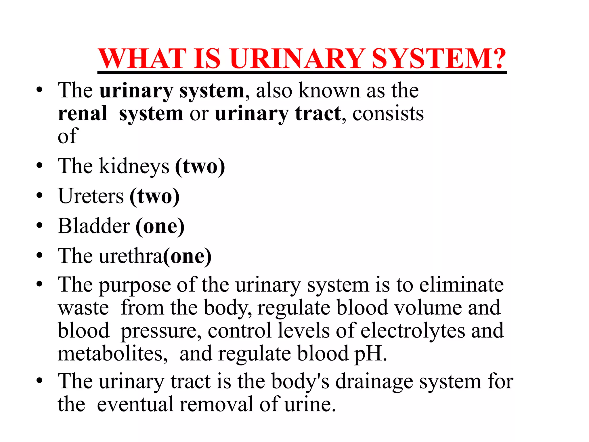
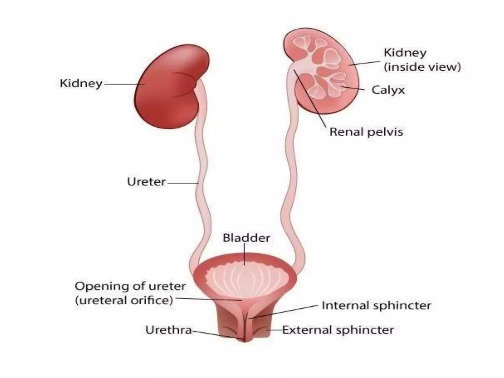
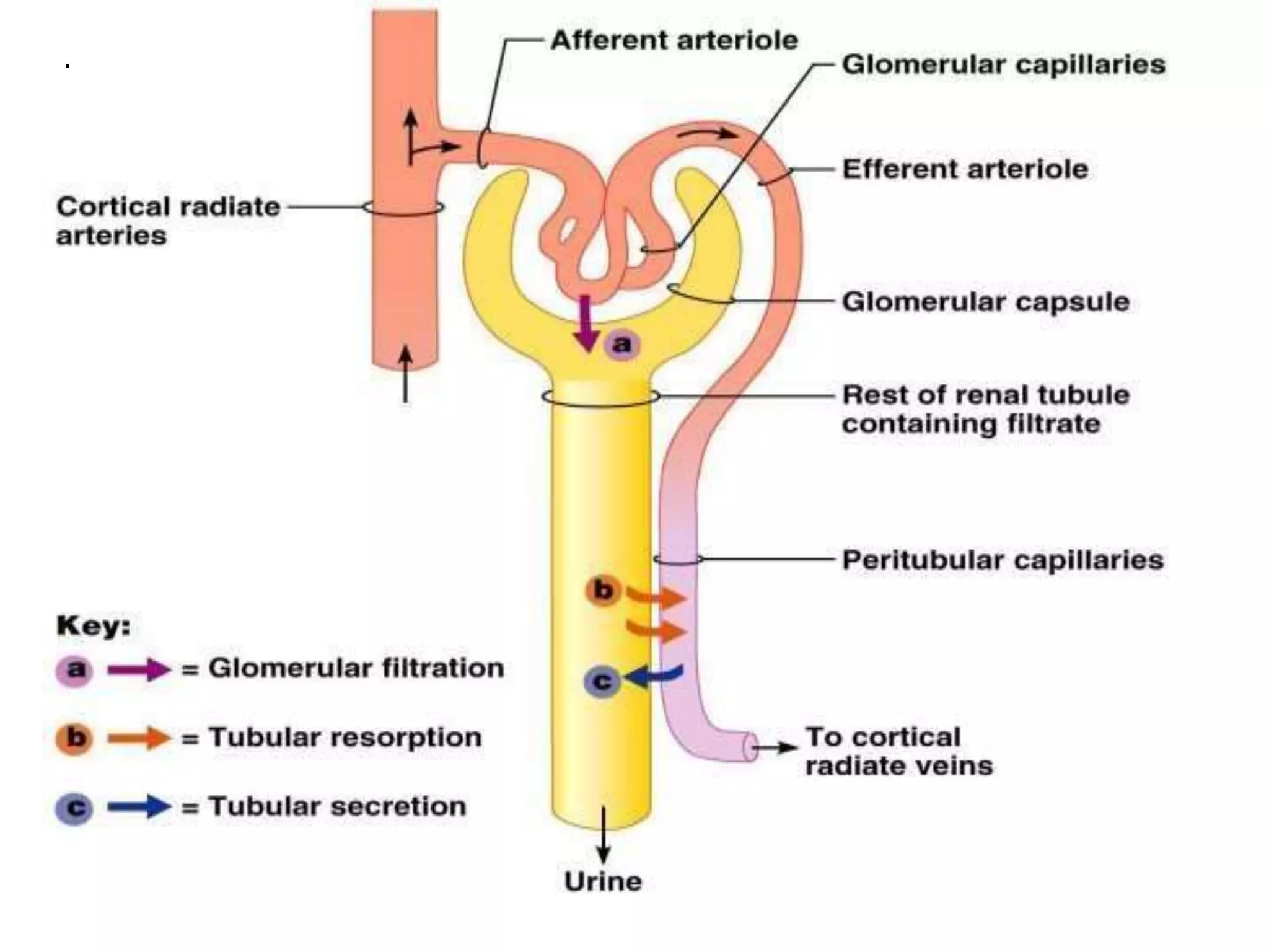
Unit-III: - Urinary System
Represented By,
Mr. Audumbar Mali,
(Asst. Prof.)
Sahyadri College of Pharmacy
Methwade
BP201T. Human Anatomy And
Physiology-II
WHAT IS URINARY SYSTEM?
• The urinary system, also known as the
renal system or urinary tract, consists
of
• The kidneys (two)
• Ureters (two)
• Bladder (one)
• The urethra(one)
• The purpose of the urinary system is to eliminate
waste from the body, regulate blood volume and
blood pressure, control levels of electrolytes and
metabolites, and regulate blood pH.
• The urinary tract is the body's drainage system for
the eventual removal of urine.
There are several functions of the
Urinary System:
• Removal of waste product from the
body (mainly urea and uric acid).
• Regulation of electrolyte balance (e.g.
sodium, potassium and calcium).
• Regulation of acid-base homeostasis.
• Controlling blood volume and
maintaining blood pressure.
THE KIDNEYS
The kidneys are two bean shaped
organs found on the left and right sides of the
body in vertebrates.
• They are located at the back of the abdominal cavity
in the retroperitoneal (i. e. behind the peritoneum)
space.
• The kidneys are located high in the abdominal cavity, one on
each side of the spine, and lie in a retroperitoneal position at
a slightly oblique angle.
• The asymmetry within the abdominal cavity, caused by the
position of the liver, typically results in the right kidney
being slightly lower and smaller than the left, and being
placed slightly more to the middle than the left kidney.
• RELATIONSHIP:-
• Right kidney
• Anteriorly:the duodenum,hepatic flexure of the
colon & right lobe of the liver.
• Posteriorly: Diaphragm,muscles of posterior
abdominal wall.
• Superiorly: the right adrenal gland.
• Left kidney
• Anteriorly: the spleen & splenic vessels, jejunum
splenic flexure of the colon,pancreas & stomach.
• Posteriorly: Diaphragm,muscles of posterior
abdominal wall.
• Superiorly: the left adrenal gland.
STRUCTURE:-
• LENGTH:11 cm
• WIDTH:6 cm
• THICK:3 cm
• WEIGHT: 150 gm.
• The left kidney is approximately at the vertebral
level T12 to L3 and the right is slightly lower.
• The upper parts of the kidneys are partially
protected by the 11th and 12th ribs.
• The kidney is a bean-shaped structure with a
convex and a concave border.
• the kidney is divided into two major structures: the
outer renal cortex and the inner renal medulla.
• The area on the concave border is
the renal hilum, where the renal artery
enters the kidney and the renal vein and
ureter leave.
• The kidney is surrounded by tough
fibrous tissue, the renal capsule.
• the kidney is divided into two major
structures: the outer renal cortex and
the inner renal medulla.
• these structures take the shape of 8 to 18
cone-shaped renal lobes, each
containing renal cortex surrounding a
portion of medulla called a renal
pyramid.
• Between the renal pyramids are
projections of cortex called renal
columns.
• The nephron is the structural and
functional unit of the kidney, span the
cortex and medulla.
• Each adult kidney contains around
one million nephrons.
• The initial filtering portion of a
nephron is the renal corpuscle
which is located in the cortex.
• This is followed by a renal tubule that
passes from the cortex deep into the
medullary pyramids.
• Part of the renal cortex, a medullary
ray is a collection of renal tubules
that drain into a single collecting
duct.
• each pyramid empties urine into a
minor calyx; minor calyces empty
into major calyces, and major
calyces empty into the renal pelvis.
• This becomes the ureter.
• At the hilum, the ureter and renal
vein exit the kidney and the renal
artery enters.
 Excrete nitrogenous waste from the body
 Urea
 Ammonia
 Creatinine
 Regulate blood volume
 Help regulate electrolyte content of the blood
 Regulate acid-base balance (pH)
 Regulate blood pressure
 Regulates red bloodcell production
Functions of the Kidneys :-
• THE NEPHRON
• The nephron is the microscopic
structural and functional unit of the
kidney.
• It is composed of a renal corpuscle
and a renal tubule.
• The renal corpuscle consists of
of capillaries called a glomerulus
and an Bowman's capsule.
• A healthy adult has 0.8 to 1.5 million nephrons
in each kidney.
• They cleanse the blood and balance the
constituents of the circulation. The afferent
arterioles form a high- pressure capillaries about
200 μm in diameter, the glomerulus.
• After passing through the renal corpuscle, the
capillaries form a second arteriole, the efferent
arteriole.
• In a dissected kidney, it is easy to identify the
cortex; it appears lighter in color compared to
the rest of the kidney.
• All of the renal corpuscles as well as both
the proximal convoluted tubules (PCTs)
and distal convoluted tubules are found
here.
• Some nephrons have a short loop of
Henle that does not dip beyond the
cortex.
URINE FORMATION
• Nephrons take a simple filtrate of the blood
and modify it into urine. Many changes
take place in the different parts of the
nephron before urine is created for disposal.
.
• The principle task of the nephron
population is to balance the plasma to
homeostatic set points and excrete
potential toxins in the urine.
• They do this by accomplishing three
principle functions—filtration,
reabsorption, and secretion.
• They also have additional secondary
functions that exert control in three
areas: blood pressure (via production of
renin), red blood cell production (via the
hormone), and calcium absorption (via
conversion of calcidiol into calcitriol,the
active form of vitamin D).
Renal Corpuscle: -
• The renal corpuscle consists of capillaries
called the glomerulus that is largely
surrounded by Bowman’s (glomerular)
capsule.
• The glomerulus is a high-pressure
capillary bed between afferent and
efferent arterioles.
• As blood passes through the glomerulus, 10
to 20 percent of the plasma filters between
these sieve- like fingers to be captured by
Bowman’s capsule and funneled to the
PCT (proximal convoluted tubules).
Proximal Convoluted Tubule (PCT): -
• Filtered fluid collected by Bowman’s capsule
enters into the PCT.
• It is called convoluted due to its tortuous (i.e.
full of twists and turns) path. Simple cuboidal
cells form this tubule with prominent
microvilli on the luminal surface, forming a
border.
• These microvilli create a large surface area to
maximize the absorption and secretion of
solutes (Na+, Cl–, glucose, etc.), the most
essential function of this portion of the
nephron.
Loop of Henle:
• The descending and ascending portions of the loop
of Henle (sometimes referred to as the nephron
loop) are, just continuations of the same tubule.
• The descending loop of Henle consists of an initial
short, thick portion and long, whereas the
ascending loop consists of an initial short, thin
portion followed by a long, thick portion.
• The descending thick portion consists of simple
cuboidal epithelium similar to that of the PCT.
The descending and ascending thin portions
consists of simple squamous epithelium.
Distal Convoluted Tubule (DCT):
• The DCT, like the PCT, is very tortuous and formed
by simple cuboidal epithelium, but it is shorter than
the PCT.
• These cells are not as active as those in the PCT; thus,
there are fewer microvilli on the apical surface.
• However, these cells must also pump ions against
their concentration gradient.
• Collecting Ducts
• The collecting ducts are continuous with the nephron
but not technically part of it.
• In fact, each duct collects filtrate from several
nephrons for final modification.
• They are lined with simple squamous epithelium with
receptors for ADH (antidiuretic hormone) or
Vasopressin.
Glomerular Filtration Rate (GFR):
• The volume of filtrate formed by both kidneys per
minute is termed the glomerular filtration rate
(GFR).
• The heart pumps about 5 L blood per min under resting
conditions.
• Approximately 20 percent or one liter enters the
kidneys to be filtered.
• On average, this liter results in the production of about
125 mL/min filtrate produced in men (range of 90 to
140 mL/min) and 105 mL/min filtrate produced in
women (range of 80 to 125 mL/min).
URETERS
• The kidneys and ureters are completely
retroperitoneal, and the bladder has a peritoneal
covering only over the dome.
• As urine is formed, it drains into the calyces of
the kidney, which merge to form the funnel-
shaped renal pelvis in the hilum of each kidney.
• The hilum narrows to become the ureter of each
kidney.
• As they approach the bladder, they turn medially
and penetrate the bladder wall obliquely.
• This is important because it creates an one-way
valve that allows urine into the bladder but
prevents reflux of urine from the bladder back
into the ureter.
• Children born lacking this oblique course of
the ureter through the bladder wall are
susceptible to “vesicoureteral reflux,” which
increases their risk of serious UTI.
• Pregnancy also increases the likelihood of
reflux and UTI.
• The ureters are approximately 30 cm long.
• The muscular layer of the ureter consists of
longitudinal and circular smooth muscles
that create the peristaltic contractions to
move the urine into the bladder without the
aid of gravity.
URINARY BLADDER
• The urinary bladder collects urine from both
ureters.
• The bladder lies anterior to the uterus in
females,posterior to the pubic bone and
anterior to the rectum.
• During pregnancy, its capacity is reduced
due to compression by the enlarging uterus,
resulting in increased frequency of urination.
• The bladder is partially
retroperitoneal (outside the
peritoneal cavity)
• Volumes in adults can range from nearly zero
to 500–600 mL.
Structure of the Urinary Bladder andUrethra
Figure23.16b
Trigon
e
Bladder neck
Internal urethral
sphincter
External urethral
sphincter
Urogenital
diaphragm
Urethra
External urethral
orifice
Peritoneum
Ureter Rugae
Detrusor muscle
Ureteric orifices
(b) Female
• The bladder’s strength diminishes with age, but voluntary
contractions of abdominal skeletal muscles can increase
intra-abdominal pressure to promote more forceful bladder
emptying.
• Such voluntary contraction is also used in forceful
defecation and childbirth.
Micturition Reflex:
• Micturition is a less-often used for urination.
• It results from an interplay of involuntary and
voluntary actions by the internal and external
urethral sphincters.
• When bladder volume reaches about 150 mL, an urge
to void is sensed but is easily overridden.
• As the bladder fills, subsequent urges become
harder to ignore.
• Ultimately, voluntary constraint fails with
resulting incontinence, which will occur as
bladder volume approaches 300 to 400 mL.
• The micturition reflex is active in infants but with
maturity, children learn to override the reflex by
asserting external sphincter control, thereby
delaying voiding (drain away).
• Urethra
• The urethra transports urine from the bladder to the
outside of the body for disposal.
• The urethra is the only urologic organ that shows
any significant anatomic difference between males
and females.
• Voiding is regulated by an involuntary autonomic nervous
system-controlled internal urinary sphincter, consisting of
smooth muscle and voluntary skeletal muscle that forms the
external urinary sphincter below it.
• Female Urethra
• The external urethral orifice is embedded in the anterior
vaginal wall inferior to the clitoris, superior to the vaginal
opening and medial to the labia minora.
• Its short length, about 4 cm, is less of a barrier to fecal
bacteria than the longer male urethra and the best
explanation for the greater incidence of UTI in women.
• Male Urethra
• The male urethra passes through the prostate gland immediately
inferior to the bladder before passing below the pubic
symphysis.
• The length of the male urethra varies between men
but averages 20 cm in length.
• Filtration, Reabsorption, Secretion: The Three
Steps of Urine Formation
• The kidneys filter unwanted substances from the
blood and produce urine to excrete them.
• There are three main steps of urine formation:
glomerular filtration, reabsorption, and secretion.
• These processes ensure that only waste and
excess water are removed from the body.
• 1. The Glomerulus Filters Water and Other
Substances from the Bloodstream.
• Each kidney contains over 1 million tiny structures
called nephrons. Each nephron has a glomerulus, the
site of blood filtration.
• The glomerulus is a network of capillaries surrounded by a cuplike
structure, the glomerular capsule (or Bowman’s capsule). As blood
flows through the glomerulus, blood pressure pushes water and
solutes from the capillaries into the capsule through a filtration
membrane.
• This glomerular filtration begins the urine formation process.
2. The Filtration Membrane Keeps Blood Cells and Large
Proteins in the Bloodstream.
• Inside the glomerulus, blood pressure pushes fluid from capillaries
into the glomerular capsule through a specialized layer of cells.
• This layer, the filtration membrane, allows water and small solutes to
pass but blocks blood cells and large proteins. Those components
remain in the bloodstream.
• The filtrate (the fluid that has passed through the membrane) flows
from the glomerular capsule further into the nephron.
• 3. Reabsorption Moves Nutrients and Water
Back into the Bloodstream
• The glomerulus filters water and small solutes out
of the bloodstream.
• The resulting filtrate contains waste, but also other
substances the body needs: essential ions,
glucose, amino acids, and smaller proteins. When
the filtrate exits the glomerulus, it flows into a duct
in the nephron called the renal tubule.
• As it moves, the needed substances and some
water are reabsorbed through the tube wall into
adjacent capillaries.
• This reabsorption of vital nutrients from the
filtrate is the second step in urine creation.
• 4. Waste Ions and Hydrogen Ions Secreted from
the Blood Complete the Formation of Urine
• The filtrate absorbed in the glomerulus flows
through the renal tubule, where nutrients and water
are reabsorbed into capillaries.
• At the same time, waste ions and hydrogen ions
pass from the capillaries into the renal tubule.
• This process is called secretion.
• The secreted ions combine with the remaining
filtrate and become urine.
• The urine flows out of the nephron tubule into a
collecting duct. It passes out of the kidney through
the renal pelvis, into the ureter, and down to the
bladder.
Physical Characteristics of Urine
• The urinary system’s ability to filter the blood resides in
about 2 to 3 million bunch of specialized capillaries—the
glomeruli—distributed more or less equally between the two
kidneys.
• The glomeruli create about 200 liters of this filtrate every
day, yet you excrete less than two liters of waste you call
urine.
• Some of the characteristics such as color and odor are
rough descriptors of your state of hydration.
• For example, if you exercise or work outside, and sweat a
great deal, your urine will turn darker and produce a slight
odor, even if you drink plenty of water.
CHARACTERISTIC NORMAL VALUES
COLOR Pale yellow to deep amber
ODOR Odorless
VOLUME 750–2000 mL/24 hour
PH 4.5–8.0
SPECIFIC GRAVITY 1.003–1.032
VOLUME
CONDITION VOLUME CAUSES
NORMAL 1–2 L/day
POLYURIA >2.5 L/day Diabetes mellitus; diabetes insipidus; excess
caffeine or alcohol; kidney disease;
certain drugs, such as diuretics; sickle cell
anemia; excessive water intake.
OLIGURIA (the
production of
abnormally small
amounts of urine.)
300–500
mL/day
Dehydration; blood loss; diarrhea; cardiogenic
shock; kidney disease; enlarged
Prostate.
ANURIA (failure of
the kidneys to
produce urine.)
<50mL/
day
Kidney failure; obstruction, such as kidney stone
or tumor; enlarged prostate.
• Specific gravity is a measure of the quantity of
solutes per unit volume of a solution.
• Urine will always have a specific gravity
greater than pure water (water = 1.0) due to
the presence of solutes.
• Endocrine Regulation of Kidney Function:
• Several hormones have specific, important
roles in regulating kidney function.
• They act to stimulate or inhibit blood flow.
• Renin–Angiotensin–Aldosterone
• Antidiuretic Hormone (ADH)
Regulation of Fluid Volume and Composition : -
• The major hormones influencing total body water
are ADH, aldosterone.
• Blood volume is important in maintaining
sufficient blood pressure.
• Diuretics and Fluid Volume
• A diuretic is a compound that increases urine
volume.
• Regulation of Extracellular Na+
• Sodium has a very strong osmotic effect and
attracts water.
• It plays a larger role in the osmolarity of the
plasma than any other circulating component of
the blood.
• If there is too much Na+ present, either due to poor
control or excess dietary consumption, a series of
metabolic problems occurs.
• There is an increase in total volume of water, which
leads to hypertension (high blood pressure).
• Over a long period, this increases the risk of
serious complications such as heart attacks,strokes,
and aneurysms (i. e. excessive localized swelling of
wall of an artery).
• It can also contribute to system-wide edema
(swelling).
• Mechanisms for regulating Na+ concentration
include the renin–angiotensin–aldosterone system
and ADH.
Disorders of the Urinary System: -
Glomerulonephritis: Glomerulonephritis is inflammation of the
tiny filters in your kidneys (glomeruli). Glomeruli remove excess
fluid, electrolytes and waste from your bloodstream and pass them
into your urine. Glomerulonephritis can come on suddenly (acute) or
gradually (chronic).
Symptoms: Hypertension
Polycystic Kidney:
Polycystic kidney disease (PKD) is an inherited disorder in which
clusters of cysts develop primarily within your kidneys, causing
your kidneys to enlarge and lose function over time. Cysts are
noncancerous round sacs containing fluid. The cysts vary in size,
and they can grow very large.
Pyelonephritis is inflammation of the kidney, typically due
to a bacterial infection. Symptoms most often include fever
and flank tenderness. Other symptoms may include nausea,
burning with urination, and frequent urination.
Complications may include pus around the kidney, sepsis,
or kidney failure.
Renal Calculi – kidney stones: Kidney stones are hard
deposits of minerals and acid salts that stick together in
concentrated urine. They can be painful when passing
through the urinary tract, but usually don't cause
permanent damage. The most common symptom is severe
pain, usually in the side of the abdomen, that's often
associated with nausea.
Treatment includes pain relievers and drinking lots of
water to help pass the stone. Medical procedures may be
required to remove or break up larger stones.
Kidney Failure:
Kidney failure happens when damage results
in loss of normal kidney function. There are 2
types of kidney failure—acute and chronic.
Acute kidney failure starts abruptly(i.e.
suddenly and unexpectedly). It can sometimes
be reversed.
Chronic kidney failure progresses slowly over
at least 3 months. It can lead to permanent
kidney failure. This is also called end stage
renal disease (ESRD).
What causes kidney failure?
Conditions that may lead to acute or chronic kidney failure
may include:
Acute kidney failure
Rhabdomyolysis (i.e. the destruction of striated muscle cells),
kidney damage that can happen from muscle breakdown due to
severe dehydration, infection, or other causes.
Decreased blood flow to the kidneys from blood loss or shock
A blockage along the urinary tract, Hemolytic uremic
syndrome, blockage of the small vessels in the kidney, often
caused by an E. Coli infection. Infection or certain medicines
that may be toxic to the kidneys. Glomerulonephritis,
inflammation of the tiny units within the kidney where blood
is cleaned. It impairs the kidney’s ability to filter urine. Any
condition that impairs the flow of oxygen and blood to the
kidneys, such as a heart attack
Chronic kidney failure : -
Diabetic nephropathy, kidney damage due to high blood
sugar, High blood pressure,
Lupus, i.e. an immune system disease,
Urinary track blockage, Alport syndrome, a genetic
disorder i. e. Nephrotic syndrome, a condition that leads to
too much protein in the urine,
Polycystic kidney disease i.e. a genetic disorder that
causes fluid-filled cysts in the kidneys,
Cystinosis i.e. a genetic problem in which the amino acid
builds up within the cells in the kidneys,
Interstitial nephritis or pyelonephritis i.e. inflammation of
the small structures in the kidney.
Reference: -
1. A Textbook of Human Anatomy and
Physiology-II, By, Dr. S. B. Bhise and Dr. A. V.
Yadav. Nirali Prakashan, Page No.:- 5.1-5.14.
2. https://www.slideshare.net/jaganlogan/general-
anatomy-of-urinary-system-ppt
3. https://www.slideshare.net/visanth/urinary-
system-anatomy-ppt
4. https://www.slideshare.net/AIDAROSLI91/urin
ary-system-36067478.
Thank
you.
THANK YOU

Urinary System.pptx

  • 1.
    Unit-III: - UrinarySystem Represented By, Mr. Audumbar Mali, (Asst. Prof.) Sahyadri College of Pharmacy Methwade BP201T. Human Anatomy And Physiology-II
  • 2.
    WHAT IS URINARYSYSTEM? • The urinary system, also known as the renal system or urinary tract, consists of • The kidneys (two) • Ureters (two) • Bladder (one) • The urethra(one) • The purpose of the urinary system is to eliminate waste from the body, regulate blood volume and blood pressure, control levels of electrolytes and metabolites, and regulate blood pH. • The urinary tract is the body's drainage system for the eventual removal of urine.
  • 4.
    There are severalfunctions of the Urinary System: • Removal of waste product from the body (mainly urea and uric acid). • Regulation of electrolyte balance (e.g. sodium, potassium and calcium). • Regulation of acid-base homeostasis. • Controlling blood volume and maintaining blood pressure.
  • 5.
    THE KIDNEYS The kidneysare two bean shaped organs found on the left and right sides of the body in vertebrates. • They are located at the back of the abdominal cavity in the retroperitoneal (i. e. behind the peritoneum) space. • The kidneys are located high in the abdominal cavity, one on each side of the spine, and lie in a retroperitoneal position at a slightly oblique angle. • The asymmetry within the abdominal cavity, caused by the position of the liver, typically results in the right kidney being slightly lower and smaller than the left, and being placed slightly more to the middle than the left kidney.
  • 6.
    • RELATIONSHIP:- • Rightkidney • Anteriorly:the duodenum,hepatic flexure of the colon & right lobe of the liver. • Posteriorly: Diaphragm,muscles of posterior abdominal wall. • Superiorly: the right adrenal gland. • Left kidney • Anteriorly: the spleen & splenic vessels, jejunum splenic flexure of the colon,pancreas & stomach. • Posteriorly: Diaphragm,muscles of posterior abdominal wall. • Superiorly: the left adrenal gland.
  • 7.
    STRUCTURE:- • LENGTH:11 cm •WIDTH:6 cm • THICK:3 cm • WEIGHT: 150 gm. • The left kidney is approximately at the vertebral level T12 to L3 and the right is slightly lower. • The upper parts of the kidneys are partially protected by the 11th and 12th ribs. • The kidney is a bean-shaped structure with a convex and a concave border. • the kidney is divided into two major structures: the outer renal cortex and the inner renal medulla.
  • 9.
    • The areaon the concave border is the renal hilum, where the renal artery enters the kidney and the renal vein and ureter leave. • The kidney is surrounded by tough fibrous tissue, the renal capsule. • the kidney is divided into two major structures: the outer renal cortex and the inner renal medulla. • these structures take the shape of 8 to 18 cone-shaped renal lobes, each containing renal cortex surrounding a portion of medulla called a renal pyramid.
  • 10.
    • Between therenal pyramids are projections of cortex called renal columns. • The nephron is the structural and functional unit of the kidney, span the cortex and medulla. • Each adult kidney contains around one million nephrons. • The initial filtering portion of a nephron is the renal corpuscle which is located in the cortex. • This is followed by a renal tubule that passes from the cortex deep into the medullary pyramids.
  • 11.
    • Part ofthe renal cortex, a medullary ray is a collection of renal tubules that drain into a single collecting duct. • each pyramid empties urine into a minor calyx; minor calyces empty into major calyces, and major calyces empty into the renal pelvis. • This becomes the ureter. • At the hilum, the ureter and renal vein exit the kidney and the renal artery enters.
  • 12.
     Excrete nitrogenouswaste from the body  Urea  Ammonia  Creatinine  Regulate blood volume  Help regulate electrolyte content of the blood  Regulate acid-base balance (pH)  Regulate blood pressure  Regulates red bloodcell production Functions of the Kidneys :-
  • 13.
    • THE NEPHRON •The nephron is the microscopic structural and functional unit of the kidney. • It is composed of a renal corpuscle and a renal tubule.
  • 14.
    • The renalcorpuscle consists of of capillaries called a glomerulus and an Bowman's capsule. • A healthy adult has 0.8 to 1.5 million nephrons in each kidney. • They cleanse the blood and balance the constituents of the circulation. The afferent arterioles form a high- pressure capillaries about 200 μm in diameter, the glomerulus. • After passing through the renal corpuscle, the capillaries form a second arteriole, the efferent arteriole.
  • 17.
    • In adissected kidney, it is easy to identify the cortex; it appears lighter in color compared to the rest of the kidney. • All of the renal corpuscles as well as both the proximal convoluted tubules (PCTs) and distal convoluted tubules are found here. • Some nephrons have a short loop of Henle that does not dip beyond the cortex. URINE FORMATION • Nephrons take a simple filtrate of the blood and modify it into urine. Many changes take place in the different parts of the nephron before urine is created for disposal.
  • 18.
  • 19.
    • The principletask of the nephron population is to balance the plasma to homeostatic set points and excrete potential toxins in the urine. • They do this by accomplishing three principle functions—filtration, reabsorption, and secretion. • They also have additional secondary functions that exert control in three areas: blood pressure (via production of renin), red blood cell production (via the hormone), and calcium absorption (via conversion of calcidiol into calcitriol,the active form of vitamin D).
  • 20.
    Renal Corpuscle: - •The renal corpuscle consists of capillaries called the glomerulus that is largely surrounded by Bowman’s (glomerular) capsule. • The glomerulus is a high-pressure capillary bed between afferent and efferent arterioles. • As blood passes through the glomerulus, 10 to 20 percent of the plasma filters between these sieve- like fingers to be captured by Bowman’s capsule and funneled to the PCT (proximal convoluted tubules).
  • 21.
    Proximal Convoluted Tubule(PCT): - • Filtered fluid collected by Bowman’s capsule enters into the PCT. • It is called convoluted due to its tortuous (i.e. full of twists and turns) path. Simple cuboidal cells form this tubule with prominent microvilli on the luminal surface, forming a border. • These microvilli create a large surface area to maximize the absorption and secretion of solutes (Na+, Cl–, glucose, etc.), the most essential function of this portion of the nephron.
  • 22.
    Loop of Henle: •The descending and ascending portions of the loop of Henle (sometimes referred to as the nephron loop) are, just continuations of the same tubule. • The descending loop of Henle consists of an initial short, thick portion and long, whereas the ascending loop consists of an initial short, thin portion followed by a long, thick portion. • The descending thick portion consists of simple cuboidal epithelium similar to that of the PCT. The descending and ascending thin portions consists of simple squamous epithelium.
  • 23.
    Distal Convoluted Tubule(DCT): • The DCT, like the PCT, is very tortuous and formed by simple cuboidal epithelium, but it is shorter than the PCT. • These cells are not as active as those in the PCT; thus, there are fewer microvilli on the apical surface. • However, these cells must also pump ions against their concentration gradient. • Collecting Ducts • The collecting ducts are continuous with the nephron but not technically part of it. • In fact, each duct collects filtrate from several nephrons for final modification.
  • 24.
    • They arelined with simple squamous epithelium with receptors for ADH (antidiuretic hormone) or Vasopressin. Glomerular Filtration Rate (GFR): • The volume of filtrate formed by both kidneys per minute is termed the glomerular filtration rate (GFR). • The heart pumps about 5 L blood per min under resting conditions. • Approximately 20 percent or one liter enters the kidneys to be filtered. • On average, this liter results in the production of about 125 mL/min filtrate produced in men (range of 90 to 140 mL/min) and 105 mL/min filtrate produced in women (range of 80 to 125 mL/min).
  • 25.
    URETERS • The kidneysand ureters are completely retroperitoneal, and the bladder has a peritoneal covering only over the dome. • As urine is formed, it drains into the calyces of the kidney, which merge to form the funnel- shaped renal pelvis in the hilum of each kidney. • The hilum narrows to become the ureter of each kidney. • As they approach the bladder, they turn medially and penetrate the bladder wall obliquely. • This is important because it creates an one-way valve that allows urine into the bladder but prevents reflux of urine from the bladder back into the ureter.
  • 26.
    • Children bornlacking this oblique course of the ureter through the bladder wall are susceptible to “vesicoureteral reflux,” which increases their risk of serious UTI. • Pregnancy also increases the likelihood of reflux and UTI. • The ureters are approximately 30 cm long. • The muscular layer of the ureter consists of longitudinal and circular smooth muscles that create the peristaltic contractions to move the urine into the bladder without the aid of gravity.
  • 27.
    URINARY BLADDER • Theurinary bladder collects urine from both ureters. • The bladder lies anterior to the uterus in females,posterior to the pubic bone and anterior to the rectum. • During pregnancy, its capacity is reduced due to compression by the enlarging uterus, resulting in increased frequency of urination. • The bladder is partially retroperitoneal (outside the peritoneal cavity) • Volumes in adults can range from nearly zero to 500–600 mL.
  • 29.
    Structure of theUrinary Bladder andUrethra Figure23.16b Trigon e Bladder neck Internal urethral sphincter External urethral sphincter Urogenital diaphragm Urethra External urethral orifice Peritoneum Ureter Rugae Detrusor muscle Ureteric orifices (b) Female
  • 30.
    • The bladder’sstrength diminishes with age, but voluntary contractions of abdominal skeletal muscles can increase intra-abdominal pressure to promote more forceful bladder emptying. • Such voluntary contraction is also used in forceful defecation and childbirth. Micturition Reflex: • Micturition is a less-often used for urination. • It results from an interplay of involuntary and voluntary actions by the internal and external urethral sphincters. • When bladder volume reaches about 150 mL, an urge to void is sensed but is easily overridden.
  • 31.
    • As thebladder fills, subsequent urges become harder to ignore. • Ultimately, voluntary constraint fails with resulting incontinence, which will occur as bladder volume approaches 300 to 400 mL. • The micturition reflex is active in infants but with maturity, children learn to override the reflex by asserting external sphincter control, thereby delaying voiding (drain away). • Urethra • The urethra transports urine from the bladder to the outside of the body for disposal. • The urethra is the only urologic organ that shows any significant anatomic difference between males and females.
  • 33.
    • Voiding isregulated by an involuntary autonomic nervous system-controlled internal urinary sphincter, consisting of smooth muscle and voluntary skeletal muscle that forms the external urinary sphincter below it. • Female Urethra • The external urethral orifice is embedded in the anterior vaginal wall inferior to the clitoris, superior to the vaginal opening and medial to the labia minora. • Its short length, about 4 cm, is less of a barrier to fecal bacteria than the longer male urethra and the best explanation for the greater incidence of UTI in women. • Male Urethra • The male urethra passes through the prostate gland immediately inferior to the bladder before passing below the pubic symphysis.
  • 34.
    • The lengthof the male urethra varies between men but averages 20 cm in length. • Filtration, Reabsorption, Secretion: The Three Steps of Urine Formation • The kidneys filter unwanted substances from the blood and produce urine to excrete them. • There are three main steps of urine formation: glomerular filtration, reabsorption, and secretion. • These processes ensure that only waste and excess water are removed from the body. • 1. The Glomerulus Filters Water and Other Substances from the Bloodstream. • Each kidney contains over 1 million tiny structures called nephrons. Each nephron has a glomerulus, the site of blood filtration.
  • 35.
    • The glomerulusis a network of capillaries surrounded by a cuplike structure, the glomerular capsule (or Bowman’s capsule). As blood flows through the glomerulus, blood pressure pushes water and solutes from the capillaries into the capsule through a filtration membrane. • This glomerular filtration begins the urine formation process. 2. The Filtration Membrane Keeps Blood Cells and Large Proteins in the Bloodstream. • Inside the glomerulus, blood pressure pushes fluid from capillaries into the glomerular capsule through a specialized layer of cells. • This layer, the filtration membrane, allows water and small solutes to pass but blocks blood cells and large proteins. Those components remain in the bloodstream. • The filtrate (the fluid that has passed through the membrane) flows from the glomerular capsule further into the nephron.
  • 37.
    • 3. ReabsorptionMoves Nutrients and Water Back into the Bloodstream • The glomerulus filters water and small solutes out of the bloodstream. • The resulting filtrate contains waste, but also other substances the body needs: essential ions, glucose, amino acids, and smaller proteins. When the filtrate exits the glomerulus, it flows into a duct in the nephron called the renal tubule. • As it moves, the needed substances and some water are reabsorbed through the tube wall into adjacent capillaries. • This reabsorption of vital nutrients from the filtrate is the second step in urine creation.
  • 38.
    • 4. WasteIons and Hydrogen Ions Secreted from the Blood Complete the Formation of Urine • The filtrate absorbed in the glomerulus flows through the renal tubule, where nutrients and water are reabsorbed into capillaries. • At the same time, waste ions and hydrogen ions pass from the capillaries into the renal tubule. • This process is called secretion. • The secreted ions combine with the remaining filtrate and become urine. • The urine flows out of the nephron tubule into a collecting duct. It passes out of the kidney through the renal pelvis, into the ureter, and down to the bladder.
  • 39.
    Physical Characteristics ofUrine • The urinary system’s ability to filter the blood resides in about 2 to 3 million bunch of specialized capillaries—the glomeruli—distributed more or less equally between the two kidneys. • The glomeruli create about 200 liters of this filtrate every day, yet you excrete less than two liters of waste you call urine. • Some of the characteristics such as color and odor are rough descriptors of your state of hydration. • For example, if you exercise or work outside, and sweat a great deal, your urine will turn darker and produce a slight odor, even if you drink plenty of water.
  • 40.
    CHARACTERISTIC NORMAL VALUES COLORPale yellow to deep amber ODOR Odorless VOLUME 750–2000 mL/24 hour PH 4.5–8.0 SPECIFIC GRAVITY 1.003–1.032
  • 41.
    VOLUME CONDITION VOLUME CAUSES NORMAL1–2 L/day POLYURIA >2.5 L/day Diabetes mellitus; diabetes insipidus; excess caffeine or alcohol; kidney disease; certain drugs, such as diuretics; sickle cell anemia; excessive water intake. OLIGURIA (the production of abnormally small amounts of urine.) 300–500 mL/day Dehydration; blood loss; diarrhea; cardiogenic shock; kidney disease; enlarged Prostate. ANURIA (failure of the kidneys to produce urine.) <50mL/ day Kidney failure; obstruction, such as kidney stone or tumor; enlarged prostate.
  • 42.
    • Specific gravityis a measure of the quantity of solutes per unit volume of a solution. • Urine will always have a specific gravity greater than pure water (water = 1.0) due to the presence of solutes. • Endocrine Regulation of Kidney Function: • Several hormones have specific, important roles in regulating kidney function. • They act to stimulate or inhibit blood flow. • Renin–Angiotensin–Aldosterone • Antidiuretic Hormone (ADH)
  • 43.
    Regulation of FluidVolume and Composition : - • The major hormones influencing total body water are ADH, aldosterone. • Blood volume is important in maintaining sufficient blood pressure. • Diuretics and Fluid Volume • A diuretic is a compound that increases urine volume. • Regulation of Extracellular Na+ • Sodium has a very strong osmotic effect and attracts water. • It plays a larger role in the osmolarity of the plasma than any other circulating component of the blood.
  • 44.
    • If thereis too much Na+ present, either due to poor control or excess dietary consumption, a series of metabolic problems occurs. • There is an increase in total volume of water, which leads to hypertension (high blood pressure). • Over a long period, this increases the risk of serious complications such as heart attacks,strokes, and aneurysms (i. e. excessive localized swelling of wall of an artery). • It can also contribute to system-wide edema (swelling). • Mechanisms for regulating Na+ concentration include the renin–angiotensin–aldosterone system and ADH.
  • 45.
    Disorders of theUrinary System: - Glomerulonephritis: Glomerulonephritis is inflammation of the tiny filters in your kidneys (glomeruli). Glomeruli remove excess fluid, electrolytes and waste from your bloodstream and pass them into your urine. Glomerulonephritis can come on suddenly (acute) or gradually (chronic). Symptoms: Hypertension Polycystic Kidney: Polycystic kidney disease (PKD) is an inherited disorder in which clusters of cysts develop primarily within your kidneys, causing your kidneys to enlarge and lose function over time. Cysts are noncancerous round sacs containing fluid. The cysts vary in size, and they can grow very large.
  • 46.
    Pyelonephritis is inflammationof the kidney, typically due to a bacterial infection. Symptoms most often include fever and flank tenderness. Other symptoms may include nausea, burning with urination, and frequent urination. Complications may include pus around the kidney, sepsis, or kidney failure. Renal Calculi – kidney stones: Kidney stones are hard deposits of minerals and acid salts that stick together in concentrated urine. They can be painful when passing through the urinary tract, but usually don't cause permanent damage. The most common symptom is severe pain, usually in the side of the abdomen, that's often associated with nausea. Treatment includes pain relievers and drinking lots of water to help pass the stone. Medical procedures may be required to remove or break up larger stones.
  • 47.
    Kidney Failure: Kidney failurehappens when damage results in loss of normal kidney function. There are 2 types of kidney failure—acute and chronic. Acute kidney failure starts abruptly(i.e. suddenly and unexpectedly). It can sometimes be reversed. Chronic kidney failure progresses slowly over at least 3 months. It can lead to permanent kidney failure. This is also called end stage renal disease (ESRD).
  • 48.
    What causes kidneyfailure? Conditions that may lead to acute or chronic kidney failure may include: Acute kidney failure Rhabdomyolysis (i.e. the destruction of striated muscle cells), kidney damage that can happen from muscle breakdown due to severe dehydration, infection, or other causes. Decreased blood flow to the kidneys from blood loss or shock A blockage along the urinary tract, Hemolytic uremic syndrome, blockage of the small vessels in the kidney, often caused by an E. Coli infection. Infection or certain medicines that may be toxic to the kidneys. Glomerulonephritis, inflammation of the tiny units within the kidney where blood is cleaned. It impairs the kidney’s ability to filter urine. Any condition that impairs the flow of oxygen and blood to the kidneys, such as a heart attack
  • 49.
    Chronic kidney failure: - Diabetic nephropathy, kidney damage due to high blood sugar, High blood pressure, Lupus, i.e. an immune system disease, Urinary track blockage, Alport syndrome, a genetic disorder i. e. Nephrotic syndrome, a condition that leads to too much protein in the urine, Polycystic kidney disease i.e. a genetic disorder that causes fluid-filled cysts in the kidneys, Cystinosis i.e. a genetic problem in which the amino acid builds up within the cells in the kidneys, Interstitial nephritis or pyelonephritis i.e. inflammation of the small structures in the kidney.
  • 50.
    Reference: - 1. ATextbook of Human Anatomy and Physiology-II, By, Dr. S. B. Bhise and Dr. A. V. Yadav. Nirali Prakashan, Page No.:- 5.1-5.14. 2. https://www.slideshare.net/jaganlogan/general- anatomy-of-urinary-system-ppt 3. https://www.slideshare.net/visanth/urinary- system-anatomy-ppt 4. https://www.slideshare.net/AIDAROSLI91/urin ary-system-36067478.
  • 51.