
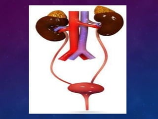
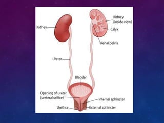


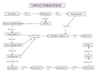
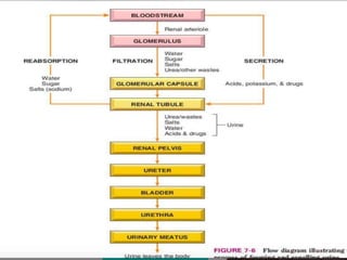


The urinary system consists of the kidneys, ureters, bladder, and urethra. The kidneys filter waste from the blood to produce urine, regulate electrolytes and acid-base balance, and control blood pressure. The nephron is the functional unit of the kidney that filters blood to form urine via glomerular filtration, reabsorption of nutrients, and secretion of wastes. Urine is transported from the kidneys to the bladder via the ureters for storage and later excretion through the urethra.


