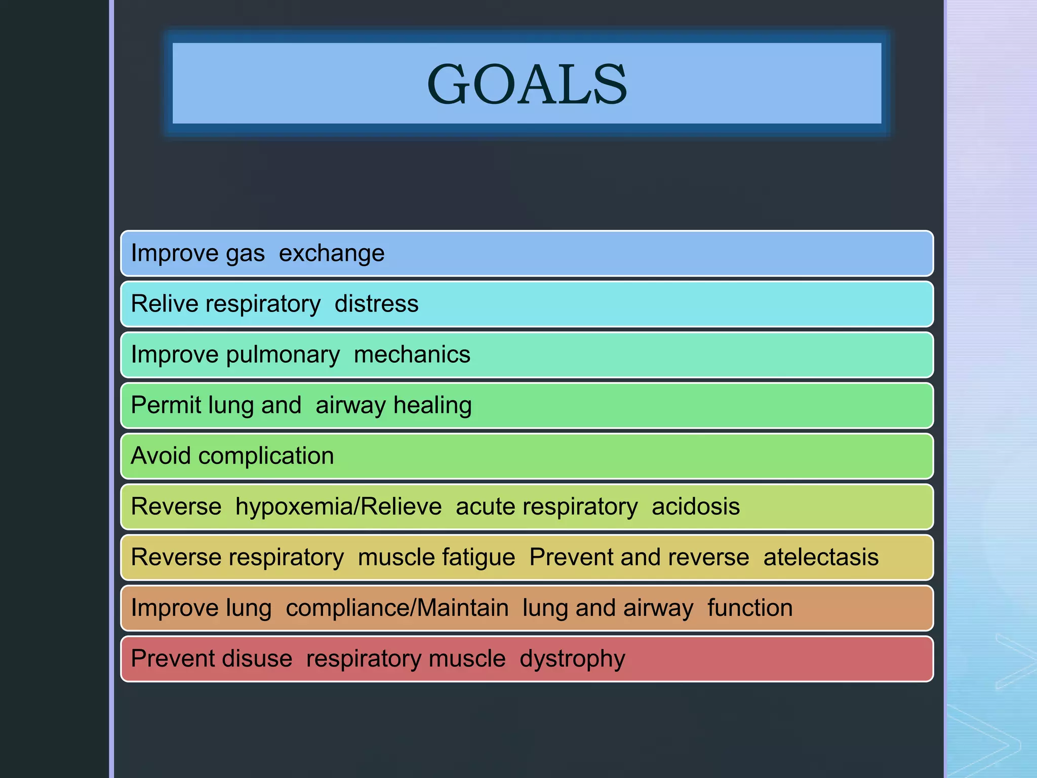
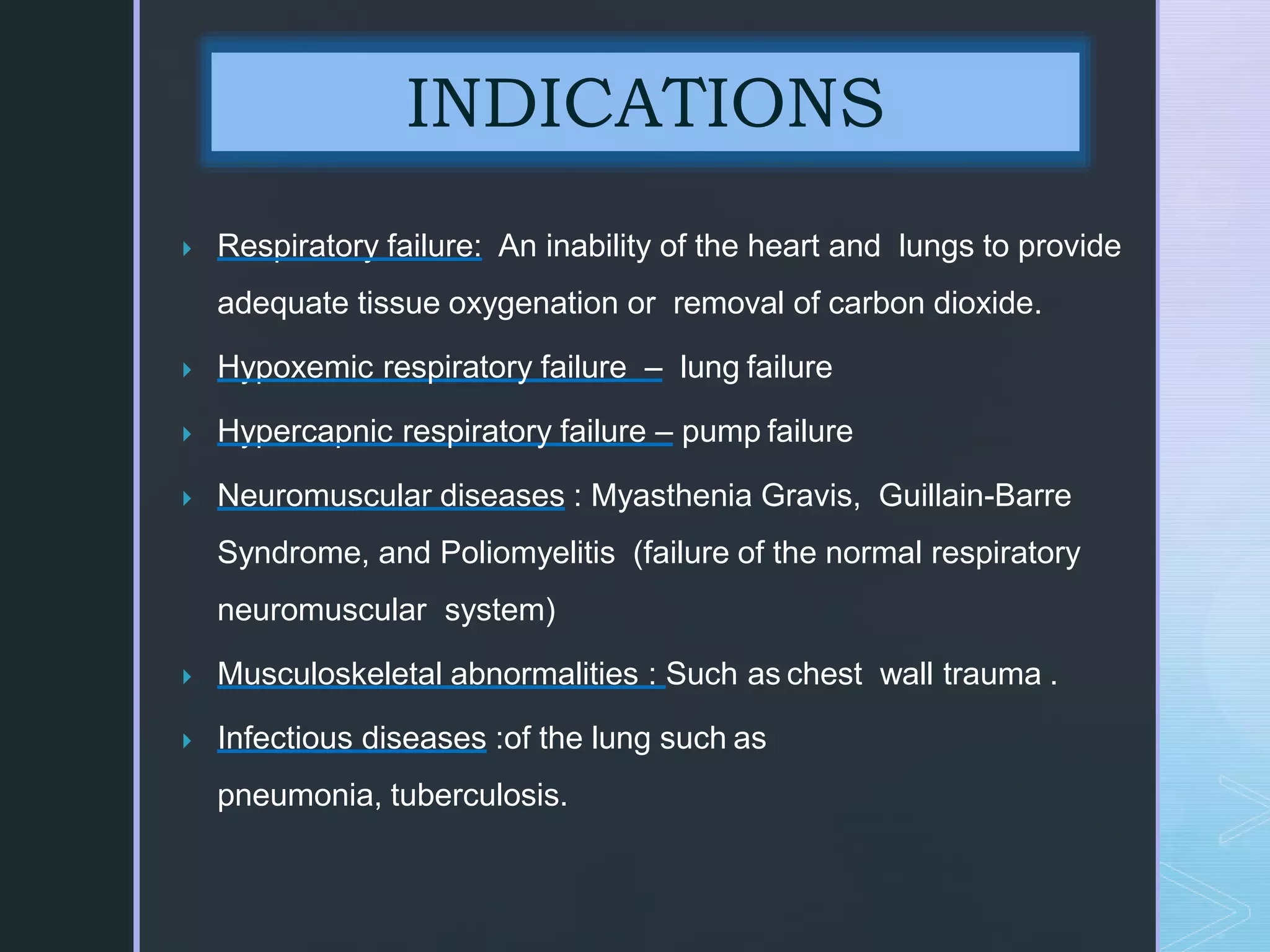
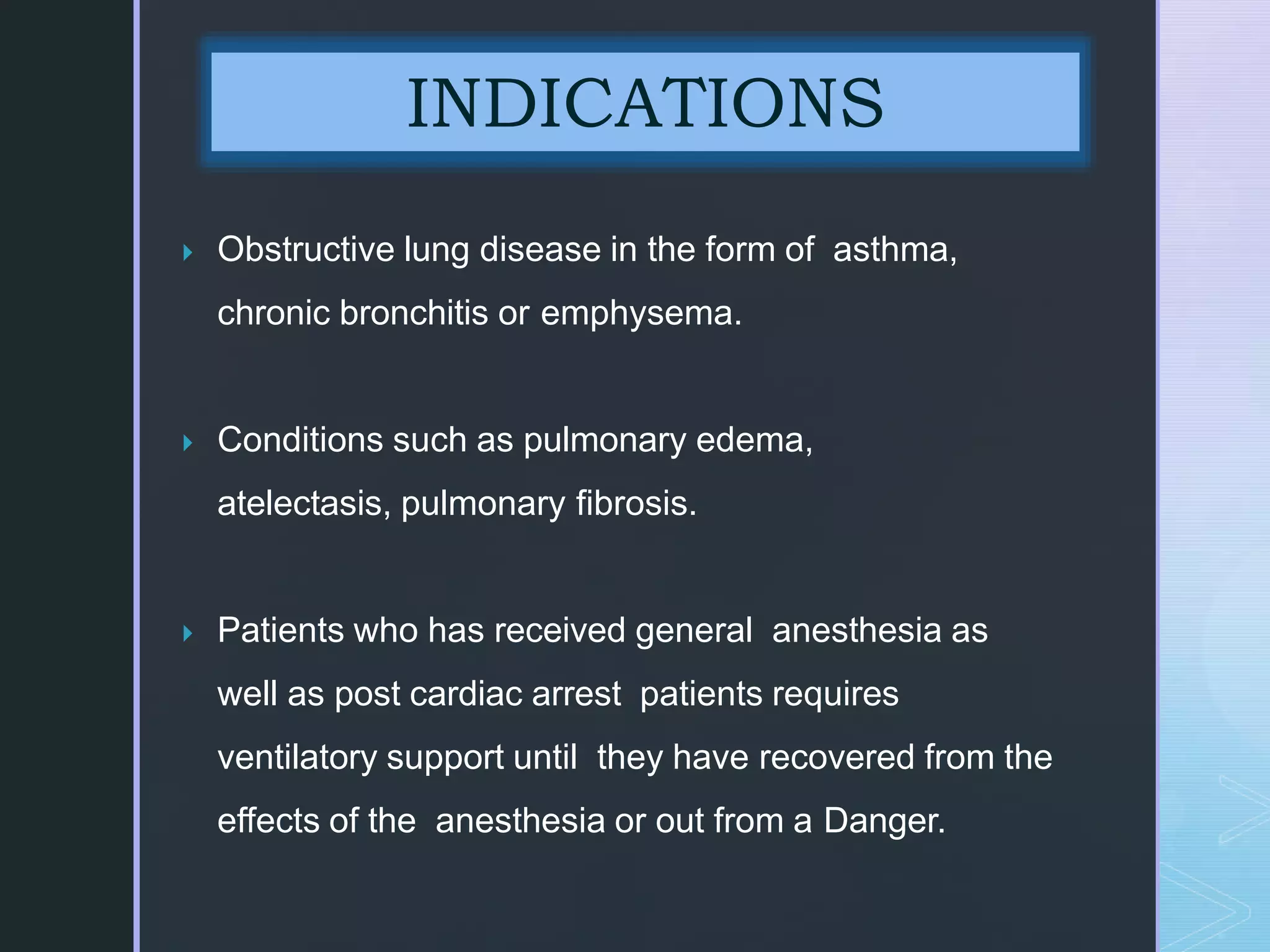
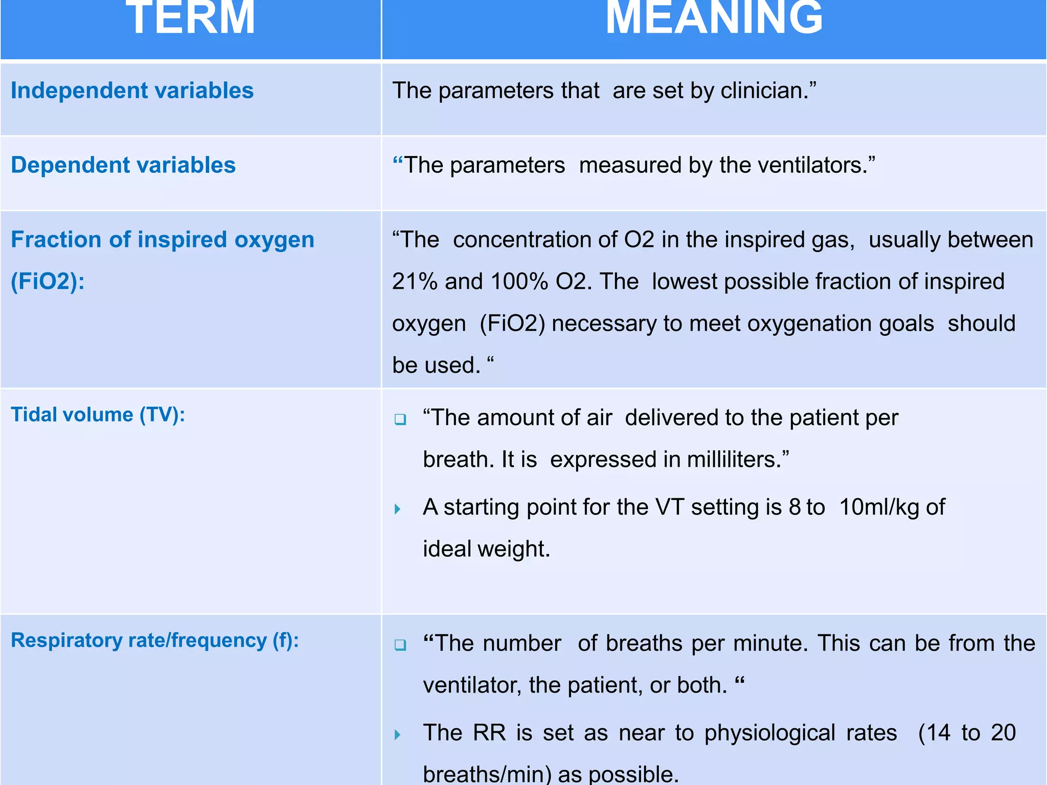
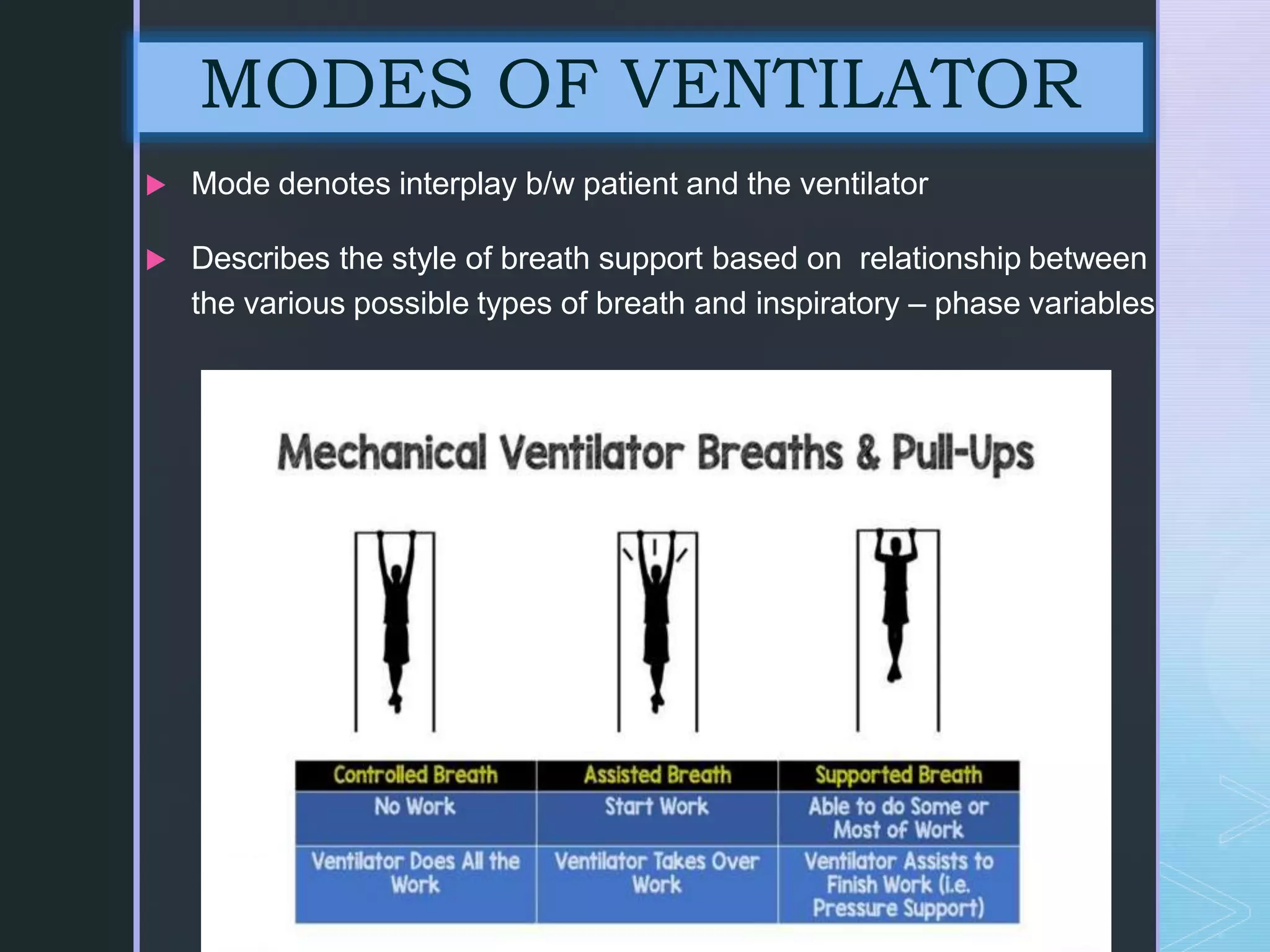
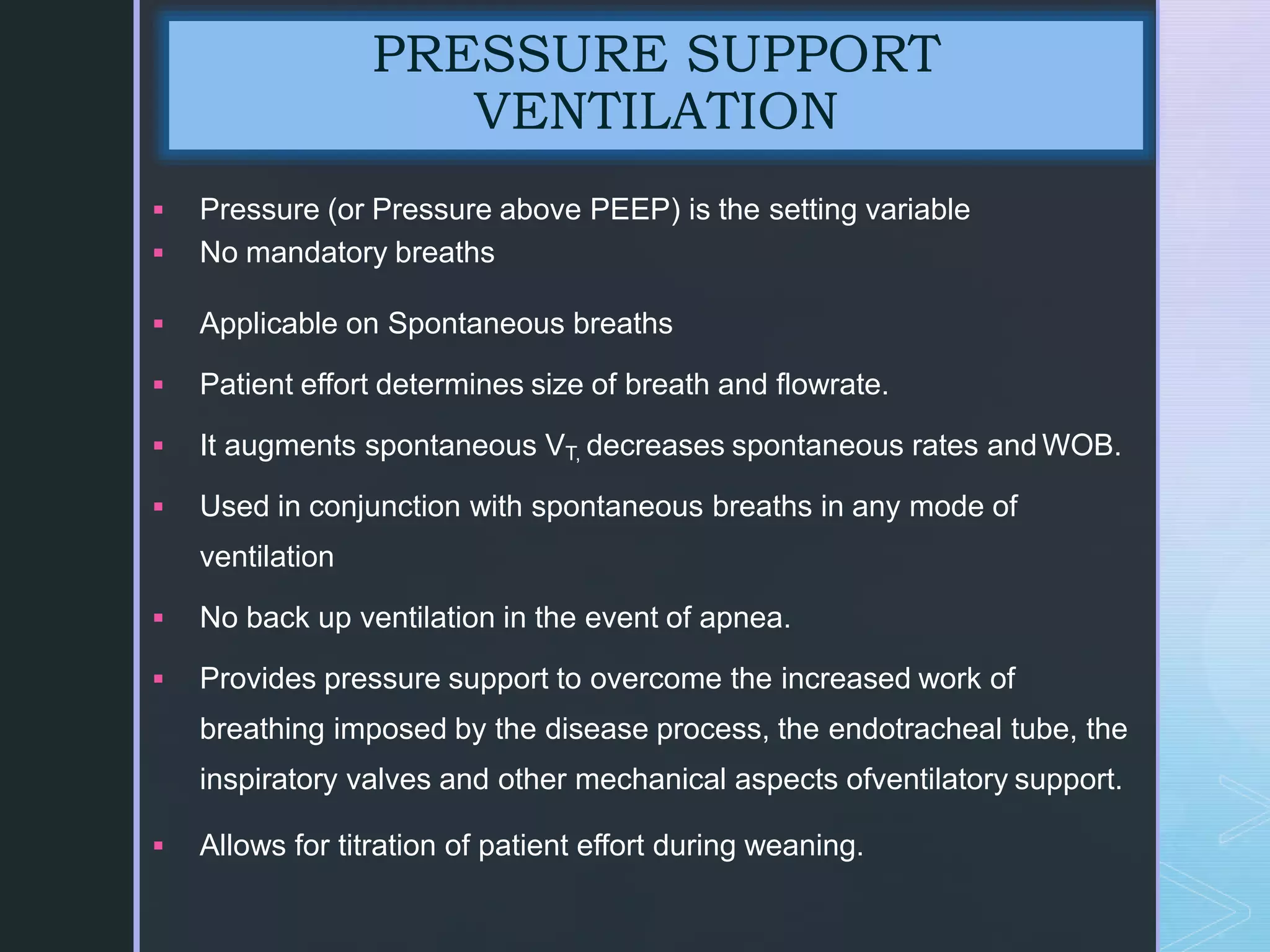
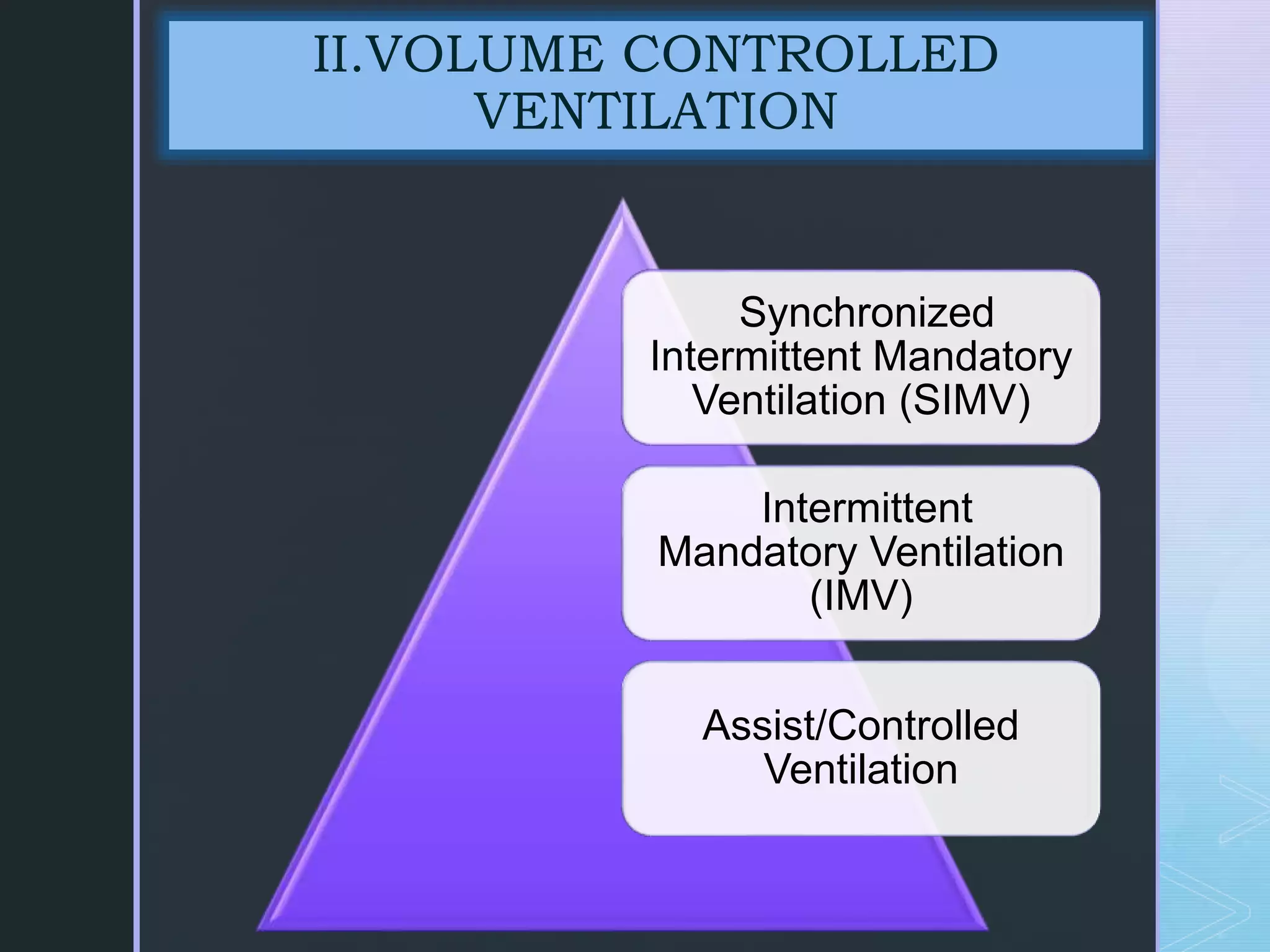
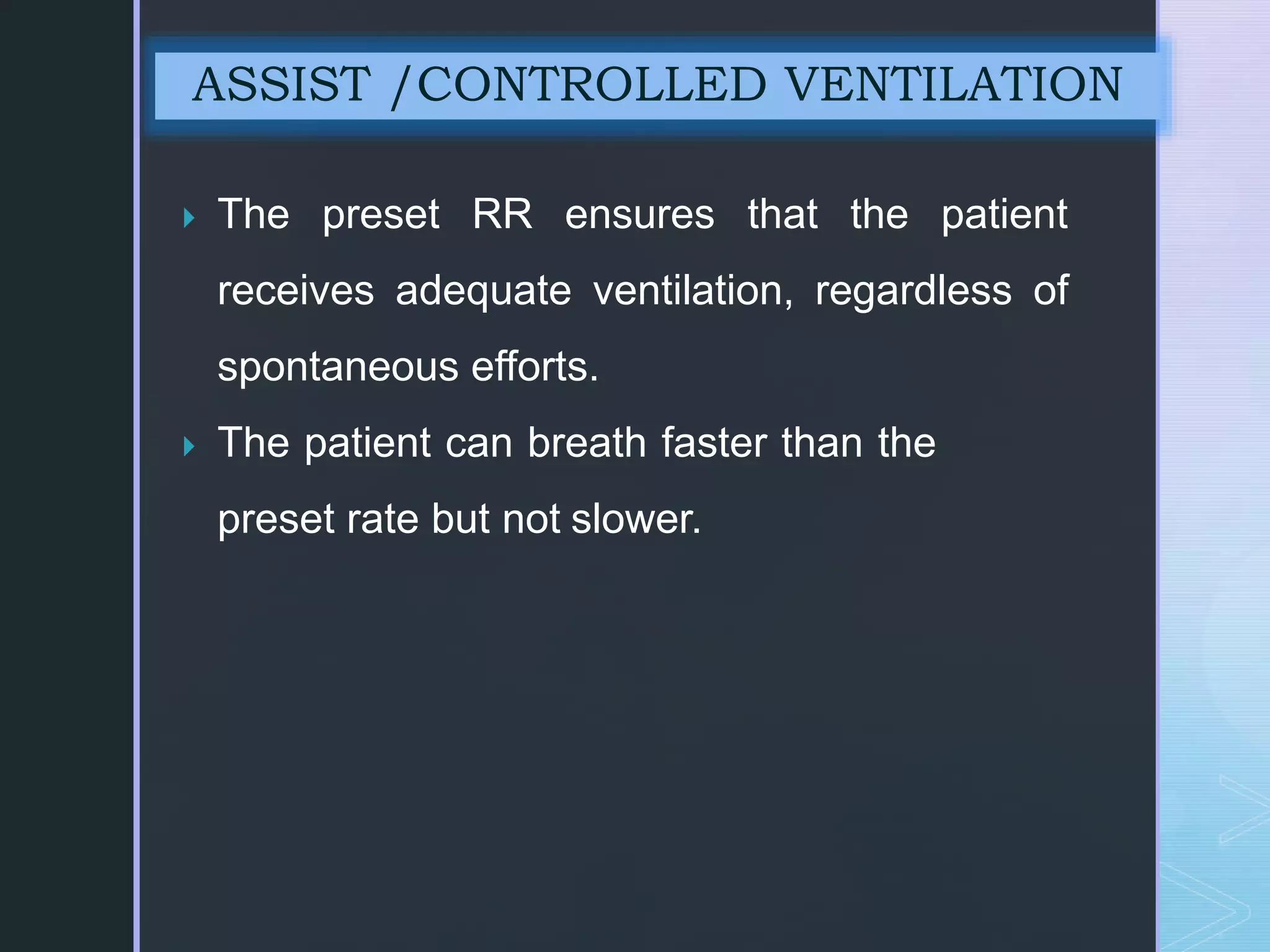
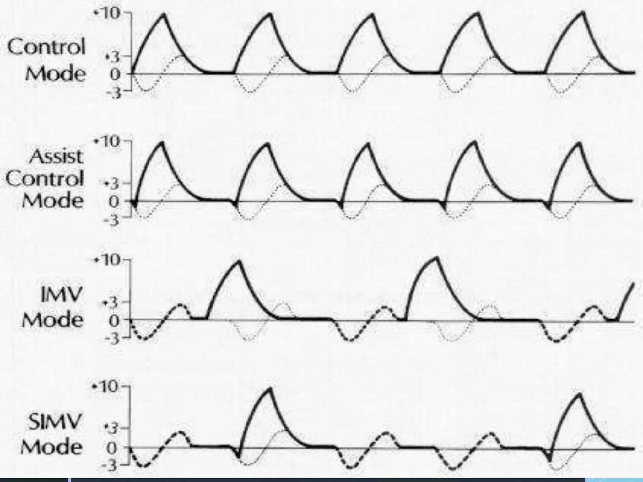
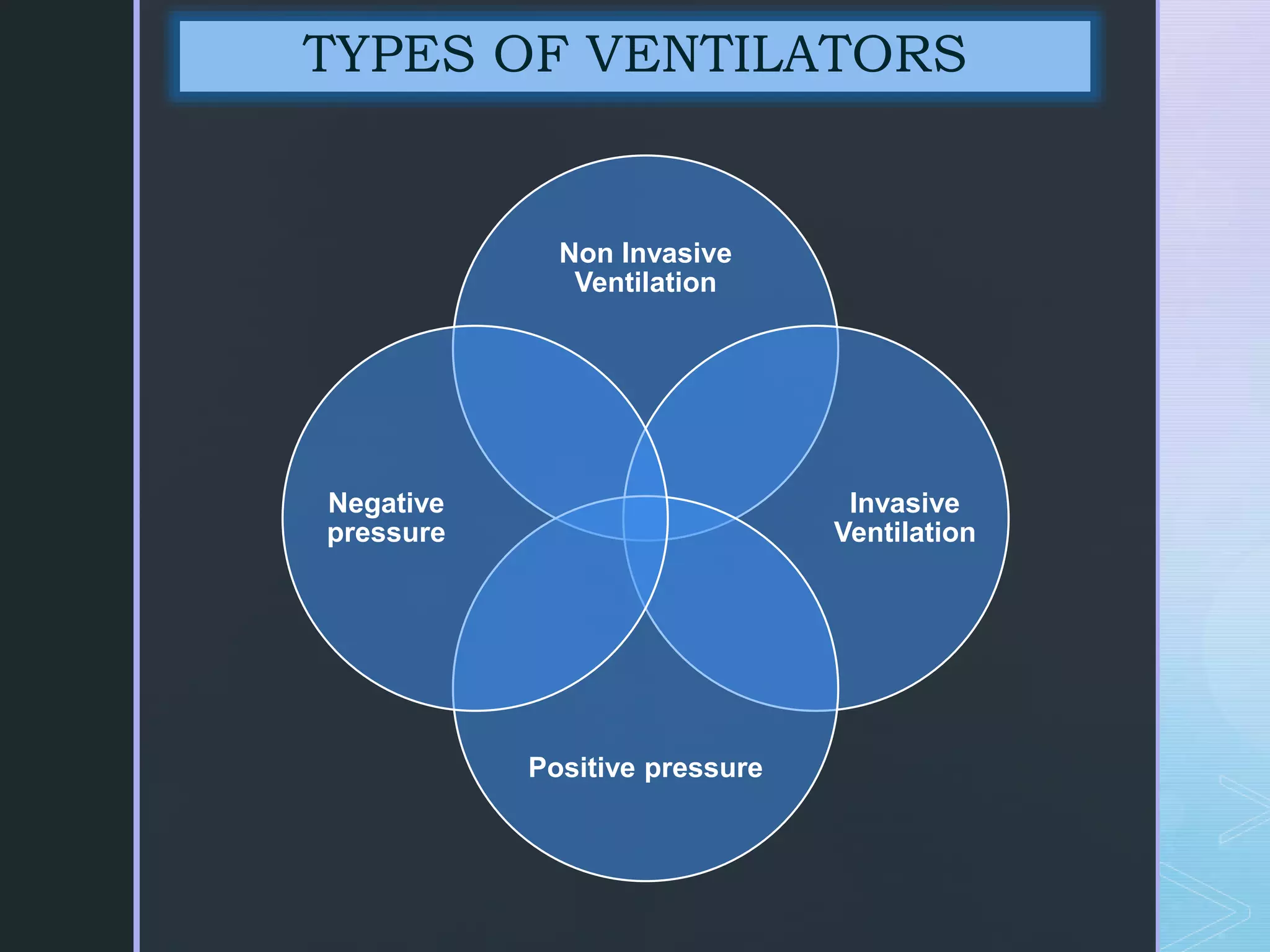
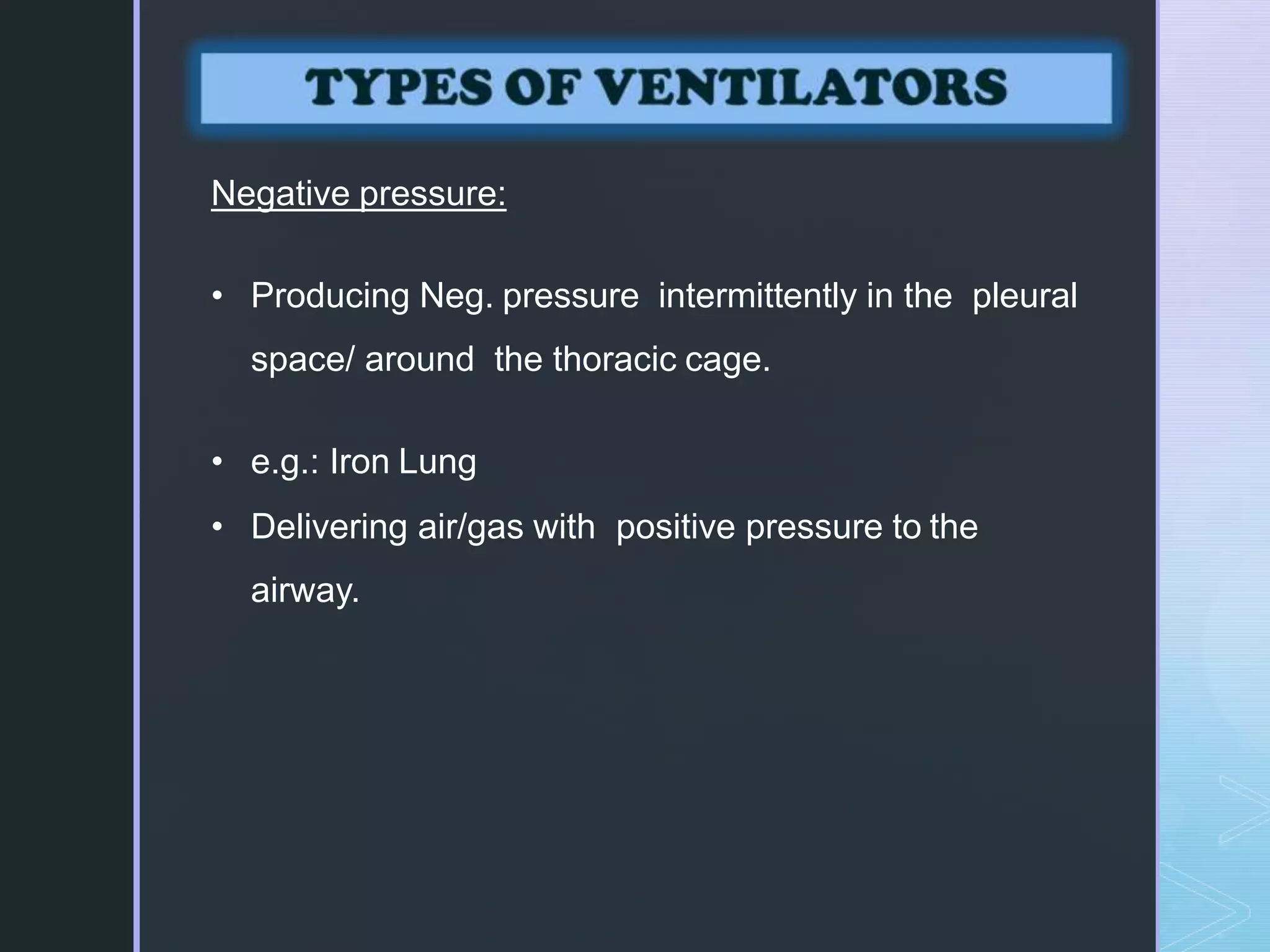
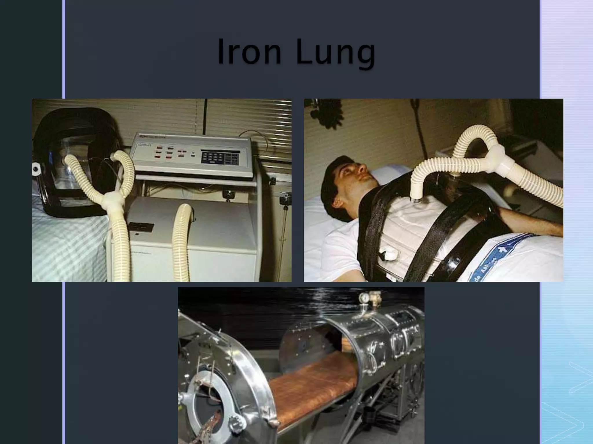
Mechanical ventilation involves using a ventilator to mechanically move air into and out of the lungs to maintain proper oxygen and carbon dioxide levels. It has several purposes including improving gas exchange, relieving respiratory distress, and avoiding complications. There are various modes of ventilation including pressure control, volume control, and time-cycled modes. Parameters like tidal volume, respiratory rate, and PEEP must be set and monitored appropriately for different patients and conditions.
Mechanical ventilation ppt including airway, ventilator, tubings and connections, nursing management, trouble shooting common problems and issues, suctioning etc.
The presentation deals with the principles of mechanical ventilation, its only for the educations purpose!
Any kind of replication, modifications and republication is strictly prohibited.
All Rights reserved to the Author. 2016
A treadmill exercise stress test is used to determine the effects of exercise on the heart. Exercise allows doctors to detect abnormal heart rhythms (arrhythmias) and diagnose the presence or absence of coronary artery disease.
This test involves walking in place on a treadmill while monitoring the electrical activity of your heart. Throughout the test, the speed and incline of the treadmill increase. The results show how well your heart responds to the stress of different levels of exercise.
Description
A technologist will explain the test to you, take a brief medical history, and answer any questions you may have. Your blood pressure, heart rate, and electrocardiogram (ECG) will be monitored before, during, and after the test.
You will be asked to sign a consent form. This form is required before the test can proceed.
You will be asked to remove all upper body clothing, and to put on a gown with the opening to the front.
Adhesive electrodes will be put onto your chest to capture an ECG. The sites where the electrodes are placed will be cleaned with alcohol and shaved if necessary. A mild abrasion may also be used to ensure a good quality ECG recording.
Your resting blood pressure, heart rate, and ECG will be recorded.
You will be asked to walk on a treadmill. The walk starts off slowly, then the speed and incline increases at set times. It is very important that you walk as long as possible because the test is effort-dependent.
You will be monitored throughout the test. If a problem occurs, the technologist will stop the test right away. It is very important for you to tell the technologist if you experience any symptoms, such as chest pain, dizziness, unusual shortness of breath, or extreme fatigue.
Following the test, you will be asked to lie down. Your blood pressure, heart rate, and ECG will be monitored for three to five minutes after exercise.
The data will be reviewed by a cardiologist after the test is completed. A report will be sent to the doctor(s) involved in your care.
Mitral valve replacement is a procedure whereby the diseased mitral valve of a patients heart is replaced by either a mechanical or tissue(bioprosthetic )valve.’
i have prepared this ppt. from various Books as a refrences as well as uses of web pages and explain and modify in simplify language which are easily understand by medical or para medical personnel..thank you..
Mechanical Ventilation (MV) is almost always a challenging topic for ICU nurses and practitioners. In this presentation we are going to review and relearn basics of MV together.
Global launch of the Healthy Ageing and Prevention Index 2nd wave – alongside...ILC- UK
The Healthy Ageing and Prevention Index is an online tool created by ILC that ranks countries on six metrics including, life span, health span, work span, income, environmental performance, and happiness. The Index helps us understand how well countries have adapted to longevity and inform decision makers on what must be done to maximise the economic benefits that comes with living well for longer.
Alongside the 77th World Health Assembly in Geneva on 28 May 2024, we launched the second version of our Index, allowing us to track progress and give new insights into what needs to be done to keep populations healthier for longer.
The speakers included:
Professor Orazio Schillaci, Minister of Health, Italy
Dr Hans Groth, Chairman of the Board, World Demographic & Ageing Forum
Professor Ilona Kickbusch, Founder and Chair, Global Health Centre, Geneva Graduate Institute and co-chair, World Health Summit Council
Dr Natasha Azzopardi Muscat, Director, Country Health Policies and Systems Division, World Health Organisation EURO
Dr Marta Lomazzi, Executive Manager, World Federation of Public Health Associations
Dr Shyam Bishen, Head, Centre for Health and Healthcare and Member of the Executive Committee, World Economic Forum
Dr Karin Tegmark Wisell, Director General, Public Health Agency of Sweden
R3 Stem Cells and Kidney Repair A New Horizon in Nephrology.pptxR3 Stem Cell
R3 Stem Cells and Kidney Repair: A New Horizon in Nephrology" explores groundbreaking advancements in the use of R3 stem cells for kidney disease treatment. This insightful piece delves into the potential of these cells to regenerate damaged kidney tissue, offering new hope for patients and reshaping the future of nephrology.
The dimensions of healthcare quality refer to various attributes or aspects that define the standard of healthcare services. These dimensions are used to evaluate, measure, and improve the quality of care provided to patients. A comprehensive understanding of these dimensions ensures that healthcare systems can address various aspects of patient care effectively and holistically. Dimensions of Healthcare Quality and Performance of care include the following; Appropriateness, Availability, Competence, Continuity, Effectiveness, Efficiency, Efficacy, Prevention, Respect and Care, Safety as well as Timeliness.
The Importance of Community Nursing Care.pdfAD Healthcare
NDIS and Community 24/7 Nursing Care is a specific type of support that may be provided under the NDIS for individuals with complex medical needs who require ongoing nursing care in a community setting, such as their home or a supported accommodation facility.
One of the most developed cities of India, the city of Chennai is the capital of Tamilnadu and many people from different parts of India come here to earn their bread and butter. Being a metropolitan, the city is filled with towering building and beaches but the sad part as with almost every Indian city
Medical Technology Tackles New Health Care Demand - Research Report - March 2...pchutichetpong
M Capital Group (“MCG”) predicts that with, against, despite, and even without the global pandemic, the medical technology (MedTech) industry shows signs of continuous healthy growth, driven by smaller, faster, and cheaper devices, growing demand for home-based applications, technological innovation, strategic acquisitions, investments, and SPAC listings. MCG predicts that this should reflects itself in annual growth of over 6%, well beyond 2028.
According to Chris Mouchabhani, Managing Partner at M Capital Group, “Despite all economic scenarios that one may consider, beyond overall economic shocks, medical technology should remain one of the most promising and robust sectors over the short to medium term and well beyond 2028.”
There is a movement towards home-based care for the elderly, next generation scanning and MRI devices, wearable technology, artificial intelligence incorporation, and online connectivity. Experts also see a focus on predictive, preventive, personalized, participatory, and precision medicine, with rising levels of integration of home care and technological innovation.
The average cost of treatment has been rising across the board, creating additional financial burdens to governments, healthcare providers and insurance companies. According to MCG, cost-per-inpatient-stay in the United States alone rose on average annually by over 13% between 2014 to 2021, leading MedTech to focus research efforts on optimized medical equipment at lower price points, whilst emphasizing portability and ease of use. Namely, 46% of the 1,008 medical technology companies in the 2021 MedTech Innovator (“MTI”) database are focusing on prevention, wellness, detection, or diagnosis, signaling a clear push for preventive care to also tackle costs.
In addition, there has also been a lasting impact on consumer and medical demand for home care, supported by the pandemic. Lockdowns, closure of care facilities, and healthcare systems subjected to capacity pressure, accelerated demand away from traditional inpatient care. Now, outpatient care solutions are driving industry production, with nearly 70% of recent diagnostics start-up companies producing products in areas such as ambulatory clinics, at-home care, and self-administered diagnostics.
Deep Leg Vein Thrombosis (DVT): Meaning, Causes, Symptoms, Treatment, and Mor...The Lifesciences Magazine
Deep Leg Vein Thrombosis occurs when a blood clot forms in one or more of the deep veins in the legs. These clots can impede blood flow, leading to severe complications.
How many patients does case series should have In comparison to case reports.pdfpubrica101
Pubrica’s team of researchers and writers create scientific and medical research articles, which may be important resources for authors and practitioners. Pubrica medical writers assist you in creating and revising the introduction by alerting the reader to gaps in the chosen study subject. Our professionals understand the order in which the hypothesis topic is followed by the broad subject, the issue, and the backdrop.
https://pubrica.com/academy/case-study-or-series/how-many-patients-does-case-series-should-have-in-comparison-to-case-reports/
Navigating Challenges: Mental Health, Legislation, and the Prison System in B...Guillermo Rivera
This conference will delve into the intricate intersections between mental health, legal frameworks, and the prison system in Bolivia. It aims to provide a comprehensive overview of the current challenges faced by mental health professionals working within the legislative and correctional landscapes. Topics of discussion will include the prevalence and impact of mental health issues among the incarcerated population, the effectiveness of existing mental health policies and legislation, and potential reforms to enhance the mental health support system within prisons.
2. “Mechanical ventilation is the use of a
ventilator to move room air or oxygen
enriched air into and out of the lungs
mechanically to maintain proper
levels of oxygen and carbon dioxide
in the blood.”
DEFINITION
3. GOALS
Improve gas exchange
Relive respiratory distress
Improve pulmonary mechanics
Permit lung and airway healing
Avoid complication
Reverse hypoxemia/Relieve acute respiratory acidosis
Reverse respiratory muscle fatigue Prevent and reverse atelectasis
Improve lung compliance/Maintain lung and airway function
Prevent disuse respiratory muscle dystrophy
4. To maintain gas exchange in case of
acute and chronic respiratory failure.
To maintain ventilator support after CPR.
To reduce pulmonary vascular resistance.
To excrete increased CO2 production.
To give general anesthesia with
muscle relaxants.
PURPOSES
5. Acute respiratory failure
Apnoea or impending inability
to breath
Severe Hypoxia/Hypoxemia
Respiratory muscle fatigue
Cardiac Insufficiency
Neurological problems
Therapeutic and prophylactic
INDICATIONS
6. Respiratory failure: An inability of the heart and lungs to provide
adequate tissue oxygenation or removal of carbon dioxide.
Hypoxemic respiratory failure – lung failure
Hypercapnic respiratory failure – pump failure
Neuromuscular diseases : Myasthenia Gravis, Guillain-Barre
Syndrome, and Poliomyelitis (failure of the normal respiratory
neuromuscular system)
Musculoskeletal abnormalities : Such as chest wall trauma .
Infectious diseases :of the lung such as
pneumonia, tuberculosis.
INDICATIONS
7. Obstructive lung disease in the form of asthma,
chronic bronchitis or emphysema.
Conditions such as pulmonary edema,
atelectasis, pulmonary fibrosis.
Patients who has received general anesthesia as
well as post cardiac arrest patients requires
ventilatory support until they have recovered from the
effects of the anesthesia or out from a Danger.
INDICATIONS
8. z
PARAMETERS VENTILATOR INDICATED NORMAL RANGE
A- Pulmonary function studies:
• Respiratory rate (breaths/min).
• Tidal volume (ml/kg body wt.)
• Vital capacity (ml/kg body wt.)
• Maximum Inspiratory Force (cm HO2)
ARTERIAL BLOOD GAS:
• PH
• PaO2 (mmHg)
• PaCO2 (mmHg)
>35
<5
<15
<-20
< 7.25
< 60
> 50
15-20
5-7
65-75
75-100
7.35-7.45
75-100
35-45
9. TERM MEANING
Independent variables The parameters that are set by clinician.”
Dependent variables “The parameters measured by the ventilators.”
Fraction of inspired oxygen
(FiO2):
“The concentration of O2 in the inspired gas, usually between
21% and 100% O2. The lowest possible fraction of inspired
oxygen (FiO2) necessary to meet oxygenation goals should
be used. “
Tidal volume (TV): “The amount of air delivered to the patient per
breath. It is expressed in milliliters.”
A starting point for the VT setting is 8 to 10ml/kg of
ideal weight.
Respiratory rate/frequency (f): “The number of breaths per minute. This can be from the
ventilator, the patient, or both. “
The RR is set as near to physiological rates (14 to 20
breaths/min) as possible.
10. TERM MEANING
Minute ventilation (V E): “The product of V and respiratory frequency (VT * f). It is
usually expressed in liters/minute.”
Exhaled Tidal Volume:(E TV): “It is the amount of gas that comes out of the patients
lungs on exhalation.”
This is the most accurate measure of the volume
received by the patient
If the ETV deviates from the set TV by 50ml or more,
troubleshoot the system to identify the source of gas loss.
Fraction of inspired oxygen
(FiO2):
“The concentration of O2 in the inspired gas, usually between
21% and 100% O2. The lowest possible fraction of inspired
oxygen (FiO2) necessary to meet oxygenation goals should
be used. “
Sigh : A deep breath , A breath that has a greater volume
than the tidal volume.
It provides hyperinflation and prevents atelectasis
11. TERM MEANING
Inverse Inspiratory to
Expiratory ratio:
“I:E ratios such as 1:1,2:1 and 3:1 arecalled inverse
I:E ratios”
Inverse I:E ratio allows unstable alveoli time to fill and also
prevents collapse by shortened expiratory phase.
Sigh volume : Usual volume is 1.5 –2 times tidal volume.
Positive end-expiratory pressure
(PEEP):
““The amount of positive pressure that is maintained at
end-expiration.”
Typical settings for PEEP are 5 to 20 cm H2O
PEEP increases oxygenation by preventing
collapse of small airways
It increases the functional residual capacity of the lungs
A typical initial applied PEEP is 5 cmH2O.
However, up to 20 cmH2O may be used in
patients undergoing low tidal volume ventilation for
acute respiratory distress syndrome (ARDS)
12. TERM MEANING
Auto PEEP: “ Auto PEEP is the spontaneous development of PEEP
caused by gas trapping in the lung resulting from insufficient
expiratory time and incomplete exhalation.”
Causes of auto PEEP formation include rapid RR, high VE
demand, airflow obstruction and inverse I:E ratio ventilation.
Auto PEEP = Total PEEP - Set PEEP
Inspiratory to Expiratory ratio
(I:E):
“The I:E ratio is usually set to mimic the pattern of
spontaneous ventilation.”
During spontaneous breathing, the normal I:E ratio is 1:2,
indicating that for normal patients the exhalation time is
about twice as long as inhalation time.
If exhalation time is too short “breath stacking” occurs
resulting in an increase in end- expiratory pressure also
called auto-PEEP.
13. TERM MEANING
Peak airway pressure (Paw): The pressure that is required to deliver the TV to the
patient. It has a unit of centimeters of water (cm H2O).”
Plateau pressure (Pplat): “The pressure that is needed to distend the lung. This
pressure can only be obtained by applying an end
inspiratory pause. It also has a unit of cm H2O.”
Mean airway pressure: “The time-weighted average pressure during the respiratory
cycle. It is expressed in cm H2O.”
Peak inspiratory Pressure
(PIP):
In adults if the peak airway pressure is persistently above
45 cmH2O, the risk of barotrauma is increased and efforts
should be made to try to reduce the peak airway pressure.
“Increasing PIP is also sign of Blockage of airway and
needed to suctioning or changeEt
/TT.”
Sensitivity ( Trigger Sensitivity)
:
: “The sensitivity function controls the amount of patient
effort needed to initiate an inspiration.”
14. z
• Inspiration: Inspiratory valve opens and expiratory
valve is closed
• Inspiratory pause: inspiratory valve and expiratory
valve closed
• Expiration: Inspiratory valve closed and expiratory
valve open
• Expiratory Pause: Inspiratory valve and expiratory
(PEEP) valve closed at the end of expiration
PHASES OF VENTILATOR
15. z
Mode denotes interplay b/w patient and the ventilator
Describes the style of breath support based on relationship between
the various possible types of breath and inspiratory – phase variables
MODES OF VENTILATOR
16. z
MODES OF VENTILATOR
PRESSURE CYCLED
VENTILATION MODES
CPAP
BiPAP
PSV
PCV
PCIRV
VOLUME CONTROLLED
MODES
ACV
IMV
SIMV
3. TIME CYCLED
VENTILATION
17. CPAP is positive pressure applied throughout the
respiratory cycle to the spontaneously breathing
patient.
A continuous level of elevated pressure is provided
through the patient circuit to maintain adequate
oxygenation, decrease the work of breathing.
CPAP may be used invasively through an
endotracheal tube or tracheostomy or
noninvasively with a face mask or nasal
prongs.
CONTINOUS POSITIVE AIRWAY
PRESSURE
18. provides pressure at end expiration, which
prevents alveolar collapse and improves the
functional residual capacity and oxygenation.
CPAP allows the nurse to observe the ability
of the patient to breath spontaneously while
still on the ventilator.
It may used as a Weaning Mode.
19. • Similar to CPAP
• Non invasive mechanical ventilation.
• Indicated for sleep apnea patients with high pressure
settings
• Used when CPAP fails
• Effective for CHF patients and often prescribed for
patients with lung disorders or neuromuscular disorders.
• CPAP offers constant singular pressure difficult to exhale
against.
• For patients with higher pressure strengths exhaling
against an incoming air can feel difficult as
•
BILEVEL POSITIVE AIRWAY PRESSURE
20. • For patients with higher pressure strengths exhaling against
an incoming air can feel difficult as if they are having to
force their breathing out.
• Bipaps can also be set to include a breath timing feature
that measures the amount of breaths per minute a person
should be taking . If the time between the breaths exceeds
the set limit , the machine can force the person to breath by
temporarily increasing the air pressure.
• BiPAP has two pressure settings: a prescribed pressure for
inhalation -Ipap and a lower pressure for exhalation Epap.
This dual settings allow the patient to get more air into the
lungs
BILEVEL POSITIVE AIRWAY PRESSURE
21. In pressure controlled ventilation the breathing
gas flows under constant pressure into the
lungs during the selected inspiratory time.
The flow is highest at the beginning of
inspiration( i.e when the volume is lowest in
the lungs).
As the pressure is constant the flow is
initially high and then decreases with
increasing filling of the lungs.
PRESSURE CONTROLLED
VENTILATION
22. z
Pressure (or Pressure above PEEP) is the setting variable
No mandatory breaths
Applicable on Spontaneous breaths
Patient effort determines size of breath and flowrate.
It augments spontaneous VT, decreases spontaneous rates and WOB.
Used in conjunction with spontaneous breaths in any mode of
ventilation
No back up ventilation in the event of apnea.
Provides pressure support to overcome the increased work of
breathing imposed by the disease process, the endotracheal tube, the
inspiratory valves and other mechanical aspects ofventilatory support.
Allows for titration of patient effort during weaning.
PRESSURE SUPPORT
VENTILATION
23. • Inverse ratio ventilation (IRV) mode reverses this
ratio so that inspiratory time is equal to, or longer
than, expiratory time (1:1 to 4:1).
• Inverse I:E ratios are used in conjunction with
pressure control to improve oxygenation by
expanding stiff alveoli by using longer distending
times, thereby providing more opportunity for gas
exchange and preventing alveolar collapse.
PC-INVERSE RATIO VENTILATION
25. The ventilator provides the patient with a pre-
set tidal volume at a pre-set rate.
The patient may initiate a breath on his own, but
the ventilator assists by delivering a specified
tidal volume to the patient .
Client can breathe at a higher rate than the
preset number of breaths/minute
ASSIST /CONTROLLED VENTILATION
26. The total respiratory rate is determined by the
number of spontaneous inspiration initiated by
the patient plus the number of breaths set on
the ventilator.
If the patient wishes to breathe faster, he or she
can trigger the ventilator and receive a full-volume
breath.
Often used as initial mode of ventilation When the
patient is too weak to perform the work of
breathing (e.g. when emerging from anesthesia)
ASSIST /CONTROLLED VENTILATION
27. The preset RR ensures that the patient
receives adequate ventilation, regardless of
spontaneous efforts.
The patient can breath faster than the
preset rate but not slower.
ASSIST /CONTROLLED VENTILATION
28. Elongated tank, which encases the patient up to the
neck. The neck is sealed with a rubber gasket, the
patient's face are exposed to the room air.
These exert negative pressure on the external chest
decreasing the intra-thoracic pressure during
inspiration, allows air to flow into the lungs, filling its
volume.
The cessation of the negative pressure causes the
chest wall to fall and exhalation to occur.
NEGATIVE PRESSURE VENTILATORS
29. A mode of mechanical ventilation in which the
patient is allowed to breath independently
except during certain prescribed intervals,
when a ventilator delivers a breath either
under positive pressure or in a measured
volume.
This mode is not use mostly in clinical practice
due to many new tech. Modes.
INTERMITTENT MANDATORY
VENTILATION
30. z
PROS CONS
• Freedom for natural
spontaneous breaths
even on the machine
• Asynchrony
• Lesser chances of
hyperventilation
• Random chances of
breath stacking
• Increased work of
breathing
• Random barotrauma and
volutrauma
INTERMITTENT MANDATORY
VENTILATION
31. z
Ventilator delivers either patient triggered assisted
breaths or time triggered mandatory breath in a
synchronized fashion so as to avoid breath stacking
If the patient breathes between mandatory breaths, the
ventilator will allow the patient to breathe a normal
breath by opening the demand (inspiratory) valve but
not offering any inspiratory assistance.
If patient does not make an inspiratory effort then ventilator will
deliver a time triggered mandatory breath
SYNCHRONIZED INTERMITTENT
MANDATORY VENTILATOR
33. The SIMV mode of ventilation delivers a set number of
breaths of a set TV, and between these mandatory
breaths the patient may initiate spontaneous breaths.
If the patient initiates a breath near the time a mandatory
breath is due, the delivery of the mandatory breath is
synchronized with the patient’s spontaneous effort to prevent
patient ventilator dys synchrony.
In between the ventilator-delivered breaths , the patient is
able to breath spontaneously at his own tidal volume and rate
with no assistance from the ventilator.
Ventilators breaths are synchronized with the patient
spontaneous breath.
SYNCHRONIZED INTERMITTENT
MANDATORY VENTILATOR
34.
35. z
Machine breaths are delivered at a set rate (volume or
pressure limit)
Patient is allowed to breath spontaneously from
either a demand valve or a continuous flow of
gases but not offering any inspiratory assistance.
Patient’s capability determines Tidal volume of
spontaneously breaths
Some freedom to breath naturally even on
mechanical ventilator
INTERMITTENT MANDATORY
VENTILATION
37. Non Invasive Ventilation: “Ventilatory
support that is given without establishing
endo- tracheal intubation or tracheostomy is
called Non invasive mechanical ventilation.”
Invasive Ventilation: “Ventilatory support that
is given through endotracheal intubation or
tracheostomy is called as Invasive mechanical
ventilation.”
TYPES OF VENTILATORS
38. Negative pressure:
• Producing Neg. pressure intermittently in the pleural
space/ around the thoracic cage.
• e.g.: Iron Lung
• Delivering air/gas with positive pressure to the
airway.
39.
40. The patient’s body was encased in an iron cylinder and negative
pressure was generated
The iron lung are still occasionally used today.
These are simple to use and do not require intubations of the
airway; consequently, they are especially adaptable for home
use.
It is used mainly in chronic respiratory failure associated with
neuromuscular conditions such as poliomyelitis, muscular
dystrophy and myasthenia gravis.
The use of negative-pressure ventilators is restricted in clinical
practice, however, because they limit positioning and movement
and they lack adaptability to large or small body torsos (chests).
41. POSITIVE PRESSURE VENTILATION:
inflate the lungs by exerting positive pressure on the
airway forcing the alveoli to expand during inspiration.
Expiration occurs passively.
Positive-pressure ventilators require an artificial airway
(Endotracheal or tracheostomy tube) in invasive ventilation
and in NIV includes BiPAP Mask , O2 mask , Nasal
mask/cannula , O2 high concentrated reservoir mask etc.
Inspiration can be triggered either by the patient
or the machine.
42. “Ventilator Alarms defined as a Voice or sound to alert
Nurse/Doctor and caused by any abnormal value of
either in client or in Ventilator.”
Check for bucking the vent
These Alarms have 3main Types as below:
(i)Pressure alarms
(ii)Volume alarms
(iii)Apnea alarms
VENTILATOR ALARMS
43.
44. z
PRESSURE ALARMS
They are triggered when
there is increased airway
resistance or decreased
lung compliance.
Low pressure alarms
and high pressure
alarms
VOLUME ALARMS
Volume alarms are
valuable for ensuring
adequate alveolar
ventilation, particularly in
the patient receiving a
pressure mode of
ventilation
VENTILATOR ALARMS
APNEA ALARMS
This alarm is very
important when the
patient is on a
spontaneous breathing
mode such as PS or
CPAP, and no
mandatory breaths are
set.
45. An alarm should never be silenced until the
cause has been investigated and corrected.
If the source of the alarm cannot be determined,
disconnect the client from the ventilator and use a
hand-held resuscitation bag for manual ventilation
with 100% oxygen until the problem can be
resolved
TWO RULES….
48. Aspiration
Decreased clearance of secretions
Nosocomial or ventilator-acquired pneumonia
(VAP)
I.AIRWAY COMPLICATIONS
49. • Hypoventilation with atelectasis with respiratory
acidosis or hypoxemia.
• Hyperventilation with hypocapnia and respiratory
alkalosis
• Barotrauma
• Closed pneumothorax,
• Tension pneumothorax,
• Subcutaneous emphysema.
• Alarm “turned off”
• Failure of alarms or ventilator
• Inadequate nebulization or humidification
• Overheated inspired air, resulting in hyperthermia
MECHANICAL COMPLICATONS
50. Fluid overload with humidified air and sodium
chloride (NaCl) retention
Depressed cardiac function and
hypotension
Stress ulcers
Paralytic ileus
Gastric distension
Starvation
Dyssynchronous breathing pattern
PHYSIOLOGICAL COMPLICATIONS
51. (A) IN ENDOTRACHEAL TUBE :
Tube kinked or plugged
Tracheal stenosis or
tracheomalacia
Main stem intubation with
contralateral (located on or
affecting the opposite side of the
lung)
lung atelectasis
Cuff failure
Sinusitis
Otitis media
Laryngeal edema
ARTIFICIAL AIRWAY COMPLICATIONS
(B) IN TRACHEOSTOMY TUBE :
Acute haemorrhage at the site
Air embolism
Aspiration
Tracheal stenosis
Failure of the tracheostomy cuff
Laryngeal nerve damage
Obstruction of tracheostomy tube
Pneumothorax
Subcutaneous and mediastina
emphysema
Infection
Accidental decannulation with loss of
airway
52. SYSTEM WISE COMPLICATIONS
SYSTEM COMPLICATIONS
CARDIIOVASCULA
R SYSTEM
◦ Increased intrathoracic pressure
◦ Reduced CO2
PULMONARY ◦ Barotrauma (trauma r/t pressure)
Pneumothorax
Subcutaneous emphysema
Alveolar
hypoventilation
Cuff leak
Ventilator settings
Secretions
Atelectasis
Alveolar
hyperventilation
Due to hypoxemia, fear, pain, anxiety → alkalosis
RX: sedate, analgesia, communication, correct
hypoxemia
Due to inappropriate ventilator settings
high tidal volume
High rate
◦ Pulmonary Infection
53. SYSTEM COMPLICATIONS
NEUROLOGICAL ◦ Positive pressure ventilation → increased
intrathoracic pressure
◦ interferes with venous drainage; increased ICP
GI: ◦ Stress ulcers and GI bleeds; Rx with H2
receptor blockers
Gastric and bowel dilation
Musculoskeletal: Muscle atrophy r/t immobilisation
Mobilise
ROM
Psychologic: Stress
Communication very important
Sedate, explain, family visits, pain management
Facilitate expression of needs
54. High Peak Pressures Low Plateau
Pressures
High Peak Pressures High Plateau
Pressures
• Mucus Plug • ARDS
• Bronchospasm • Pulmonary Edema
• ET tube blockage • Pneumothorax
• Biting • ET tube migration to a single
bronchus
• Effusion
55. Weaning is the process of withdrawing
mechanical ventilator support and
transferring the work of breathing from
the ventilator to the patient which is done
only when patient is free from the cause
to be kept on mechanical ventilation.
“Weaning success is defined as effective
spontaneous breathing without any
mechanical ventilation for 24 hours or
more.” or “The process of going OFF
from ventilator dependence to
spontaneous breathing”
WEANING
57. Tidal volume greater than 5 ml/kg),
Respiratory frequency less than 30
breaths/min
Oxygen partial pressure be above PaO2
greater than 60mm Hg and FIO2 <40%
Vital capacity 10 to 15 ml/kg.
CRITERIA FOR WEANING
58. It consists of removing the patient from the ventilator and
having him / her breathe spontaneously on a T-piece
connected to oxygen source.
During T-piece weaning, periods of ventilator support are
alternated with spontaneous breathing.
The goal is to progressively increase the time spent of f the
ventilator.
1.T-PIECE TRIAL
59. Observe for signs & Symptoms of….
Hypoxia,
increasing fatigue,
Tachy cardia- Ischemic ECG changes
Restlessness
RR > 35/min
Use of accessory muscles for breathing
Paradoxical chest movement
1.T-PIECE TRIAL
60. ET/TT removed only if following criterion
met…
Spontaneous ventilation is adequate
Pharyngeal and laryngeal reflexes are active
Pt maintain adequate airway and can swallow,
move the jaw clench teeth , voluntary cough is
effective to bring out secretion
Before the tube is removed—a trail with
nose/mouth breathing is done – Deflating cuff,
using fenestrated tube etc
1.T-PIECE TRIAL
61. SIMV is the most common method of
weaning.
It consists of gradually decreasing the number of
breaths delivered by the ventilator to allow the
patient to increase number of spontaneous
breaths.
In pt’s who – satisfies all criteria for weaning
but cannot have spontaneous breathing for long
time.
2.WEANING FROM VENTILATOR-SIMV
62. When placed on CPAP, the patient does all the
work of breathing without the aid of a back up
rate or tidal volume.
No mandatory (ventilator-initiated) breaths are
delivered in this mode i.e. all ventilation is
spontaneously initiated by the patient.
Weaning by gradual decrease in
pressure value
2.WEANING FROM VENTILATOR-CPAP
63. The patient must initiate all pressure support
breaths.
During weaning using the PSV mode the level of
pressure support is gradually decreased based on
the patient maintaining an adequate tidal volume (8
to 12 mL/kg) and a respirator y rate of less than 25
breaths/minute.
PSV weaning is indicated for :-
- Difficult to wean patients
- Small spontaneous tidal volume.
2.WEANING FROM VENTILATOR-PSV
64. Diaphoresis
Dyspnea & Labored respirator y pattern
Increased anxiety ,Restlessness, Decrease in
level of consciousness
Dysrhythmia , Increase or decrease in heart rate
of > 20 beats /min. or heart rate > 110b/m ,
Sustained heart rate >20% higher or lower than
baseline.
Tidal volume ≤5 mL/kg, Sustained minute
ventilation <200 mL/kg/minute
SIGNS OF WEANING INTOLERANCE
65. Pt successfully weaned---- and has adequate
respiratory function – weaned from O2
FIO2 is gradually reduced until SPO2 is in
range of 80-100 mmHg while breathing in
Room air
If air SPO2 less than 70 supplementary O2
recommended
2.WEANING FROM OXYGEN
66.
67. Ineffective breathing pattern
Potential for pulmonary infection
Impaired water and fluid regulation
Oral hygiene
Potential altered nutritional status: less than
body requirement related to NPO status
Potential for complications related to
immobility
NURSING DIAGNOSIS
68. Knowledge deficit related to intubation and
mechanical ventilation
Elimination care
Promoting coping ability
Preventing trauma and infections
Promoting rest and sleep
Safety and security needs.
NURSING DIAGNOSIS