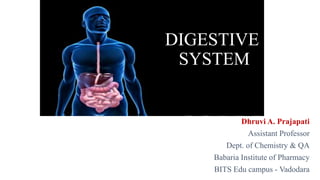
The digestive system includes the organs of the alimentary canal and accessory structures. The alimentary canal forms a continuous tube that is open to the outside environment at both ends. The organs of the alimentary canal are the mouth, pharynx, esophagus, stomach, small intestine, and large intestine.
THIS PRESENTATION INCLUDES DETAILED INFORMATION ABOUT ACCESSORY ORGANS OF DIGESTIVE SYSTEM..i,e TEETH, TONGUE, SALIVARY GLANDS, PANCREAS, LIVER AND GALL BLADDER
Digestion is the breakdown of large insoluble food molecules into small water-soluble food molecules so that they can be absorbed into the watery blood plasma. In certain organisms, these smaller substances are absorbed through the small intestine into the blood stream.
Skeletal system. anatomy and physiology of skeletal system. appendicular skel...mamtabisht10
SKELETAL SYSTEM
bones, cartilage and ligaments are tightly joined to form a strong, flexible framework called skeletal system
anatomy and physiology of axial and appendicular skeletal system
Axial Skeleton: The axial skeleton includes the skull, spine, ribs and sternum.
Appendicular Skeleton:
The appendicular skeleton includes the appendages of the body, which are the shoulders, arms, hips, and legs.
special sense organs (anatomy and physiology) - a brief discussion Pallab Nath
brief discussion on special senses, Basic level class for technicians. topics discussed include eyes and vision, nose and sense of smell, tongue and sense of taste and ears and hearing
1st Semester Anatomy - Digestive System - GIT - By thirumurugan.pptxthiru murugan
Digestive System:
Also known as gastrointestinal tract, digestive tract, digestional tract, GI tract, GIT, gut, or alimentary canal.
Consist of alimentary tract and Accessory organs
It involve in Ingestion, Digestion, Absorption and Excretion
It is started from mouth and ended at anal canal, that is assisted and supported by many parts.
Parts of Digestive System: Primary digestive organs & Accessory organs
Primary digestive organs: Mouth, Pharynx, Esophagus, Stomach, Small Intestine, Large Intestine, Rectum, Anal canal
Accessory organs: Teeth, Tongue, Salivary glands, Liver, Gallbladder, Pancreas.
Mouth
First part of GIT, Also known as oral or buccal cavity, It consist of muscles & bones
Tongue or Lingua:Voluntary muscular structure, Occupies floor of mouth
Superior surface consist of numerous papillae contains taste bud for the sense of taste. Attached inferiorly with hyoid bone, Highly mobile muscular part of GIT.
Teeth: Teeth are the hard and whitish substances present in the mouth Which is essential for chewing & speech. They fixed in socket of alveolar ridge in mandible & maxilla (Jaw). Diphyodont is a type of dentition in which two successive sets of teeth are developed during the lifetime. The first set of teeth is temporary or deciduous or milk and the other set is permanent teeth
Salivary glands: These are exocrine glands found in oral cavity that secrete complex fluid known as saliva
Types: Major & Minor salivary glands
Major salivary gland: Parotid, Submandibular & Sublingual
Minor salivary glands: There are 450 minor salivary glands present in oral cavity, lips, cheeks, palate and floor of the mouth
Pharynx: Wide, muscular tube situated behind the nose, mouth & larynx
Cavity of pharynx divided into nasopharynx, oropharynx and laryngopharynx.
Esophagus: The esophagus is a fibromuscular tube, approximately 25cm in length,
It transports food from the pharynx to the stomach.
Stomach: The stomach is a hollow organ in the GIT.
It is the “J” shaped dilated part, situated in the upper part of the abdomen.
Gross Anatomy of stomach: The stomach has four main anatomical divisions; the cardia, fundus, body and pylorus:
Cardia: surrounds the superior opening of the stomach. it consist cardiac sphincter
Fundus: the rounded, often gas filled portion superior to and left of the cardia.
Body: the large central portion inferior to the fundus.
Pylorus: This area connects the stomach to the duodenum. It is divided into the pyloric antrum, pyloric canal and pyloric sphincter.
Pancreas: The pancreas is a soft, finely lobulated, elongated dual (Exo Endocrine) gland.
Liver: It is the largest gland of the body. It involve metabolic activities
The liver is one of the vital organs of the body, responsible for chemical actions that the body needs to survive.
Small intestine: The intestine which is the longest part of the digestive tube is divided into small intestine and large intestine.
Large Intestine or Colon: It is a last part of the GIT
HOT NEW PRODUCT! BIG SALES FAST SHIPPING NOW FROM CHINA!! EU KU DB BK substit...GL Anaacs
Contact us if you are interested:
Email / Skype : kefaya1771@gmail.com
Threema: PXHY5PDH
New BATCH Ku !!! MUCH IN DEMAND FAST SALE EVERY BATCH HAPPY GOOD EFFECT BIG BATCH !
Contact me on Threema or skype to start big business!!
Hot-sale products:
NEW HOT EUTYLONE WHITE CRYSTAL!!
5cl-adba precursor (semi finished )
5cl-adba raw materials
ADBB precursor (semi finished )
ADBB raw materials
APVP powder
5fadb/4f-adb
Jwh018 / Jwh210
Eutylone crystal
Protonitazene (hydrochloride) CAS: 119276-01-6
Flubrotizolam CAS: 57801-95-3
Metonitazene CAS: 14680-51-4
Payment terms: Western Union,MoneyGram,Bitcoin or USDT.
Deliver Time: Usually 7-15days
Shipping method: FedEx, TNT, DHL,UPS etc.Our deliveries are 100% safe, fast, reliable and discreet.
Samples will be sent for your evaluation!If you are interested in, please contact me, let's talk details.
We specializes in exporting high quality Research chemical, medical intermediate, Pharmaceutical chemicals and so on. Products are exported to USA, Canada, France, Korea, Japan,Russia, Southeast Asia and other countries.
Explore natural remedies for syphilis treatment in Singapore. Discover alternative therapies, herbal remedies, and lifestyle changes that may complement conventional treatments. Learn about holistic approaches to managing syphilis symptoms and supporting overall health.
MANAGEMENT OF ATRIOVENTRICULAR CONDUCTION BLOCK.pdfJim Jacob Roy
Cardiac conduction defects can occur due to various causes.
Atrioventricular conduction blocks ( AV blocks ) are classified into 3 types.
This document describes the acute management of AV block.
Report Back from SGO 2024: What’s the Latest in Cervical Cancer?bkling
Are you curious about what’s new in cervical cancer research or unsure what the findings mean? Join Dr. Emily Ko, a gynecologic oncologist at Penn Medicine, to learn about the latest updates from the Society of Gynecologic Oncology (SGO) 2024 Annual Meeting on Women’s Cancer. Dr. Ko will discuss what the research presented at the conference means for you and answer your questions about the new developments.
Title: Sense of Taste
Presenter: Dr. Faiza, Assistant Professor of Physiology
Qualifications:
MBBS (Best Graduate, AIMC Lahore)
FCPS Physiology
ICMT, CHPE, DHPE (STMU)
MPH (GC University, Faisalabad)
MBA (Virtual University of Pakistan)
Learning Objectives:
Describe the structure and function of taste buds.
Describe the relationship between the taste threshold and taste index of common substances.
Explain the chemical basis and signal transduction of taste perception for each type of primary taste sensation.
Recognize different abnormalities of taste perception and their causes.
Key Topics:
Significance of Taste Sensation:
Differentiation between pleasant and harmful food
Influence on behavior
Selection of food based on metabolic needs
Receptors of Taste:
Taste buds on the tongue
Influence of sense of smell, texture of food, and pain stimulation (e.g., by pepper)
Primary and Secondary Taste Sensations:
Primary taste sensations: Sweet, Sour, Salty, Bitter, Umami
Chemical basis and signal transduction mechanisms for each taste
Taste Threshold and Index:
Taste threshold values for Sweet (sucrose), Salty (NaCl), Sour (HCl), and Bitter (Quinine)
Taste index relationship: Inversely proportional to taste threshold
Taste Blindness:
Inability to taste certain substances, particularly thiourea compounds
Example: Phenylthiocarbamide
Structure and Function of Taste Buds:
Composition: Epithelial cells, Sustentacular/Supporting cells, Taste cells, Basal cells
Features: Taste pores, Taste hairs/microvilli, and Taste nerve fibers
Location of Taste Buds:
Found in papillae of the tongue (Fungiform, Circumvallate, Foliate)
Also present on the palate, tonsillar pillars, epiglottis, and proximal esophagus
Mechanism of Taste Stimulation:
Interaction of taste substances with receptors on microvilli
Signal transduction pathways for Umami, Sweet, Bitter, Sour, and Salty tastes
Taste Sensitivity and Adaptation:
Decrease in sensitivity with age
Rapid adaptation of taste sensation
Role of Saliva in Taste:
Dissolution of tastants to reach receptors
Washing away the stimulus
Taste Preferences and Aversions:
Mechanisms behind taste preference and aversion
Influence of receptors and neural pathways
Impact of Sensory Nerve Damage:
Degeneration of taste buds if the sensory nerve fiber is cut
Abnormalities of Taste Detection:
Conditions: Ageusia, Hypogeusia, Dysgeusia (parageusia)
Causes: Nerve damage, neurological disorders, infections, poor oral hygiene, adverse drug effects, deficiencies, aging, tobacco use, altered neurotransmitter levels
Neurotransmitters and Taste Threshold:
Effects of serotonin (5-HT) and norepinephrine (NE) on taste sensitivity
Supertasters:
25% of the population with heightened sensitivity to taste, especially bitterness
Increased number of fungiform papillae
Ethanol (CH3CH2OH), or beverage alcohol, is a two-carbon alcohol
that is rapidly distributed in the body and brain. Ethanol alters many
neurochemical systems and has rewarding and addictive properties. It
is the oldest recreational drug and likely contributes to more morbidity,
mortality, and public health costs than all illicit drugs combined. The
5th edition of the Diagnostic and Statistical Manual of Mental Disorders
(DSM-5) integrates alcohol abuse and alcohol dependence into a single
disorder called alcohol use disorder (AUD), with mild, moderate,
and severe subclassifications (American Psychiatric Association, 2013).
In the DSM-5, all types of substance abuse and dependence have been
combined into a single substance use disorder (SUD) on a continuum
from mild to severe. A diagnosis of AUD requires that at least two of
the 11 DSM-5 behaviors be present within a 12-month period (mild
AUD: 2–3 criteria; moderate AUD: 4–5 criteria; severe AUD: 6–11 criteria).
The four main behavioral effects of AUD are impaired control over
drinking, negative social consequences, risky use, and altered physiological
effects (tolerance, withdrawal). This chapter presents an overview
of the prevalence and harmful consequences of AUD in the U.S.,
the systemic nature of the disease, neurocircuitry and stages of AUD,
comorbidities, fetal alcohol spectrum disorders, genetic risk factors, and
pharmacotherapies for AUD.
Anti ulcer drugs and their Advance pharmacology ||
Anti-ulcer drugs are medications used to prevent and treat ulcers in the stomach and upper part of the small intestine (duodenal ulcers). These ulcers are often caused by an imbalance between stomach acid and the mucosal lining, which protects the stomach lining.
||Scope: Overview of various classes of anti-ulcer drugs, their mechanisms of action, indications, side effects, and clinical considerations.
2. The digestive system
comprises of the
gastrointestinal tract with
various glands attached to
it.
The tract starts from the
mouth and ends at the anus.
The tract is 8-10 meters in
length.
Dhruvi A. Prajapati 2
3. WHAT IS THE DIGESTIVE SYSTEM ?
The gastrointestinal tract (digestive tract, digestional tract, GI tract,
GITor alimentary canal) is an organ system within humans and other
animals which takes in food, digests it and absorb energy and nutrients, and
expels the remaining waste as feces.
• Major digestive organs:
Mouth.
Pharynx.
Esophagus.
Stomach.
Small Intestine.
Large Intestine.
Rectum.
• Accessory digestive organs:
Liver
Gallbladder
pancreas
Salivary gland
Dhruvi A. Prajapati 3
4. Functions of GI tract
• Ingestion: taking of food into the alimentary tract. i.e. eating & drinking.
• Propulsion: mixes & moves the contents along the alimentary tract.
• Digestion: consist of:
• Mechanical Phase – involves the breaking up of food into small pieces,
pushing the food down the food tube & mixing with it digestive juices
• Chemical Phase – involves the further breaking up of the larger molecules
of food into smaller molecules by the action of digestive enzymes
• Absorption:this is the process by which digested food substances pass through
the walls of some organs of the walls of some organs of the alimentary canal into
the blood for circulation.
• Elimination:food substances that have been eaten but cannot be digested &
absorbed are excreted from the alimentary canal as faeces by the process of
defaecation.
Dhruvi A. Prajapati 4
5. MOUTH
Relations:
• Anteriorly-lips
• Posteriorly-continue with the oropharynx
• Laterally-muscles of cheeks
• Superiorly-bony hard palate
• Inferiorly-muscular tongue & the soft tissues of the floor of the mouth
• The mouth is the first portion of the alimentary canal that receives food and
produces saliva.
• The oral mucosa is the mucous membrane epithelium lining the inside of the
mouth.
• The palate forms the roof of the mouth & is divided into the anterior hard
palate & posterior soft palate.
• The uvula is a curved fold of muscle covered with mucousmembrane,
hanging down from the middle.
Dhruvi A. Prajapati 5
7. TONGUE
• The tongue is a muscular organ in the mouth, that manipulates food
for mastication, and is used in the act of swallowing.
• It is of importance in the digestive system and is the primary organ of
taste in the gustatory system.
• The tongue's upper surface (dorsum) is covered by taste buds housed in
numerous lingual papillae.
• The human tongue is divided into two parts, an oral part at the front
and a pharyngeal part at the back.
Dhruvi A. Prajapati 7
9. TEETH
• The human teeth function to mechanically break down items of food by cutting
and crushing them in preparation for swallowing and digesting.
• Humans have four types of teeth: incisors, canines, premolars & molars, each
with a specific function.
Dhruvi A. Prajapati 9
10. PRIMARY TEETH
Among deciduous (primary) teeth, ten are found in the maxilla (upper
jaw) and ten in the mandible (lower jaw), for a total of 20.
Start to come in (erupt) at about 6 months of age
In the primary set of teeth,
two types of incisors – centrals and laterals, one canine & two types
of molars – first and second.
All primary teeth are normally later replaced with their permanent
counterparts.
Dhruvi A. Prajapati 10
11. PERMANENT TEETH
Among permanent teeth, 16 are found in the maxilla and 16 in the mandible,
for a total of 32.
Age 21, all 32 of the permanent teeth have usually erupted.
The permanent teeth are the:
Two incisor (for cutting)-central incisor, lateral incisor
One canine (for tearing)
Two premolar (for crushing)-first premolar, second premolar,
Three molar (for grinding)-first molar, second molar, and third molar.
Dhruvi A. Prajapati 11
12. PARTS
• ENAMEL
Enamel is the hardest & most highly mineralized substance of the
body.
It is one of the four major tissues which make up the tooth, along with
dentin, cementum, and dental pulp.
96% of enamel consists of mineral, with water and organic material
comprising the rest.
The normal color of enamel varies from light yellow to grayish white.
• DENTIN
Dentin is the substance between enamel or cementum and the pulp
chamber.
The porous, yellow-hued material is made up of 70% inorganic
materials, 20% organic materials, and 10% water by weight.
Dentin is a mineralized connective tissue with an organic matrix of
collagenous proteins.
Dhruvi A. Prajapati 12
13. • CEMENTUM
Cementum is a specialized
bone like substance covering
the root of a tooth.
Its coloration is yellowish
and it is softer than dentin
and enamel.
• DENTAL PULP
The dental pulp is the central
part of the tooth filled with
soft connective tissue.
This tissue contains blood vessels
& nerves that enter the tooth from
a hole at the apex of the root.
Dhruvi A. Prajapati 13
14. FUNCTIONS OF TEETH
• Two incisor -for cutting
• One canine -for tearing
• Two premolar-for crushing
• Three molar-for grinding
• ERUPTION
• Tooth eruption in humans is a process in tooth development in which the teeth
enter the mouth and become visible.
• Primary teeth erupt into the mouth from around six months until 2 years
of age.
Dhruvi A. Prajapati 14
16. SALIVARY GLANDS
• The salivary glands in are exocrine glands that produce saliva through a
system of ducts.
• Humans have 3 paired major salivary glands:
Parotid
submandibular and
Sublingual as well hundreds of minor salivary glands.
PAROTID GLANDS
• The two parotid glands are major salivary glands wrapped around the mandibular ramus
in humans.
• The largest of the salivary glands.
• They secrete saliva to facilitate mastication and swallowing, and amylase to begin the
digestion of starches.
• It enters the oral cavity via the parotid duct.Dhruvi A. Prajapati 16
18. SUBMANDIBULAR GLANDS
• The submandibular glands are a pair of major salivary glands located beneath the
lower jaws, superior to the digastric muscles.
• The secretion produced is a mixture of both serous fluid & mucus, and enters the
oral cavity via the submandibular duct.
SUBLINGUAL GLANDS
• The sublingual glands are a pair of major salivary glands located inferior to the
tongue, anterior to the submandibular glands.
• Approximately 5% of saliva entering the oral cavity comes from these glands.
• The secretion produced is mainly mucous in nature.
MINOR SALIVARY GLANDS
• There are 800 to 1,000 minor salivary glands located throughout the oral cavity
within the submucosa of the oral mucosa in the tissue of the buccal, and lingual
mucosa. Dhruvi A. Prajapati 18
19. • BLOOD SUPPLY: External carotid artery
• VENOUS DRAINAGE: Jugular veins
COMPOSITION OF SALIVA
• About 1.5 litres of saliva is produced daily & it consists of:
Water
Mineral salts
An enzyme
Mucus
Lysozyme
Immunoglobulins
Dhruvi A. Prajapati 19
20. FUNCTION OF SALIVA
• Saliva contributes to the digestion of food & to the maintenance of oral
hygiene.
• Without normal salivary function the frequency of dental caries, gum disease
and other oral problems increases significantly.
Chemical digestion of polysaccharides
• Saliva contains the enzyme amylase that begins the breakdown of complex
sugars, including starches, reducing them to the disaccharide maltose.
• The optimum pH for the action of salivary amylase is 6.8 (slightly acid).
• Salivary pH ranges from 5.8 to 7.4 depending on the rate of flow; the higher
the flow rate, the higher is the pH.
• Enzyme action continues during swallowing until terminated by the strongly
acidic pH (1.5-1.8) of the gastric juices, which degrades the amylase.Dhruvi A. Prajapati 20
21. Lubricant
• Saliva, coats the oral mucosa, mechanically protecting it from trauma during
eating, swallowing and speaking.
• In people with little saliva soreness of the mouth is very common, and the food
(especially dry food) sticks to the inside of the mouth.
Digestion
• The digestive functions of saliva include moistening food and helping to create a
food bolus.
• This lubricative function of saliva allows the food bolus to be passed easily from
the mouth into the esophagus.
Role in taste
• Saliva is very important in the sense of taste.
• It is the liquid medium in which chemicals are carried to taste receptor cells
(mostly associated with lingual papillae).Dhruvi A. Prajapati 21
22. THE PHARYNX
• The pharynx is the part of the throat that is behind the mouth and nasal
cavity and above the esophagus and the larynx, or the tubes going down to
the stomach and the lungs.
• The pharynx is the portion of the digestive tract that receives the food from
your mouth.
• Branching off the pharynx is the esophagus, which carries food to the
stomach.
Dhruvi A. Prajapati 22
23. • The walls of the pharynx consist of three layers of tissue.
• The lining membrane (mucosa) is stratified squamous epithelium,
continuous with the lining of the mouth at one end and the oesophagus at
the other. Stratified epithelial tissue provides a lining well suited to the
wear and tear of swallowing.
• The middle layer consists of connective tissue which becomes thinner
towards the lower end and contains blood and lymph vessels and nerves.
• The outer layer consists of a number of involuntary muscles that are
involved in swallowing.
• When food reaches the pharynx swallowing is no longer under voluntary
control.
Dhruvi A. Prajapati 23
24. ESOPHAGUS
• The esophagus or oesophagus,commonly
known as the food pipe or gullet, The
esophagus is a muscular tube connecting
the throat (pharynx) with the stomach.
• The esophagus runs behind the
windpipe (trachea) and heart, and in
front of the spine.
• Length: 25 cm
• Diameter: 2 cm
Dhruvi A. Prajapati 24
25. STRUCTURE
• The wall of the esophagus from the lumen outwards consists of mucosa, submucosa
(connective tissue), layers of muscle fibers between layers of fibrous tissue, and an
outer layer of connective tissue.
• The mucosa is a stratified squamous epithelium of around 3 layers of squamous
cells, which contrasts to the single layer of columnar cells of the stomach.
• Most of the muscle is smooth muscle although striated muscle predominates in its
upper third.
• It has two muscular rings or sphincters in its wall, one at the top and one at the
bottom.
• A sphincter is a circular muscle that normally maintains constriction of a natural
body passage or orifice and which relaxes as required by normal physiological
functioning.
• The lower sphincter helps to prevent reflux of acidic stomach content.Dhruvi A. Prajapati 25
26. FUNCTIONS
• Formation of a bolus
• Swallowing
• Food is ingested through the mouth and when swallowed passes first
into the pharynx and then into the esophagus.
• Reducing gastric reflux
• Constriction of the upper and lower esophageal sphincters help to
prevent reflux (backflow) of gastric contents and acid into the
esophagus, protecting the esophageal mucosa.
Blood supply: Oesophageal arteries, Inferior phrenic arteries
Venous drainage: Left gastric vein
Dhruvi A. Prajapati 26
27. STOMACH
• The stomach is a muscular organ located on the left side of the upper
abdomen. The stomach receives food from the esophagus.
• As food reaches the end of the esophagus, it enters the stomach through a
muscular valve called the lower esophageal sphincter.
Relations:
• Anteriorly-left lobe of liver & anterior abdominal wall
• Posteriorly-abdominal aorta, pancreas, spleen, left kidney
• Superiorly-diaphragm, oesophagus & left lobe of liver
• Inferiorly-transverse colon & small intestine
• Left side-diaphragm & spleen
• Right side-liver & duodenum Dhruvi A. Prajapati 27
29. • A pouch-like organ primarily designed for food storage (for 2-4 hours),
some mechanical and chemical digestion also occur .
• Contains two sphincters at both ends to regulate food movement :
• cardiac sphincter near the esophagus
• pyloric sphincter near the small intestine .
• Divided into 4 regions :
• cardiac stomach (or cardiac),
• fundic stomach (or funded) ,
• body of stomach
• pyloric stomach (or Pylorus).
• Contain thick folds called rugae at its layer, for providing larger surface
area for expansion, secretion, digestion and some absorption.
Dhruvi A. Prajapati 29
31. FUNCTIONS
Digestion
• The stomach releases proteases (protein-digesting enzymes such as pepsin)
and hydrochloric acid, which kills or inhibits bacteria and provides the acidic
pH of 2 for the proteases to work.
• Food is churned by the stomach through muscular contractions of the wall
called peristalsis
Absorption
• Some absorption of certain small molecules nevertheless does occur in the
stomach through its lining
Dhruvi A. Prajapati 31
32. Gastric Juice
• Gastric acid, gastric juice or stomach acid, is a digestive fluid formed in the
stomach and is composed of hydrochloric acid (HCl), potassium chloride
(KCl) and sodium chloride (NaCl).
• The acid plays a key role in digestion of proteins, by activating digestive
enzymes, and making ingested proteins unravel so that digestive enzymes
break down the long chains of amino acids.
Gastric Secretory Cells
• Chief cells: secrete pepsinogen (an inactive enzyme).
• Parietal cells: secrete hydrochloric and (HCl) and "intrinsic factor" (which
helps absorption of vitamin B12 in the intestines).
• Mucous cells: secrete mucus and alkaline substances to help neutralize HCl
in the gastric juice .
• G cells: secrete a hormone called gastrin, which stimulates the parietal cells
and overall gastric. Dhruvi A. Prajapati 32
33. • Blood supply
• right gastroepiploic artery
• left gastroepiploic artery
• gastric artery
• Venous drainage
• gastric vein
Dhruvi A. Prajapati 33
34. Gastric juice
• Stomach size varies with the volume of food it contains, which may be 1.5 litres or
more in an adult.
• When a meal has been eaten the food accumulates in the stomach in layers, the last
part of the meal remaining in the fundus for some time.
• Mixing with the gastric juice takes place gradually and it may be some time before the
food is sufficiently acidified to stop the action of salivary amylase.
• The activity of gastric muscle consists of a churning movement that breaks down the
bolus and mixes it with gastric juice, and peristaltic waves that propel the stomach
contents towards the pylorus.
• When the stomach is active the pyloric sphincter closes. Strong peristaltic contraction
of the pylorus forces chyme, gastric contents after they are sufficiently liquefied,
through the pyloric sphincter into the duodenum in small spurts.
• Parasympathetic stimulation increases the motility of the stomach and secretion of
gastric juice; sympathetic stimulation has the opposite effect.
Dhruvi A. Prajapati 34
35. • About 2 litres of gastric juice are secreted daily by specialised
secretory glands in the mucosa.
• It consists of:
Water Secreted by gastric glands
Mineral salts
Mucus secreted by mucous neck cells in the glands & surface mucous cells on
the stomach surface
Hydrochloric acid Secreted by parietal cells in the gastric glands
Intrinsic factor
Inactive enzyme precursors: pepsinogens secreted by chief cells in the glands.
Dhruvi A. Prajapati 35
37. Functions of gastric juice
• Water further liquefies the food swallowed.
• Hydrochloric acid:
– acidifies the food and stops the action of salivary amylase
– kills ingested microbes
– provides the acid environment needed for effective digestion by pepsins.
• Pepsinogens are activated to pepsins by hydrochloric acid & by pepsins already present
in the stomach. These enzymes begin the digestion of proteins, breaking them into
smaller molecules. Pepsins act most effectively at a very low pH, between 1.5 and 3.5.
• Intrinsic factor (a protein) is necessary for the absorption of vitamin B12 from the ileum.
• Mucus prevents mechanical injury to the stomach wall by lubricating the contents. It
prevents chemical injury by acting as a barrier between the stomach wall and the
corrosive gastric juice. Hydrochloric acid is present in potentially damaging
concentrations and pepsins would digest the gastric tissues.Dhruvi A. Prajapati 37
38. Secretion of gastric juice
• There is always a small quantity of gastric juice present in the
stomach, even when it contains no food. This is known as fasting
juice.
• Secretion reaches its maximum level about 1 hour after a meal then
declines to the fasting level after about 4 hours.
• There are three phases of secretion of gastric juice
i. Cephalic phase
ii. Gastric phase
iii. Intestinal phase
Dhruvi A. Prajapati 38
39. i. Cephalic Phase
This flow of juice occurs before food reaches the stomach and is due
to reflex stimulation of the vagus (parasympathetic) nerves initiated by
the sight, smell or taste of food.
When the vagus nerves have been cut (vagotomy), this phase of
gastric secretion stops. Sympathetic stimulation, e.g. during emotional
states, also inhibits gastric activity.
Dhruvi A. Prajapati 39
40. ii. Gastric Phase
When stimulated by the presence of food the enteroendocrine cells in
the pylorus and duodenum secrete gastrin, a hormone which passes
directly into the circulating blood.
Gastrin, circulating in the blood which supplies the stomach,
stimulates the gastric glands to produce more gastric juice.
In this way secretion of digestive juice is continued after completion
of a meal and the end of the cephalic phase.
Gastrin secretion is suppressed when the pH in the pylorus falls to
about 1.5.
Dhruvi A. Prajapati 40
41. iii. Intestinal phase
When the partially digested contents of the stomach reach the small
intestine, two hormones, secretin and cholecystokinin, are produced by
endocrine cells in the intestinal mucosa.
They slow down the secretion of gastric juice and reduce gastric motility.
By slowing the emptying rate of the stomach, the chyme in the duodenum
becomes more thoroughly mixed with bile and pancreatic juice.
This phase of gastric secretion is most marked following a meal with a high
fat content.
The rate at which the stomach empties depends largely on the type of food
eaten.
A carbohydrate meal leaves the stomach in 2 to 3 hours, a protein meal
remains longer and a fatty meal remains in the stomach longest.
Dhruvi A. Prajapati 41
43. THE PANCREAS
• The pancreas is a glandular organ in the digestive system and endocrine system
of vertebrates.
• It is located in the abdominal cavity behind the stomach.
• It is an endocrine gland producing several important hormones, including
insulin, glucagon, somatostatin, and pancreatic polypeptide, all of which
circulate in the blood.
• Length: 15 cm or 6 inch
Dhruvi A. Prajapati 43
45. Structure
• Anatomically, the pancreas is divided into the head of
pancreas,
• the neck of pancreas,
• the body of pancreas,
• and the tail of pancreas.
• The neck is about 2.5 cm or 1 inch long & lies between
the head & the body
• The body is the largest part of the pancreas & lies behind
the pylorus.
• The tail ends by abutting the spleen.
BLOOD SUPPLY: Superior Mesenteric Artery, Splenic Artery
VENOUS DRAINAGE: Superior Mesenteric Veins, Splenic VeinsDhruvi A. Prajapati 45
46. FUNCTION
The pancreas is involved in blood sugar control & metabolism
within the body.
Sugar control and metabolism
pancreatic islets are present in the pancreas.
Within these islets are 4 main types of cells which are involved in the
regulation of blood glucose levels.
Each type of cell secretes a different type of hormone:
α alpha cells secrete glucagon (increase glucose in blood)
β beta cells secrete insulin (decrease glucose in blood)
δ delta cells secrete somatostatin (regulates/stops α and β cells) and
γ (gamma) cells, secrete pancreatic polypeptide.
Dhruvi A. Prajapati 46
47. THE LIVER
• The liver, an organ only found in vertebrates, detoxifies various metabolites,
synthesizes proteins, and produces biochemical necessary for digestion
Relations:
Anteriorly-diaphragm & anterior abdominal wall
Posteriorly-oesophagus, inferior vena cava, aorta, gall bladder, vertebral
column & diaphragm
Laterally-lower ribs & diaphragm
Superiorly-diaphragm & anterior abdominal wall
Inferiorly- stomach, bile ducts, duodenum, hepatic flexure of colon, right
kidney
Dhruvi A. Prajapati 47
48. Structure:
• The liver is a reddish-brown
wedge-shaped organ with 4 lobes
of unequal size & shape.
• weighs 1.44–1.66 kg
• width -15 cm.
• It is both the heaviest internal organ
& the largest gland in the human
body.
• The liver is grossly divided into 2
parts when viewed from above – a
right and a left lobe.
• The falciform ligament, divides the
liver into a left and right lobe.
Dhruvi A. Prajapati 48
49. FUNCTIONS
Synthesis
• Proteins produced and secreted by the liver.
• The liver plays a major role in carbohydrate, protein, amino acid & lipid metabolism.
Breakdown
• The liver is responsible for the breakdown of insulin and other hormones.
• The liver breaks down bilirubin via glucuronidation, facilitating its excretion into bile.
Other
• The liver stores a multitude of substances, including glucose (in the form of
glycogen)
• vitamin A (1–2 years' supply)
• vitamin D (1–4 months' supply)
• vitamin B12 (3–5 years' supply)
• vitamin K, iron, and copper.
• The liver produces albumin, the most abundant protein in blood serum.Dhruvi A. Prajapati 49
50. • Contains phagocytes to destroy damaged erythrocytes and foreign
substances, using phagocytosis.
BLOOD SUPPLY
• Hepatic artery
VENOUS DRAINAGE
• Hepatic veins
Dhruvi A. Prajapati 50
51. GALL BLADDER
• The gallbladder is a small hollow organ where bile is stored & concentrated
before it is released into the small intestine.
• In humans, the pear-shaped gallbladder lies beneath the liver.
Dhruvi A. Prajapati 51
53. STRUCTURE
• sits in a shallow depression below the right lobe of the liver, that is grey-blue
in life.
• Length-7 to 10 cm or 2.8 to 3.9 inches
• Diameter-4 cm or 1.6 inch
• The gallbladder has a capacity of about 50 millilitres
• The gallbladder is shaped like a pear, with its tip opening into the
cystic duct.
• Gallbladder is divided into three sections:
the fundus,
The body,
and the neck.
Dhruvi A. Prajapati 53
54. FUNCTIONS
• The main purpose of the gallbladder is to store bile, also called gall,
needed for the digestion of fats in food.
• bile flows through small vessels into the larger hepatic ducts and ultimately
though the cystic duct into the gallbladder, where it is stored.
BLOOD SUPPLY
• the cystic artery
VENOUS DRAINAGE
• the cystic veins
Dhruvi A. Prajapati 54
55. SMALL INTESTINE
• The small intestine or small
bowel is the part of the gastro
intestinal tract between the
stomach & the large intestine
and is where most of the end
absorption of foof takes place.
Structure:
• Length: 3-5 m
• Diameter: 2.5-3 cm or 1 inch
Dhruvi A. Prajapati 55
56. PARTS
• The small intestine is divided into three structural parts:
(I)The duodenum
(II)The jejunum
(III)The ileum
• The duodenum is a short structure ranging from 20 cm to 25 cm in length,
and shaped like a "C".
• The jejunum is the midsection of the small intestine, connecting the
duodenum to the ileum. It is about 2.5 m long.
• The ileum is the final section of the small intestine. It is about 3 m long, and
contains villi similar to the jejunum.
Dhruvi A. Prajapati 56
57. FUNCTIONS
Digestion
• The small intestine is where most chemical digestion takes place.
• Many of the digestive enzymes that act in the small intestine are secreted by the
pancreas and liver and enter the small intestine via the pancreatic duct.
• Digestion of proteins & carbohydrate
Absorption
• Digested food is now able to pass into the blood vessels in the wall of the intestine
through either diffusion or active transport.
• The small intestine is the site where most of the nutrients from ingested food are
absorbed.
Immunological
• The small intestine supports the body's immune system.
• The presence of gut flora appears to contribute positively to the host's immune system.
Dhruvi A. Prajapati 57
58. BLOOD SUPPLY
• the coeliac trunk
• the superior mesenteric artery
VENOUS DRAINAGE
• the superior mesenteric veins
Dhruvi A. Prajapati 58
59. THE LARGE INTESTINE
• The large intestine, also known as
the large bowel or colon, is the
last part of the gastrointestinal tract
and of the digestive system in
vertebrates.
• Water is absorbed here and the
remaining waste material is stored as
feces before being removed by
defecation.
Dhruvi A. Prajapati 59
60. STRUCTURE
• The length of male colon is 166 cm .
female colon 155 cm
• The colon consists of five sections:
the cecum
ascending colon,
the transverse colon,
the descending colon,
the sigmoid colon and the rectum.
Dhruvi A. Prajapati 60
61. Sections of the colon are:
• The ascending colon including the cecum and appendix
• The transverse colon including the colic flexures
and transverse mesocolon
• The descending colon
• The sigmoid colon – the s-shaped region of the large
intestine
Dhruvi A. Prajapati 61
62. • The average inner diameter of sections of the colon in
centimeters
• cecum 8.7 cm
• ascending colon 6.6 cm
• transverse colon 5.8 cm
• descending/sigmoid colon 6.3 cm
• and rectum near rectal/sigmoid junction 5.7 cm
Dhruvi A. Prajapati 62
63. The cecum
The cecum is the first section of the colon & involved in the digestion, while the
appendix is a structure of the colon, not involved in digestion.
The function of the appendix is uncertain.
Containing Ileocecal valve
The ileocecal valve is a sphincter muscle valve that separates the small intestine & the
large intestine.
Its critical function is to limit the reflux of colonic contents into the ileum.
The ascending colon
It is connected to the small intestine by a section of bowel called the cecum.
The ascending colon runs upwards through the abdominal cavity toward the transverse
colon for approximately 8 inches or 20 cm.
The unwanted waste material is moved upwards toward the transverse colon by the
action of peristalsis.
Peristalsis is a radially symmetrical contraction & relaxation of muscles that propagates in
a wave down a tube.
Dhruvi A. Prajapati 63
64. Transverse colon
The transverse colon is the part of the colon from the hepatic flexure to the
splenic flexure.
Descending colon
The descending colon is the part of the colon from the splenic flexure to the
beginning of the sigmoid colon,descending colon is also called the distal gut.
One function of the descending colon in the digestive system is to store feces
that will be emptied into the rectum.
Sigmoid colon
The sigmoid colon is the part of the large intestine after the descending colon
and before the rectum.
The name sigmoid means S-shaped .
The walls of the sigmoid colon are muscular, and contract to increase the
pressure inside the colon, causing the stool to move into the rectum.Dhruvi A. Prajapati 64
65. Rectum
• The rectum is the last section of the large intestine. It holds the formed
feces awaiting elimination via defecation.
The anus
• The anus is the external opening of the rectum.
• Its function is to control the expulsion of feces.
• Two sphincters control the exit of feces from the body during an act of
defecation.
• These are the internal anal sphincter and the external anal sphincter,
which are circular muscles that normally maintain constriction of the
orifice & which relaxes as required by normal physiological functioning.
Dhruvi A. Prajapati 65
66. FUNCTIONS
• The large intestine absorbs water and any remaining absorbable nutrients
from the food before sending the indigestible matter to the rectum.
• The colon absorbs vitamins that are created by the colonic bacteria, such
as vitamin K.
Gut flora
• The large intestine houses over 700 species of bacteria that perform a
variety of functions.
• The large intestine absorbs some of the products formed by the bacteria
inhabiting this region.
• Undigested polysaccharides (fiber) are metabolized to short-chain fatty
acids by bacteria in the large intestine.
Dhruvi A. Prajapati 66
67. BLOOD SUPPLY
• The Superior Mesenteric Artery (SMA)
• Inferior Mesenteric Artery
VENOUS DRAINAGE
• The Inferior Mesenteric Vein
• The Superior Mesenteric Vein
Dhruvi A. Prajapati 67
68. PHYSIOLOGY OF DIGESTION
• The mouth is the beginning of the digestive tract.
• Chewing breaks the food into pieces that are more easily digested, while saliva
mixes with food to begin the process of breaking it down into a form your body
can absorb and use.
• From pharynx food travels to the esophagus or swallowing tube.
• By means of a series of contractions, called peristalsis, the esophagus
delivers food to the stomach.
• The lower esophageal sphincter keep food from passing backwards into the
esophagus.
• The stomach secretes acid and powerful enzymes that continue the process
of breaking down the food.
• When it leaves the stomach, food is the consistency of a liquid or paste.
Dhruvi A. Prajapati 68
69. • From there the food moves to the small intestine.
• The small intestine continues the process of breaking down food by using
enzymes released by the pancreas and bile from the liver.
• Bile is a compound that aids in the digestion of fat & eliminates waste
products from the blood.
• Peristalsis is also at work in this organ, moving food through & mixing it
up with digestive secretions.
• The duodenum is largely responsible for continuing the process of
breaking down food, with the jejunum & ileum being mainly responsible
for the absorption of nutrients into the bloodstream.
• Pancreas secretes enzymes into the small intestine.
• These enzymes break down protein, fat & carbohydrates from the food we
eat.
Dhruvi A. Prajapati 69
70. • Stool, or waste left over from the digestive process, is passed through the colon
by means of peristalsis, first in a liquid state & ultimately in solid form as the
water is removed from the stool.
• A stool is stored in the sigmoid colon until a "mass movement" empties it into
the rectum once or twice a day.
Dhruvi A. Prajapati 70