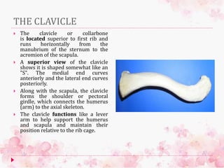
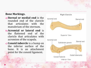
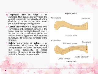
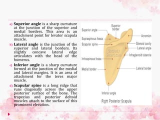
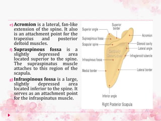
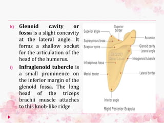
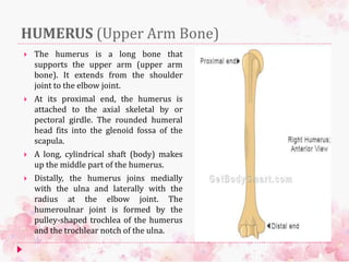
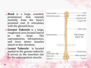
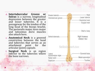
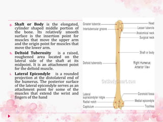
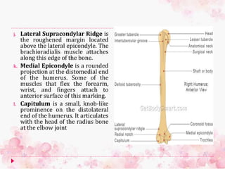
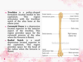
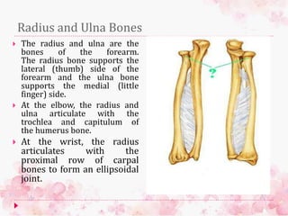
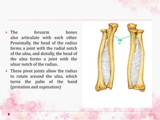
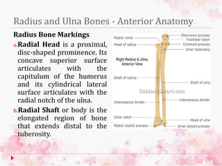
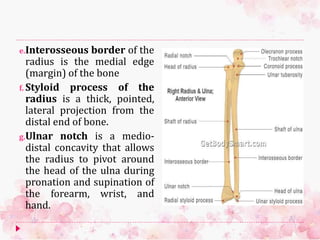
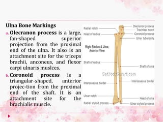
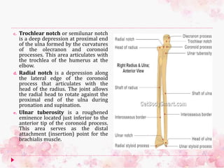
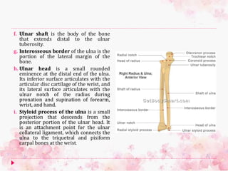
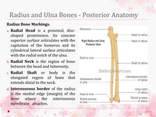
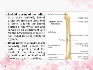
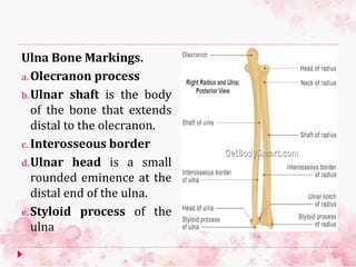
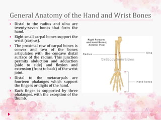
The document summarizes the bones of the upper limb, including the clavicle, scapula, humerus, radius, and ulna. It describes the location and markings of each bone, as well as their articulations with other bones and attachments of muscles. Key points include that the clavicle and scapula form the shoulder girdle, connecting the humerus to the axial skeleton. The humerus extends from the shoulder to elbow joints. The radius and ulna articulate at the elbow and wrist, allowing rotation of the forearm.
2. THE CLAVICLE
The clavicle or collarbone
is located superior to first rib and
runs horizontally from the
manubrium of the sternum to the
acromion of the scapula.
A superior view of the clavicle
shows it is shaped somewhat like an
"S". The medial end curves
anteriorly and the lateral end curves
posteriorly.
Along with the scapula, the clavicle
forms the shoulder or pectoral
girdle, which connects the humerus
(arm) to the axial skeleton.
The clavicle functions like a lever
arm to help support the humerus
and scapula and maintain their
position relative to the rib cage.
3. Bone Markings.
a) Sternal or medial end is the
rounded end of the clavicle
that articulates with the
manubrium of the sternum.
b)Acromial or lateral end is
the flattened end of the
clavicle that articulates with
acromion of the scapula.
c) Conoid tubercle is a bump on
the inferior surface of the
bone. It is an attachment
point for the conoid ligament.
4. d) Trapezoid line or ridge is an
elevation that runs obliquely from the
conoid tubercle to the lateral end of the
clavicle. It serves as an attachment
point for the trapezoid ligament.
e) Costal tuberosity is a roughened oval
elevation on the inferior surface of the
bone, near the medial (sternal) end. It
serves as an attachment point for
costoclavicular ligament, that attaches
the clavicle to the costal cartilage of the
first rib.
f) Subclavian groove or sulcus is an
indentation that runs horizontally
along inferior surface of the bone, from
the costal tuberosity to conoid
tubercle. It serves as an attachment
point for the subclavius muscle.
5. THE SCAPULA.
The scapula or shoulder blade is a flat,
triangular-shaped bone that lies
adjacent to the posterior surface of ribs
2-7.
It has three borders (superior, lateral,
medial), three angles (superior, lateral,
medial) and two surfaces (costal,
dorsal). A prominent ridge or spine
divides the dorsal surface into two,
unequal parts called the supraspinous
fossa and infraspinous fossa.
The scapula also articulates with the
clavicle to form the pectoral girdle,
which supports movements of the
humerus.
Seventeen muscles attach to the
borders, angles, ridges, bumps,
processes, and fossae found on the
surface of the scapula.
6. a) Superior border or margin is
the upper edge of the scapula
that runs next to the clavicle. The
omohyoid muscle attaches along
this surface.
b) Medial border or margin is the
medial edge of the scapula and is
also called the vetebral border.
This area is an attachment point
for the rhomboid major,
rhomboid minor and serratus
anterior muscles.
c) Lateral border or margin is the
lateral edge of the scapula. The
teres minor muscle attaches
along this surface, which is also
called the axillary border.
7. d) Superior angle is a sharp curvature
at the junction of the superior and
medial borders. This area is an
attachment point for levator scapula
muscle.
e) Lateral angle is the junction of the
superior and lateral borders. Its
slightly concave lateral edge
articulates with the head of the
humerus.
f) Inferior angle is a sharp curvature
formed at the junction of the medial
and lateral margins. It is an area of
attachment for the teres major
muscle.
g) Coracoid process is a curved, hook-
like anterolateral projection located
above the lateral angle
8. h) Suprascapular (scapular) notch is
a small groove located medial to the
coracoid process. It forms a
passageway for the suprascapular
nerve.
i) Glenoid cavity or fossa is a slight
concavity at the lateral angle. It
forms a shallow socket for the
articulation of the head of the
humerus.
j) Infraglenoid tubercle is a small
prominence on the inferior margin of
the glenoid fossa. It serves as an
attachment point for the long head of
the triceps brachii muscle
k) Subscapular fossa is a large, slightly
depressed region in the middle of
scapula's anterior surface. The
subscapularis muscle attaches to this
region of the bone.
9. a) Superior angle is a sharp curvature
at the junction of the superior and
medial borders. This area is an
attachment point for levator scapula
muscle.
b) Lateral angle is the junction of the
superior and lateral borders. Its
slightly concave lateral edge
articulates with the head of the
humerus.
c) Inferior angle is a sharp curvature
formed at the junction of the medial
and lateral margins. It is an area of
attachment for the teres major
muscle.
d) Scapular spine is a long ridge that
runs diagonally across the upper
posterior surface of the bone. The
trapezius and posterior deltoid
muscles attach to the surface of this
prominent elevation.
10. e) Acromion is a lateral, fan-like
extension of the spine. It also
is an attachment point for the
trapezius and posterior
deltoid muscles.
f) Supraspinous fossa is a
slightly depressed area
located superior to the spine.
The supraspinatus muscle
attaches to this region of the
scapula.
g) Infraspinous fossa is a large,
slightly depressed area
located inferior to the spine. It
serves as an attachment point
for the infraspinatus muscle.
11. h) Glenoid cavity or
fossa is a slight concavity
at the lateral angle. It
forms a shallow socket
for the articulation of the
head of the humerus.
i) Infraglenoid tubercle is
a small prominence on
the inferior margin of the
glenoid fossa. The long
head of the triceps
brachii muscle attaches
to this knob-like ridge
12. HUMERUS (Upper Arm Bone)
The humerus is a long bone that
supports the upper arm (upper arm
bone). It extends from the shoulder
joint to the elbow joint.
At its proximal end, the humerus is
attached to the axial skeletal by or
pectoral girdle. The rounded humeral
head fits into the glenoid fossa of the
scapula.
A long, cylindrical shaft (body) makes
up the middle part of the humerus.
Distally, the humerus joins medially
with the ulna and laterally with the
radius at the elbow joint. The
humeroulnar joint is formed by the
pulley-shaped trochlea of the humerus
and the trochlear notch of the ulna.
13. a. Head is a large, rounded,
prominence that extends
medially from the bone's
proximal end. It articulates
with the glenoid fossa
b. Greater Tubercle is a large,
roughened area located lateral
to the head. The
supraspinatus, infraspinatus,
and teres minor muscles
attach to this elevation.
c. Lesser Tubercle is located
medial to the greater tubercle.
It serves as the insertion site
for the subscapularis muscle.
14. d. Intertubercular Groove or
Sulcus is a narrow, longitudinal
depression between the greater
and lesser tubercles. It is a
passageway for the tendon of the
long head of the biceps brachii.
The pectoralis major, teres major
and latissimus dorsi muscles
also attach here.
e. Anatomical Neck is a grooved
constriction between the head
and tubercles that serves as an
attachment point for the
articular (joint) capsule.
f. Surgical Neck is an region
inferior to the tubercles where
the bone narrows.
15. g. Shaft or Body is the elongated,
cylinder shaped middle portion of
the bone. Its relatively smooth
surface is the insertion point for
muscles that move the upper arm
and the origin point for muscles that
move the lower arm.
h. Deltoid Tuberosity is a raised,
roughened area located on the
lateral side of the shaft at its
midpoint. It is an attachment point
for the deltoid muscle.
i. Lateral Epicondyle is a rounded
projection at the distolateral end of
the humerus. The posterior surface
of the lateral epicondyle serves as an
attachment point for some of the
muscles that extend the wrist and
fingers of the hand
16. j. Lateral Supracondylar Ridge is
the roughened margin located
above the lateral epicondyle. The
brachioradialis muscle attaches
along this edge of the bone.
k. Medial Epicondyle is a rounded
projection at the distomedial end
of the humerus. Some of the
muscles that flex the forearm,
wrist, and fingers attach to
anterior surface of this marking.
l. Capitulum is a small, knob-like
prominence on the distolateral
end of the humerus. It articulates
with the head of the radius bone
at the elbow joint
17. m. Trochlea is a pulley-shaped
formation located medial to the
capitulum. This region
articulates with the trochlear
notch of the ulna bone at the
elbow joint.
n. Coronoid Fossa is a depression
located superior to the anterior
aspect of the trochlea. This
region provides space for the
coronoid process of the ulna
when the forearm is flexed.
o. Radial Notch is a small
depression located superior to
the capitulum. This region
provides space for the head of
the radius when the forearm is
flexed.
18. Radius and Ulna Bones
The radius and ulna are the
bones of the forearm.
The radius bone supports the
lateral (thumb) side of the
forearm and the ulna bone
supports the medial (little
finger) side.
At the elbow, the radius and
ulna articulate with the
trochlea and capitulum of
the humerus bone.
At the wrist, the radius
articulates with the
proximal row of carpal
bones to form an ellipsoidal
joint.
19. The forearm bones
also articulate with each other.
Proximally, the head of the radius
forms a joint with the radial notch
of the ulna, and distally, the head of
the ulna forms a joint with the
ulnar notch of the radius.
These pivot joints allow the radius
to rotate around the ulna, which
turns the palm of the hand
(protation and supination)
20. Radius and Ulna Bones - Anterior Anatomy
Radius Bone Markings
a.Radial Head is a proximal,
disc-shaped prominence. Its
concave superior surface
articulates with the
capitulum of the humerus
and its cylindrical lateral
surface articulates with the
radial notch of the ulna.
b.Radial Shaft or body is the
elongated region of bone
that extends distal to the
tuberosity.
21. e.Interosseous border of the
radius is the medial edge
(margin) of the bone
f. Styloid process of the
radius is a thick, pointed,
lateral projection from the
distal end of bone.
g.Ulnar notch is a medio-
distal concavity that allows
the radius to pivot around
the head of the ulna during
pronation and supination of
the forearm, wrist, and
hand.
22. Ulna Bone Markings
a. Olecranon process is a large,
fan-shaped superior
projection from the proximal
end of the ulna. It also is an
attachment site for the triceps
brachii, anconeus, and flexor
carpi ulnaris muslces.
b. Coronoid process is a
triangular-shaped, anterior
projec-tion from the proximal
end of the shaft. It is an
attachment site for the
brachialis muscle.
23. c. Trochlear notch or semilunar notch
is a deep depression at proximal end
of the ulna formed by the curvatures
of the olecranon and coronoid
processes. This area articulates with
the trochlea of the humerus at the
elbow.
d. Radial notch is a depression along
the lateral edge of the coronoid
process that articulates with the
head of the radius. The joint allows
the radial head to rotate against the
proximal end of the ulna during
pronation and supination.
e. Ulnar tuberosity is a roughened
eminence located just inferior to the
anterior tip of the coronoid process.
This area serves as the distal
attachment (insertion) point for the
brachialis muscle.
24. f. Ulnar shaft is the body of the bone
that extends distal to the ulnar
tuberosity.
g. Interosseous border of the ulna is the
portion of the lateral margin of the
bone.
h. Ulnar head is a small rounded
eminence at the distal end of the ulna.
Its inferior surface articulates with the
articular disc cartilage of the wrist, and
its lateral surface articulates with the
ulnar notch of the radius during
pronation and supination of forearm,
wrist, and hand.
i. Styloid process of the ulna is a small
projection that descends from the
posterior portion of the ulnar head. It
is an attachment point for the ulnar
collateral ligament, which connects the
ulna to the triquetral and pisiform
carpal bones at the wrist.
25. Radius and Ulna Bones - Posterior Anatomy
Radius Bone Markings.
a. Radial Head is a proximal, disc-
shaped prominence. Its concave
superior surface articulates with the
capitulum of the humerus and its
cylindrical lateral surface articulates
with the radial notch of the ulna.
b. Radial Neck is the region of bone
between the head and tuberosity.
c. Radial Shaft or body is the
elongated region of bone that
extends distal to the neck.
d. Interosseous border of the radius
is the medial edge (margin) of the
bone where the interosseous
membrane attaches.
26. e. Styloid process of the radius
is a thick, pointed, lateral
projection from the distal end
of bone. It forms the lateral
portion of the wrist joint and
serves as an attachment site
for the brachioradialis muscle
and radial (lateral) collateral
ligament.
f. Ulnar notch is a medio distal
concavity that allows the
radius to pivot around the
head of the ulna during
pronation and supination of
the forearm, wrist, and hand.
27. Ulna Bone Markings.
a.Olecranon process
b.Ulnar shaft is the body
of the bone that extends
distal to the olecranon.
c. Interosseous border
d.Ulnar head is a small
rounded eminence at the
distal end of the ulna.
e.Styloid process of the
ulna
28. General Anatomy of the Hand and Wrist Bones
Distal to the radius and ulna are
twenty-seven bones that form the
hand.
Eight small carpal bones support the
wrist (carpus).
The proximal row of carpal bones is
convex and two of the bones
articulates with the concave distal
surface of the radius. This junction
permits abduction and adduction
(side to side) and flexion and
extension (front to back) of the wrist
joint.
Distal to the metacarpals are
fourteen phalanges which support
the fingers or digits of the hand.
Each finger is supported by three
phalanges, with the exception of the
thumb.
29. Carpal Bones: Proximal Row.
a. Scaphoid bone is a boat-shaped bone
located on the lateral (thumb) side of
the carpus (wrist). It articulates with
the distal end of the radius bone.
b. Lunate bone is cresent-shaped bone
located medial to the scaphoid bone.
Its concave proximal surface also
articulates with the distal end of the
radius bone.
c. Triquetral bone is a pyramid-shaped
bone located medial to the lunate
bone on the ulnar side of the carpus.
d. Pisiform bone is a small, pea-shaped
bone that articulates with the
triquetral bone on the ulnar side of
the carpus. It is embedded in the
tendon of the flexor carpi ulnaris
muscle and increases tension during
muscle contraction.
30. Carpal Bones: Distal Row.
a. Trapezium bone is an irregular-shaped bone
located distal to the scaphoid bone on the
lateral (thumb) side of the carpus. It is the
radial (lateral) border of the carpal tunnel,
and its distal surface articulates with the
proximal surface of the first metacarpal
(thumb) bone.
b. Trapezoid bone is a small, four-sided bone
that is wedged between the trapezium and
capitate bones. It articulates with the
proximal end of the second metacarpal bone.
c. Capitate bone is located medial to the
trapezium in the center of the distal row of
the carpus. The name of the bone is based on
its rounded proximal head which fits into the
concavities of scaphoid and lunate bones.
Distally, the capitate bone articulates with the
proximal end of the third metacarpal bone.
d. Hamate bone is located on the ulnar side of
the carpus. Projecting from its palmar surface
is a hook-like process that forms the ulnar
(medial) border of the carpal tunnel (L.,
hamat, hooked). The hamate bone articulates
distally with the fourth and fifth metacarpal
bones.
31. Metacarpal Bones
Metacarpals I - V (ossa
metacarpi) are the bones
that support the palm of
the hand. Each consists of a
elongated body (shaft),
cuboid base, and convex
head. The bases articulate
with the distal row of the
carpus and the heads
articulate with the
proximal phalanges. Each
bone is assigned a number
from 1 to 5, starting on the
lateral side (thumb side) of
the hand.
32. Phalanges.
The phalanges (singular: phalanx) are the
fourteen bones that support the fingers or
digits of the hand. Each finger is supported
by three phalanges, exception of the thumb,
which is supported by two.
a. Proximal: I - V are the phalanges located
directly anterior to metatarsal bones. The
proximal phalanges articulate anteriorly
with the middle phalanges, except the
first proxmial phalanx, which articulates
with first distal phalanx.
b. Middle: II - V are the phalanges located
between the proximal and distal
phalanges.
c. Distal: I – V are the phalanges that
support the tips of the fingers. All
articulate posteriorly with a middle
phalanx, except the first distal phalanx,
which articulates with the first proximal
phalanx