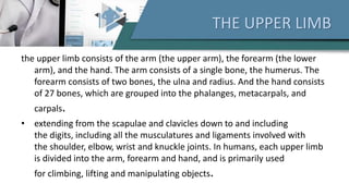
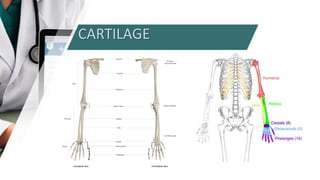
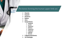
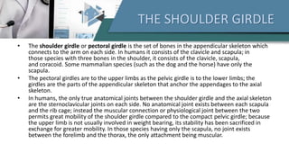
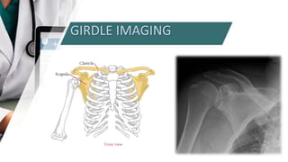
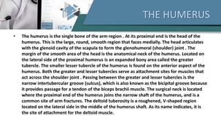
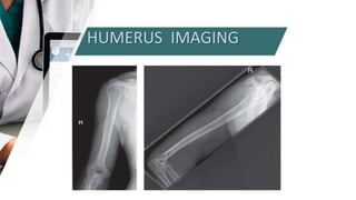
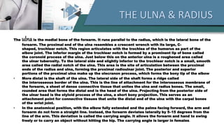
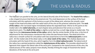
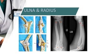
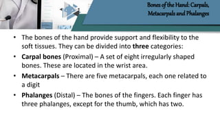
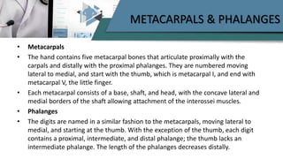
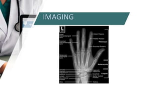
2. THE UPPER LIMB
the upper limb consists of the arm (the upper arm), the forearm (the lower
arm), and the hand. The arm consists of a single bone, the humerus. The
forearm consists of two bones, the ulna and radius. And the hand consists
of 27 bones, which are grouped into the phalanges, metacarpals, and
carpals.
• extending from the scapulae and clavicles down to and including
the digits, including all the musculatures and ligaments involved with
the shoulder, elbow, wrist and knuckle joints. In humans, each upper limb
is divided into the arm, forearm and hand, and is primarily used
for climbing, lifting and manipulating objects.
5. THE SHOULDER GIRDLE
• The shoulder girdle or pectoral girdle is the set of bones in the appendicular skeleton which
connects to the arm on each side. In humans it consists of the clavicle and scapula; in
those species with three bones in the shoulder, it consists of the clavicle, scapula,
and coracoid. Some mammalian species (such as the dog and the horse) have only the
scapula.
• The pectoral girdles are to the upper limbs as the pelvic girdle is to the lower limbs; the
girdles are the parts of the appendicular skeleton that anchor the appendages to the axial
skeleton.
• In humans, the only true anatomical joints between the shoulder girdle and the axial skeleton
are the sternoclavicular joints on each side. No anatomical joint exists between each scapula
and the rib cage; instead the muscular connection or physiological joint between the two
permits great mobility of the shoulder girdle compared to the compact pelvic girdle; because
the upper limb is not usually involved in weight bearing, its stability has been sacrificed in
exchange for greater mobility. In those species having only the scapula, no joint exists
between the forelimb and the thorax, the only attachment being muscular.
7. THE HUMERUS
• The humerus is the single bone of the arm region . At its proximal end is the head of the
humerus. This is the large, round, smooth region that faces medially. The head articulates
with the glenoid cavity of the scapula to form the glenohumeral (shoulder) joint . The
margin of the smooth area of the head is the anatomical neck of the humerus. Located on
the lateral side of the proximal humerus is an expanded bony area called the greater
tubercle. The smaller lesser tubercle of the humerus is found on the anterior aspect of the
humerus. Both the greater and lesser tubercles serve as attachment sites for muscles that
act across the shoulder joint . Passing between the greater and lesser tubercles is the
narrow intertubercular groove (sulcus), which is also known as the bicipital groove because
it provides passage for a tendon of the biceps brachii muscle. The surgical neck is located
where the proximal end of the humerus joins the narrow shaft of the humerus, and is a
common site of arm fractures. The deltoid tuberosity is a roughened, V-shaped region
located on the lateral side in the middle of the humerus shaft. As its name indicates, it is
the site of attachment for the deltoid muscle.
9. THE ULNA & RADIUS
The ulnais the medial bone of the forearm. It runs parallel to the radius, which is the lateral bone of the
forearm. The proximal end of the ulna resembles a crescent wrench with its large, C-
shaped, trochlear notch. This region articulates with the trochlea of the humerus as part of the
elbow joint. The inferior margin of the trochlear notch is formed by a prominent lip of bone called
the coronoid process of the ulna. Just below this on the anterior ulna is a roughened area called
the ulnar tuberosity. To the lateral side and slightly inferior to the trochlear notch is a small, smooth
area called the radial notch of the ulna. This area is the site of articulation between the proximal
ends of the radius and ulna, forming the proximal radioulnar joint. The posterior and superior
portions of the proximal ulna make up the olecranon process, which forms the bony tip of the elbow
• More distal is the shaft of the ulna. The lateral side of the shaft forms a ridge called
the interosseous border of the ulna. This is the line of attachment for the interosseous membrane of
the forearm, a sheet of dense connective tissue that unites the ulna and radius bones. The small,
rounded area that forms the distal end is the head of the ulna. Projecting from the posterior side of
the ulnar head is the styloid process of the ulna, a short bony projection. This serves as an
attachment point for connective tissues that unite the distal end of the ulna with the carpal bones
of the wrist joint.
• In the anatomical position, with the elbow fully extended and the palms facing forward, the arm and
forearm do not form a straight line. Instead, the forearm deviates laterally by 5–15 degrees from the
line of the arm. This deviation is called the carrying angle. It allows the forearm and hand to swing
freely or to carry an object without hitting the hip. The carrying angle is larger in females.
10. THE ULNA & RADIUS
• The radiusruns parallel to the ulna, on the lateral (thumb) side of the forearm . The head of the radius is
a disc-shaped structure that forms the proximal end. The small depression on the surface of the head
articulates with the capitulum of the humerus as part of the elbow joint, whereas the smooth, outer
margin of the head articulates with the radial notch of the ulna at the proximal radioulnar joint. The neck
of the radius is the narrowed region immediately below the expanded head. Inferior to this point on the
medial side is the radial tuberosity, an oval-shaped, bony protuberance that serves as a muscle
attachment point. The shaft of the radius is slightly curved and has a small ridge along its medial side. This
ridge forms the interosseous border of the radius, which, like the similar border of the ulna, is the line of
attachment for the interosseous membrane that unites the two forearm bones. The distal end of the
radius has a smooth surface for articulation with two carpal bones to form the radiocarpal joint or wrist
joint . On the medial side of the distal radius is the ulnar notch of the radius. This shallow depression
articulates with the head of the ulna, which together form the distal radioulnar joint. The lateral end of
the radius has a pointed projection called the styloid process of the radius. This provides attachment for
ligaments that support the lateral side of the wrist joint. Compared to the styloid process of the ulna, the
styloid process of the radius projects more distally, thereby limiting the range of movement for lateral
deviations of the hand at the wrist joint
12. Bones of the Hand: Carpals,
Metacarpals and Phalanges
• The bones of the hand provide support and flexibility to the
soft tissues. They can be divided into three categories:
• Carpal bones (Proximal) – A set of eight irregularly shaped
bones. These are located in the wrist area.
• Metacarpals – There are five metacarpals, each one related to
a digit
• Phalanges (Distal) – The bones of the fingers. Each finger has
three phalanges, except for the thumb, which has two.
13. CARPALS
• The eight, irregularly shaped carpals are the most proximal bones of the hand. The
carpals are often split into two rows, the proximal row containing the scaphoid,
lunate, triquetrum, and pisiform, moving lateral to medial.
• The scaphoid and lunate articulate with the radius, and the lunate and triquetrum
articulate with the articular disk of the wrist. The pisiform carpal is a sesamoid
bone, located within a tendon and is not involved in movement at the wrist.
• The distal row contains the trapezium, trapezoid, capitate, and hamate, moving
lateral to medial. The trapezium articulates with the scaphoid proximally and the
first, thumb, and second metacarpal distally. The trapezoid articulates with the
scaphoid proximally and the second metacarpal distally.
14. METACARPALS & PHALANGES
• Metacarpals
• The hand contains five metacarpal bones that articulate proximally with the
carpals and distally with the proximal phalanges. They are numbered moving
lateral to medial, and start with the thumb, which is metacarpal I, and end with
metacarpal V, the little finger.
• Each metacarpal consists of a base, shaft, and head, with the concave lateral and
medial borders of the shaft allowing attachment of the interossei muscles.
• Phalanges
• The digits are named in a similar fashion to the metacarpals, moving lateral to
medial, and starting at the thumb. With the exception of the thumb, each digit
contains a proximal, intermediate, and distal phalange; the thumb lacks an
intermediate phalange. The length of the phalanges decreases distally.
16. UPPER LIMB MUSCLES
• The upper limb comprises many muscles which are organized into anatomical
compartments. These muscles act on the various joints of the hand, arm, and
shoulder, maintaining tone, providing stability and allowing precise fluid
movement.
• Axioappendicular groups of muscles arise from the axial skeleton to act upon the
pectoral girdle. Scapulohumeral muscles originate from the scapula and insert
into the proximal humerus. Included in this category are the rotator cuff muscles
which provide stability to the glenohumeral joint. In the arm, the muscles of the
anterior compartment are involved in flexion of the forearm, and the posterior
comprises of the forearm extensors. Similarly, the anterior compartment of the
forearm contains the flexors of the hand and posterior has extensors. The hand is
divided into the thenar, the hypothenar, the adductor compartment, as well as
the short muscles of the hand
17. UPPER LIMB MUSCLES
• Shoulder and Upper limb muscles are specialized to perform functions of
pressure and manipulation of objects. Skeletal muscle is formed by
myofibers which contain millions of myofibrils, and each of them is
formed by sarcomeres. Sarcomeres are the contractile unit, therefore
skeletal muscle fibers are completely dedicated to generating force.
Skeletal muscle also carries out multiple functions such as voluntary
locomotion, the protection of internal organs, generation of heat, and
assisting in postural behavior.
• The muscle mass relies on the balance between synthesis and
degradation of proteins, the regulation of this process is sensitive to
several factors such as nutritional status, hormone levels, physical
activity, underlying diseases, injuries, among others