Downloaded 290 times
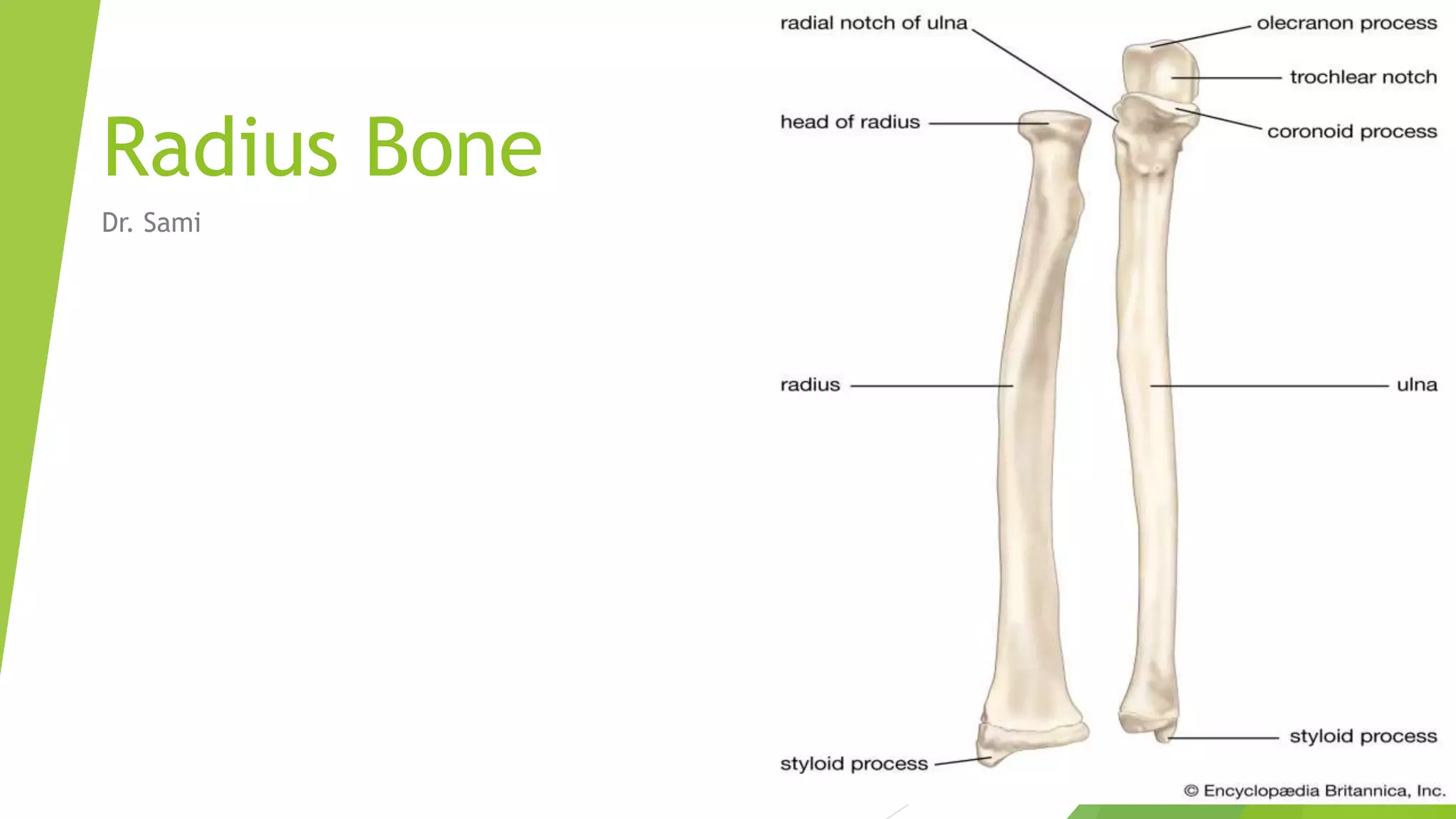
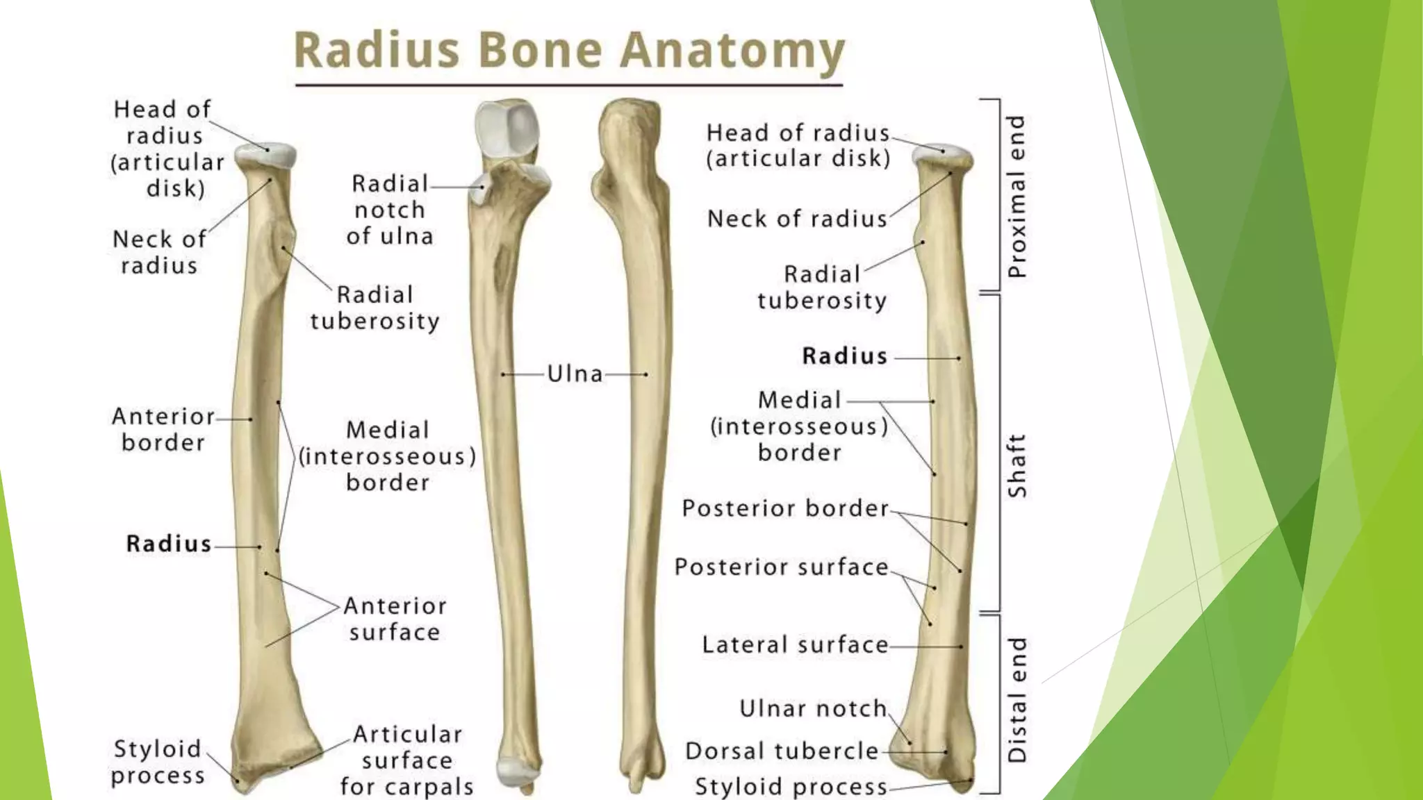

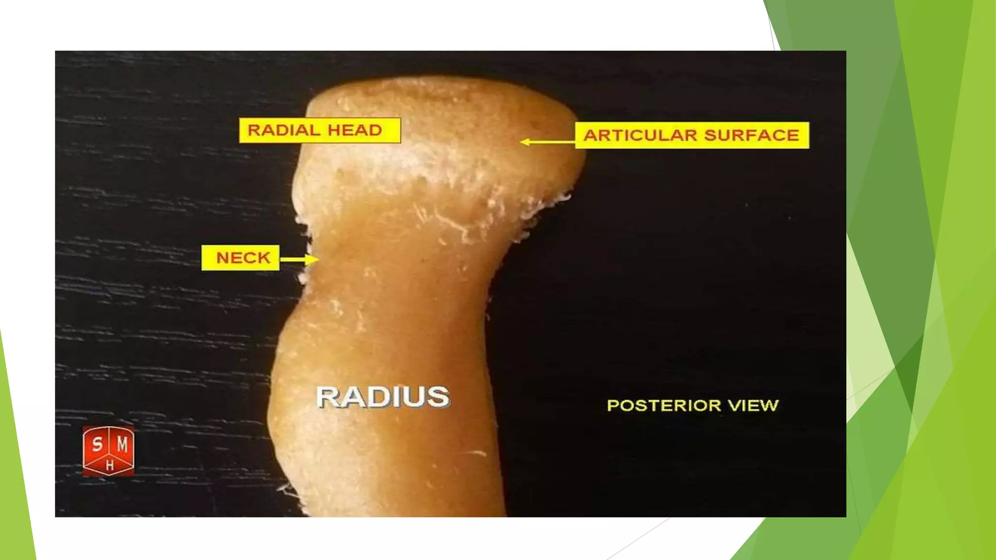
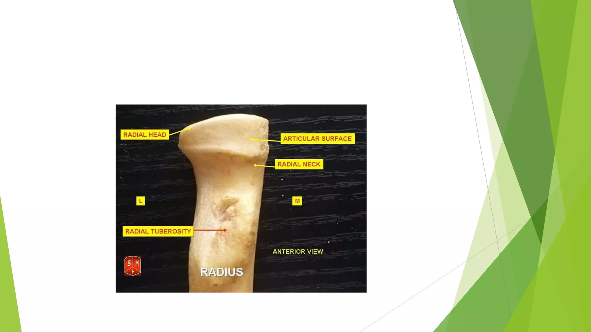

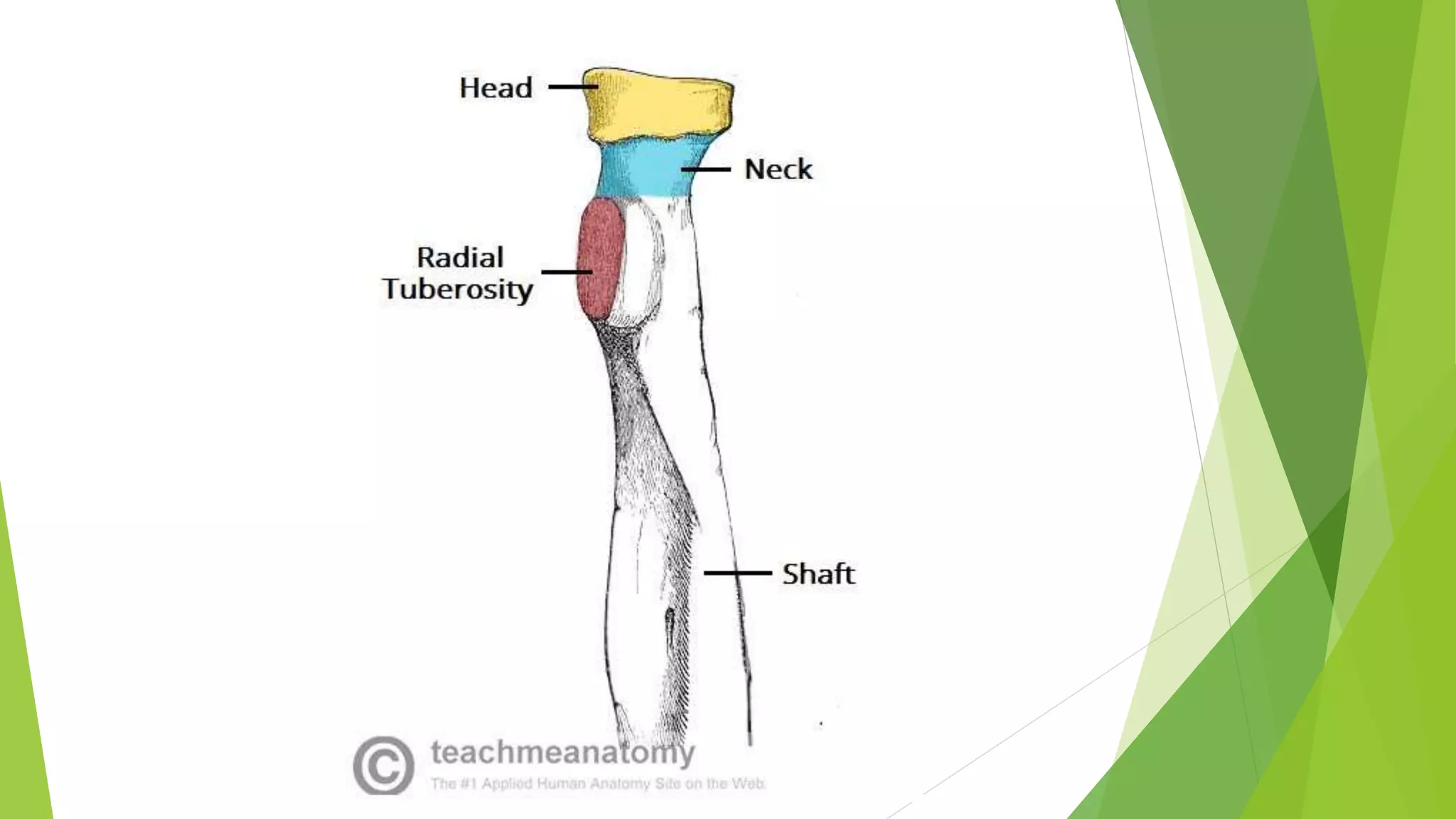

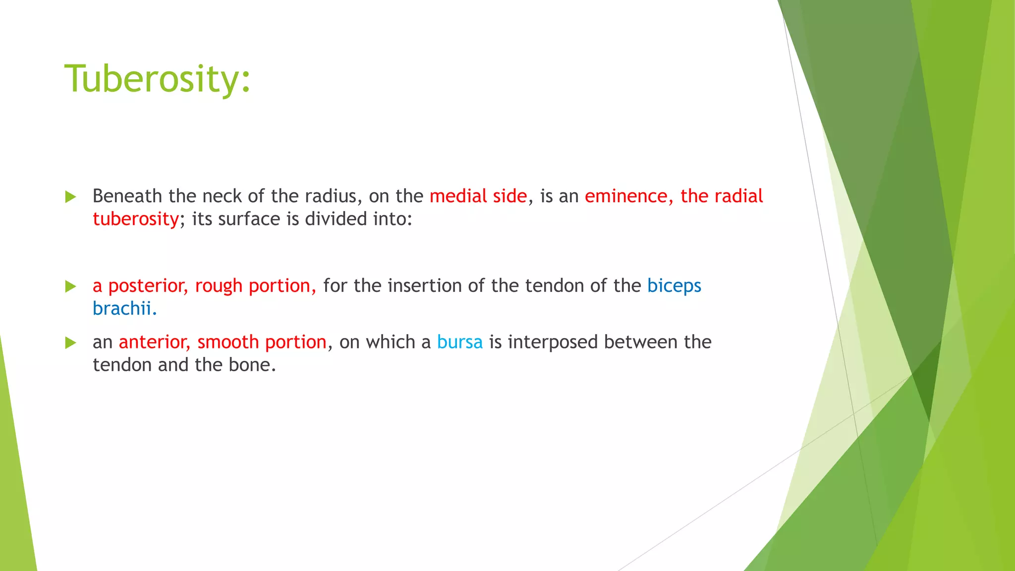
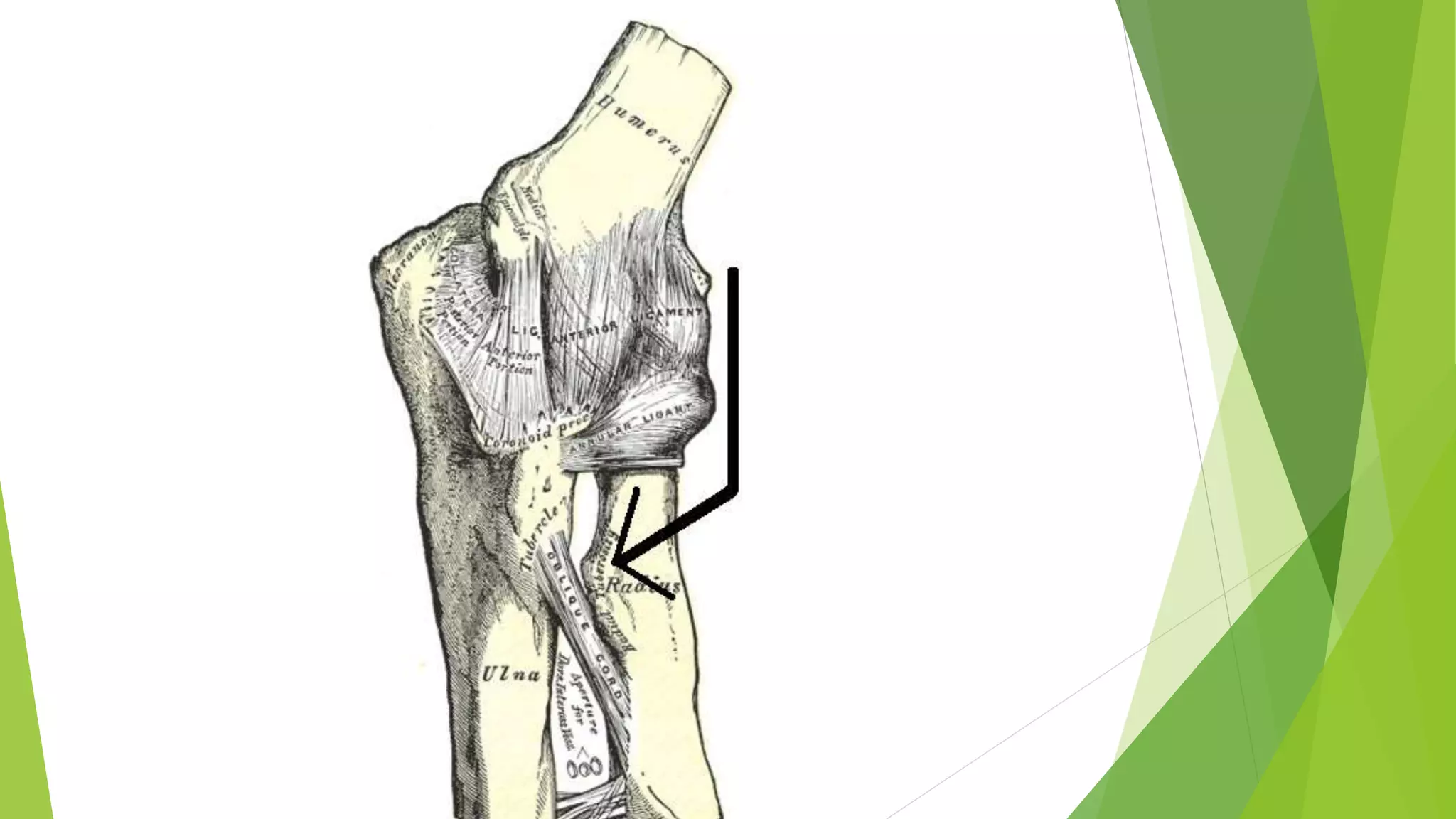
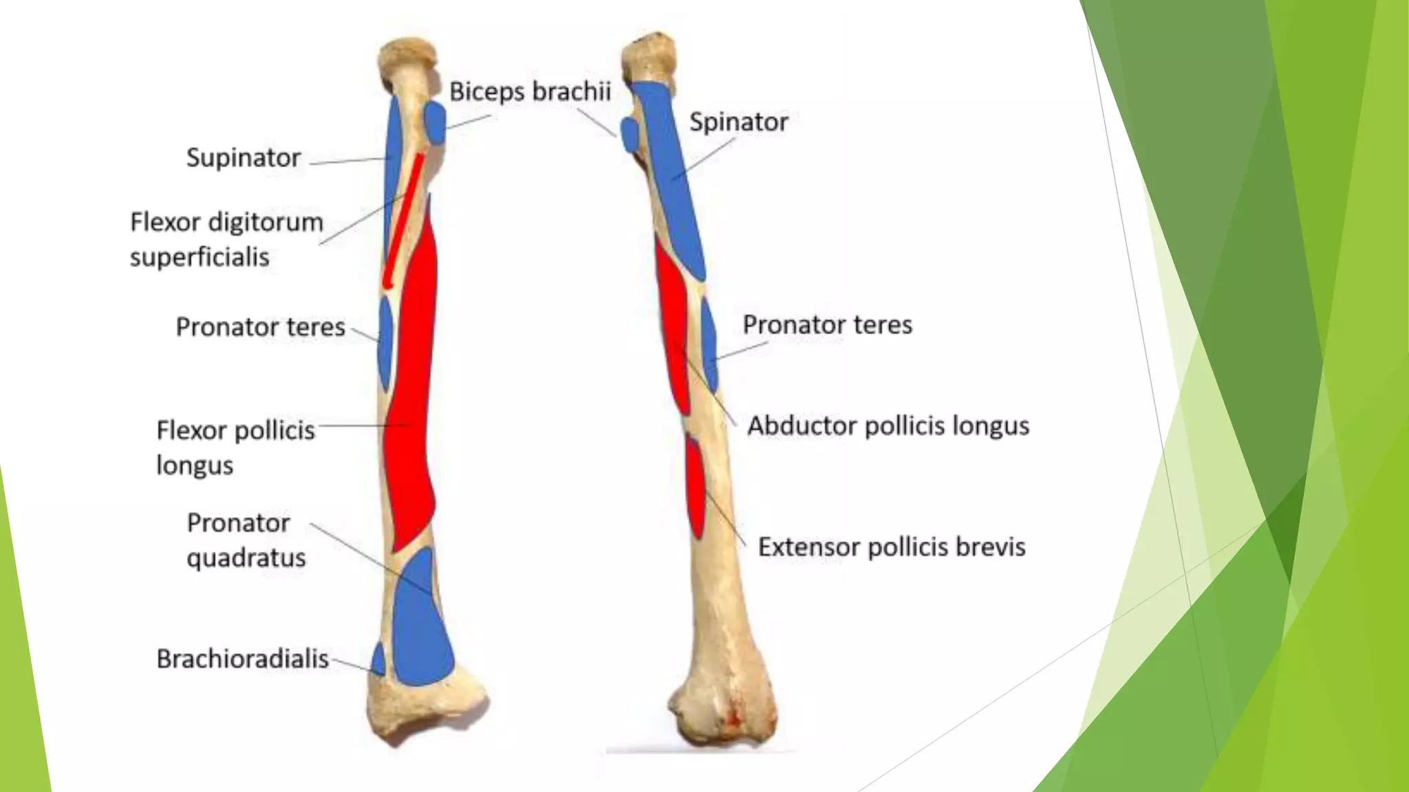

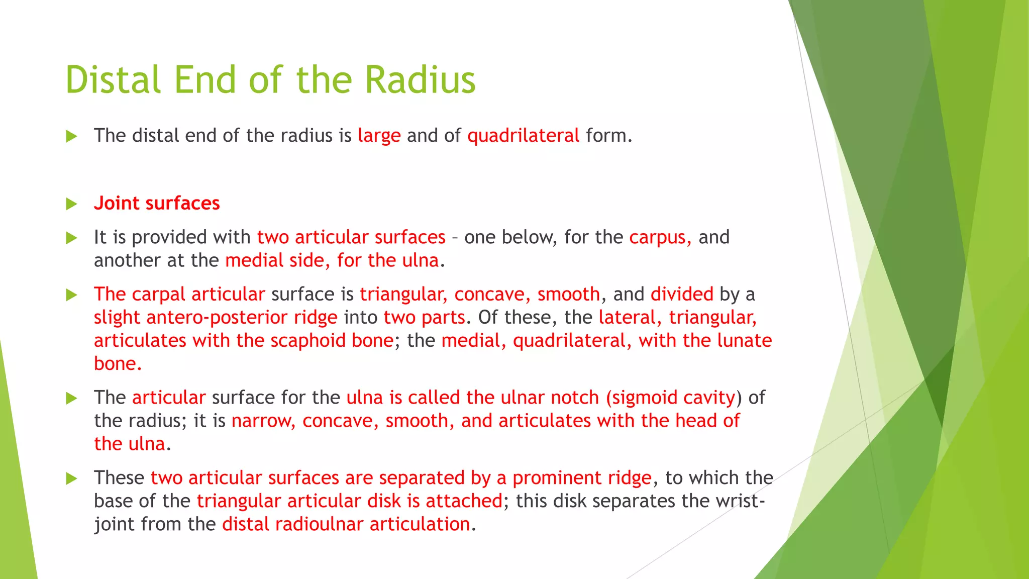
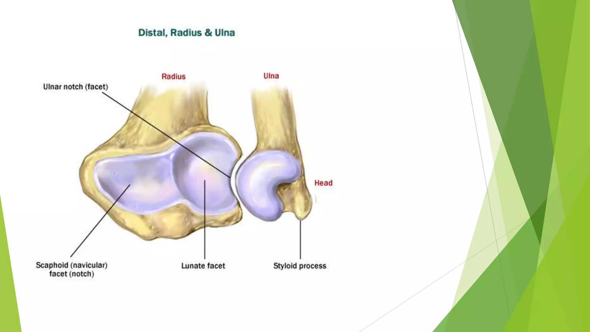


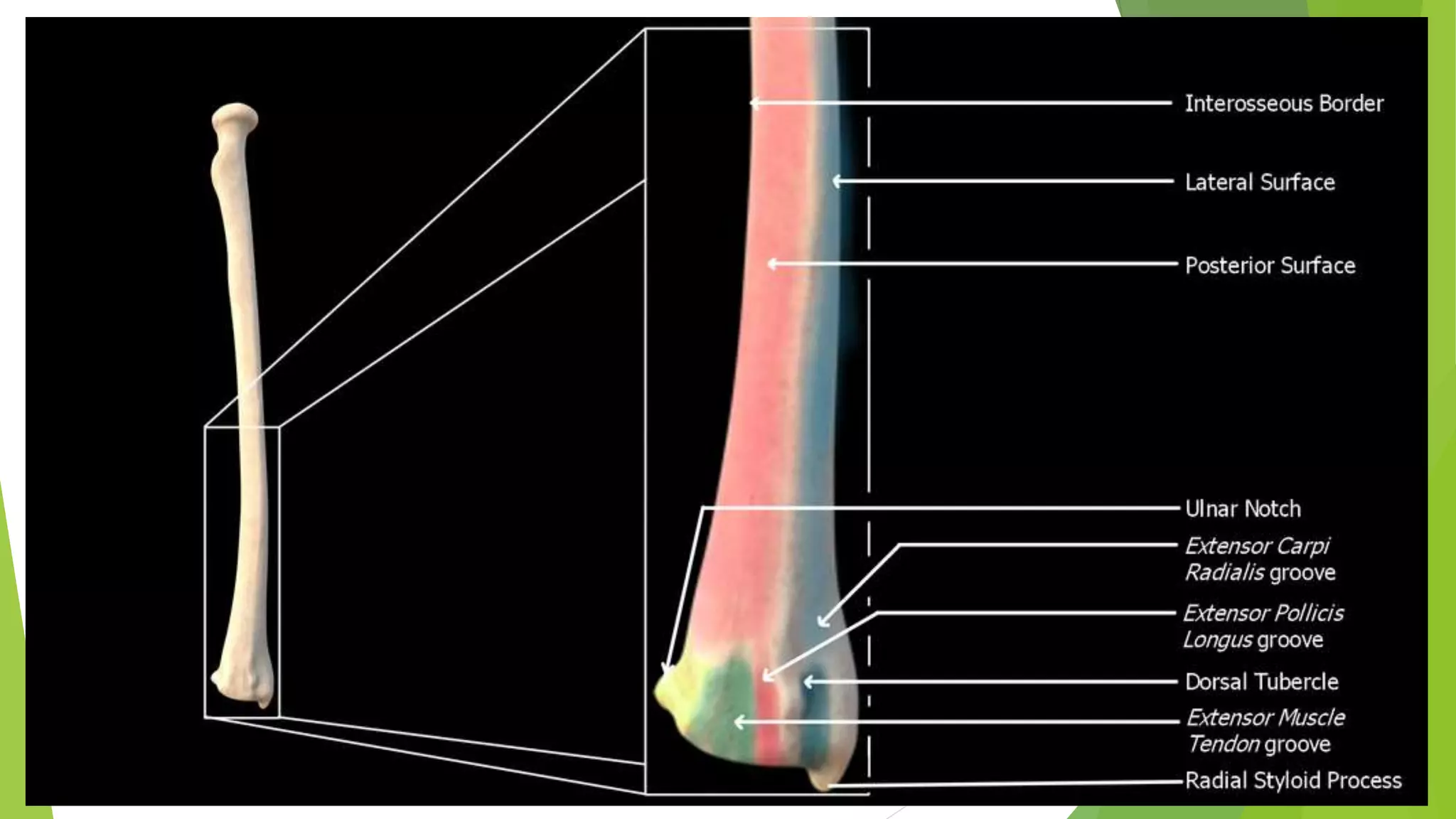
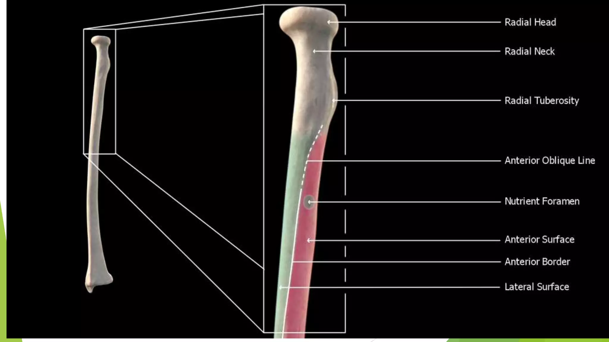
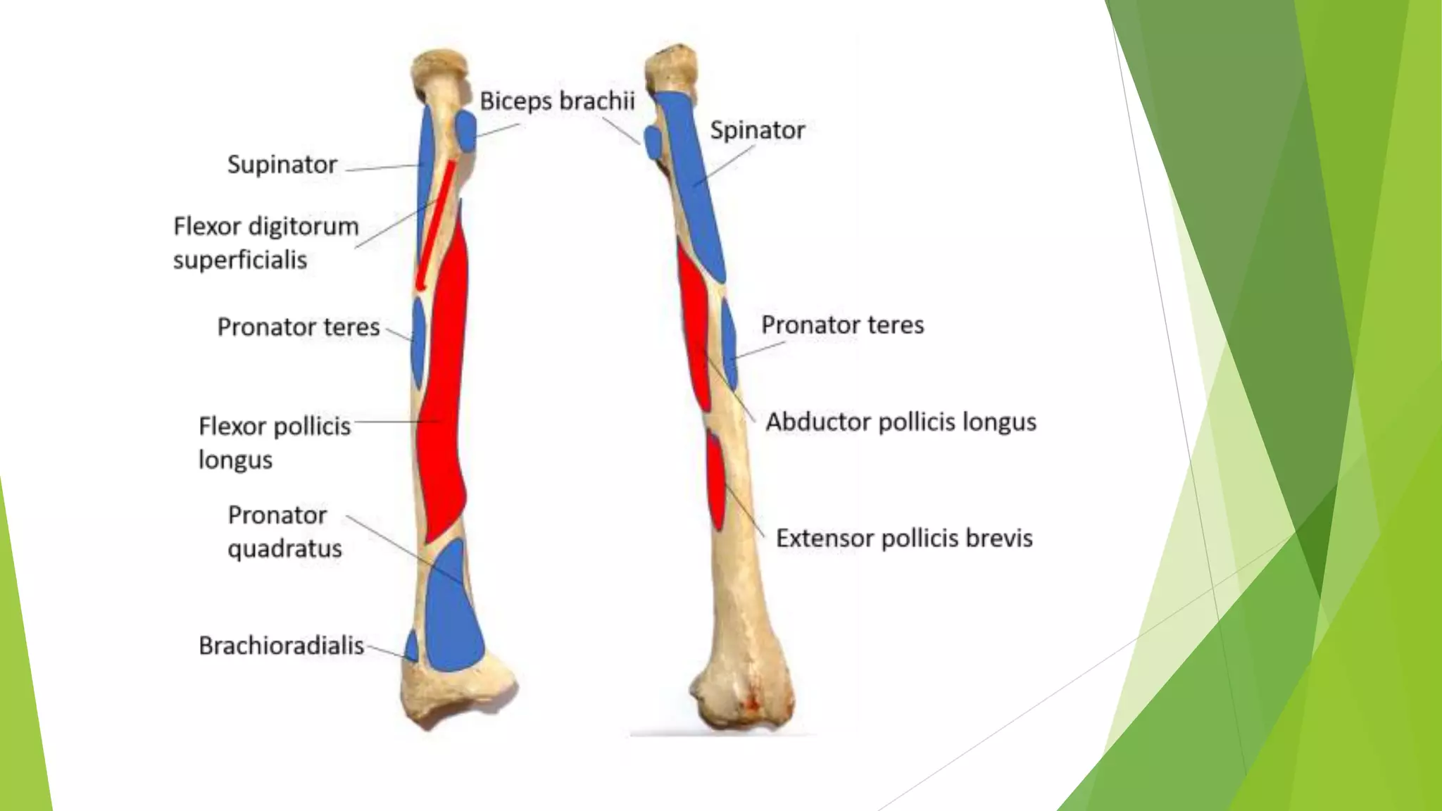
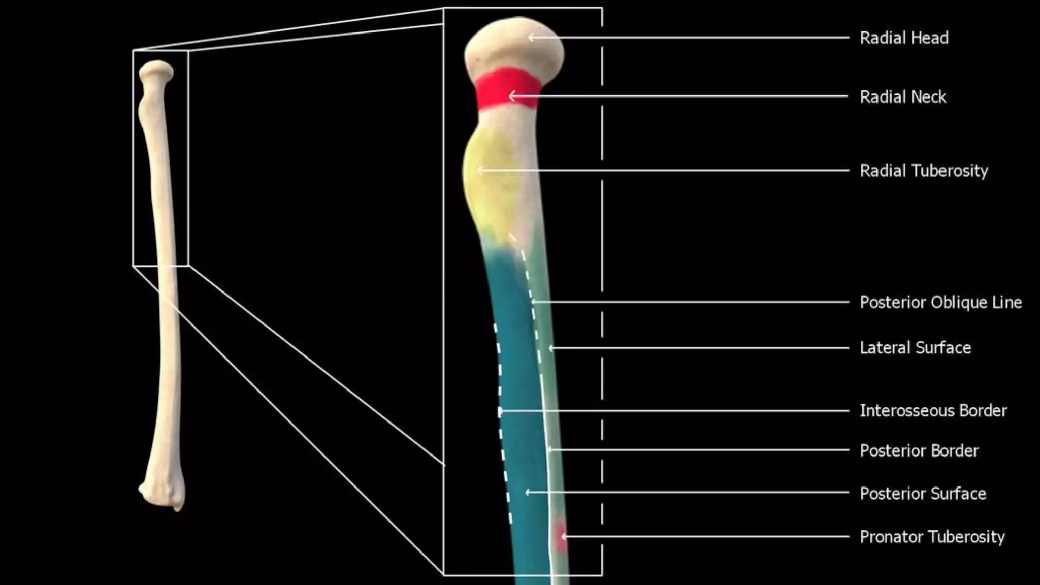


The radius has an upper and lower extremity. The upper extremity includes a head, neck, and tuberosity. The head articulates with the humerus and ulna. The lower extremity has triangular articular surfaces for the carpus and ulna. It also has grooves on its dorsal surface for tendons. The shaft is narrower proximally than distally and has borders for muscle attachments.