SlideShare a Scribd company logo
1 of 24
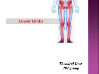
Thendral Doss
204 group
The femur is the only bone in
the thigh. It is classed as a
long bone, and is the longest
bone in the body. The main
function of the femur is to
transmit forces from the tibia
to the hip joint.
It acts as the site of origin and
attachment of many muscles
and ligaments, and can be
divided into three areas;
1. proximal epiphysis - upper
end
2. shaft (body)
3. distal epiphysis - lower
end.
The femur
1
2
3
The proximal epiphysis of femur
 Head of femur, caput femoris, constitutes the proximal
epiphysis of the bone and bears a wide articular surface for the
articulation with the lunate surface of the acetabulum;
 Fovea for ligament of head, fovea capitis femoris, resides
on the head of the femur;
 Neck of femur, collum femoris, narrow area below the head.
 Greater trochanter, trochanter major, prominent projection
located on the border between the neck and the shaft;
 Trochanteric fossa, fossa trochanterica, lies on the medial
surface of the greater trochanter;
 Lesser trochanter, trochanter minor, situated medially on
the border between the neck and the shaft;
 Intertrochanteric line, linea intertrochanterica, connects
the trochanters in the front and runs obliquely.
 Intertrochanteric crest, crista intertrochanterica, connects
the trochanters in the back; runs obliquely;
Anterior view Posterior view
fossa trochanterica
The shaft of the femur:
Linea aspera, linea aspera,
extends along the posterior
surface; consists of two lips:
•lateral lip, labium laterale;
•medial lip, labium mediale;
Gluteal tuberosity,
tuberositas glutea, resides at
the upper portion of the lateral
lip of the linea aspera;
Pectineal line, linea pectinea,
lies at the proximal end of the
medial lip of the linea aspera
below the lesser trochanter;
Popliteal surface, facies
poplitea, flat, triangular area,
which resides in the back on the
posterior surface.
The distal epiphysis:
Medial condyle, condylus medialis,
rounded projection, which resides
medially and bears the articular
surface;
Lateral condyle, condylus lateralis,
rounded projection with the articular
surface located laterally. Both condyles
form the distal epiphysis of the femur;
Intercondylar fossa, fossa
intercondylaris, lies between the two
condyles;
Medial epicondyle, epicondylus
medialis, resides above the medial
condyle;
Lateral epicondyle, epicondylus
lateralis, lies above the lateral
condyle;
Patellar surface, facies patellaris,
located in the front between the two
condyles.
Ant view
Post view
The patella (knee-cap) is located at the front
of the knee joint. It attaches superiorly to
the quadriceps tendon and inferiorly to
the patellar ligament.It is classified as
a sesamoid type bone due to its position
within the quadriceps tendon, and is the
largest sesamoid bone in the body.
It is triangular in shape
 A base (upper border )
 An apex (rounded lower tip )
 2 borders (medial & lateral)
 2 surfaces (ant. & post.)
The posterior surface of the patella
articulates with the femur, and is marked
by two facets:
•Medial facet – articulates with the medial
condyle of the femur.
•Lateral facet – articulates with the lateral
condyle of the femur.
The patella
The tibia and fibula are
the bones of the leg.
The shafts of the tibia
and fibula are connected by a
dense interosseous membrane
Bones of the leg
Tibia is situated on the medial side
of the foreleg. The fibula, syno-nys;
os perone (from Greek) — slender
bone. The fibula is situated laterally
in the foreleg.Each of them are a long
tubular bones with the shaft and two
epiphyses — proximal and distal.
The tibia
The proximal epiphysis:
Medial condyle, condylus medialis, medial
expansion with the concave articular surface (facies
articularis superior), which provides articulation
with the medial condyle of the femur;
Lateral condyle, condylus lateralis, lateral
expansion of the bone with the concave articular
surface, which provides articulation with the lateral
condyle of the femur;
Intercondylar eminence, eminentia
intercondylaris, located between the articular
surfaces of the condyles; it consists of the medial
and lateral intercondylar tubercles (tuberculum
intercondylare mediale et latcrale);
Anterior intercondylar area, area
intercondylaris anterior, resides in the front of the
eminence;
Posterior intercondylar area, area
intercondylaris posterior, lies in the back of the
eminence;
Fibular articular facet, facies articularis
fibularis, located on the posterior, inferior surface
of the lateral condyle.
eminentia intercondylaris
area intercondylaris posterior
area
intercondylar
is ant
The shaft of the tibia:
Tibial tuberosity, tuberositas
tibiae, resides superiorly on the
anterior surface of the bone;
Anterior border, margo
anterior, sharp; extends
downward away from the tibial
tuberosity;
Interosseous border, margo
interosseus, faces the fibula;
Medial border, margo medialis;
Medial, lateral, and posterior
surfaces, facies medialis, facies
lateralis, facies posterior;
Soleal line, linea musculi solei,
runs obliquely on the posterior
surface of the upper one-third of
the bone.
Post. view
The distal epiphysis:
 Inferior articular surface, facies
articularis inferior, resides
inferiorly; gives articulation with
the talus;
 Medial malleolus, malleolus
medialis, prominently projecting
process located medially; it bears
an articular surface to provide
articulation with the talus;
 Fibular notch, incisura fibularis,
lies laterally.
The fibula bears the following
structures:
The proximal epiphysis:
 Head of fibula, caput fibulae,
represents the proximal epiphysis of
the bone; it bears the articular
surface, which forms the articular
facet for the tibia (facies articularis
capitis fibulae);
The shaft of the fibula:
The distal epiphysis:
 Lateral malleolus, malleolus
lateralis, expanded distal
epiphysis of the bone, which bears
the articular surface for the talus;
The fibula
The bones of the foot, ossa
pedis, are subdivided into the
three segments —
Tarsus (ossa tarsi)
Metatarsus (ossa metatarsi)
Phalanges (ossa digitorum).
The bones of the
foot
The tarsus, ossa tarsi, is
made up of seven
independent bones, which
are arranged in two rows
— proximal and distal. The
proximal row consists of
two large bones — the
talus and the calcaneus.
The talus, talus, bears the following
structures
Trochlea of talus, trochlea tali, the upper
portion of the bone with the articular surfaces
for the articulation with the shin bones (facies
superior, facies malleolares lateralis et
medialis);
 Head of talus, caput tali, the anterior
convex portion of the bone with the
articular surface for the articulation with
the navicular;
 Neck of talus, collum tali, a narrowing
area behind the head of the bone;
 Lateral process, processus lateralis tali,
directed laterally;
Medial process processus medialis tali
Posterior process, processus posterior tali,
directed backward;
Anterior, medial and posterior facets for
calcaneus, facies, resides interiorly and gives
articulation with the calcaneus.
The calcaneus, calcaneus resides below the talus
and have the following structures:
 Calcaneal tuberosity, tuber calcanei, relatively large projection directed
backward and downward;
 Sustentaculum tali, sustentaculum tali, a process directed medially;
 Anterior, middle, and posterior talar articular surfaces, facies
articulares talares, reside on the superior surface and provide
articulation with the talus;
 Articular surface for cuboid, facies articularis cuboidea, lies at the
distal (anterior) end of the bone.
The distal row of tarsal
bones
 Navicular, os naviculare, situated
between the head of the talus and
cuneiform bones;
 Medial cuneiform, os cuneiforme
mediale, sits in the front and medially
from the navicular and articulates with
the first metatarsal bone;
 Intermediate cuneiform, os cuneiforme
intermedium, resides in the front and
laterally from the navicular; it articulates
with the second
 Lateral cuneiform, os cuneiforme
laterale, sits in front of the navicular and
lateral to the former bone; it articulates
with the third metatarsal;
 Cuboid, os cuboideum, lies on the lateral
side of the foot in front of the calcaneus
and articulates with the forth and fifth
metatarsals.
THE METATARSALS
The metatarsals, ossa metatarsi, include five short tubular bones (I-V),
slightly convex on the plantar surface. Each metatarsal bone consists of the
base, the body, and the head:
 base, basis, faces the tarsus and contains the articular surface for the
articulation with the tarsal bones of the distal row;
 body, corpus, the middle portion of the bone;
 head, caput, articulates with the phalanges. The head of the metatarsal
represents a single epiphysis of the bone (monoepiphysial bones).
THE PHALANGES
Each digital bone, digitus pedis, (except the thumb) consists of three
short tubular bones, which are called phalanges, phalanges. The following
phalanges are distinguished:
 proximal phalanx, phalanx proximalis;
 middle phalanx, phalanx media;
 distal phalanx, phalanx distalis.
The proximal and distal phalanges
possess the base with the articular surface. The head represents a single
epiphysis (monoepiphysial bones). Distal phalanges have flat distal ends
with the tuberosity on it. The great toe, hallux, consists of two phalanges —
proximal and distal.
free lower limb.pptx

More Related Content

Similar to free lower limb.pptx

The Skull and Vertebral column
The Skull and Vertebral columnThe Skull and Vertebral column
The Skull and Vertebral columnSeddie Chitamu
 
skeletal system
skeletal systemskeletal system
skeletal systemDevu67
 
Purvi shah femur bone ppt
Purvi shah femur bone  pptPurvi shah femur bone  ppt
Purvi shah femur bone pptPurvi Shah
 
Appendicular Skeleton.ppt
Appendicular Skeleton.pptAppendicular Skeleton.ppt
Appendicular Skeleton.pptlovekeshSingh12
 
Femur (Gross Anatomy)
Femur (Gross Anatomy)Femur (Gross Anatomy)
Femur (Gross Anatomy)AtifRaza11
 
Dr. anurag applied anatomy of knee
Dr. anurag   applied anatomy of kneeDr. anurag   applied anatomy of knee
Dr. anurag applied anatomy of kneemrinal joshi
 
UPPER LIMB 4 ppt of human body..........
UPPER LIMB 4 ppt of human body..........UPPER LIMB 4 ppt of human body..........
UPPER LIMB 4 ppt of human body..........aahanak787
 
Anatomy of the Lower Limb.pdf
Anatomy of the Lower Limb.pdfAnatomy of the Lower Limb.pdf
Anatomy of the Lower Limb.pdfWalubitaWalubita1
 
Topic 5 bone of skull neck
Topic 5 bone of skull neckTopic 5 bone of skull neck
Topic 5 bone of skull neckSado Anatomist
 
ANTERIOR AND LATERAL LEG
ANTERIOR AND LATERAL LEGANTERIOR AND LATERAL LEG
ANTERIOR AND LATERAL LEGDante Mercado
 
Myology and ostrology of upper limb
Myology and ostrology of upper limbMyology and ostrology of upper limb
Myology and ostrology of upper limbzarabano1
 

Similar to free lower limb.pptx (20)

The Skull and Vertebral column
The Skull and Vertebral columnThe Skull and Vertebral column
The Skull and Vertebral column
 
Femur
FemurFemur
Femur
 
skeletal system
skeletal systemskeletal system
skeletal system
 
Anatomy of Humerus
Anatomy of HumerusAnatomy of Humerus
Anatomy of Humerus
 
Purvi shah femur bone ppt
Purvi shah femur bone  pptPurvi shah femur bone  ppt
Purvi shah femur bone ppt
 
Lower Limb Foot
Lower Limb  FootLower Limb  Foot
Lower Limb Foot
 
Appendicular Skeleton.ppt
Appendicular Skeleton.pptAppendicular Skeleton.ppt
Appendicular Skeleton.ppt
 
Tibia and Fibula
Tibia and FibulaTibia and Fibula
Tibia and Fibula
 
Lp 5 bones 2008 no path
Lp 5 bones 2008 no pathLp 5 bones 2008 no path
Lp 5 bones 2008 no path
 
Femur (Gross Anatomy)
Femur (Gross Anatomy)Femur (Gross Anatomy)
Femur (Gross Anatomy)
 
Dr. anurag applied anatomy of knee
Dr. anurag   applied anatomy of kneeDr. anurag   applied anatomy of knee
Dr. anurag applied anatomy of knee
 
Femur
FemurFemur
Femur
 
UPPER LIMB 4 ppt of human body..........
UPPER LIMB 4 ppt of human body..........UPPER LIMB 4 ppt of human body..........
UPPER LIMB 4 ppt of human body..........
 
knee complex.pptx
knee complex.pptxknee complex.pptx
knee complex.pptx
 
Femur
FemurFemur
Femur
 
Anatomy of the Lower Limb.pdf
Anatomy of the Lower Limb.pdfAnatomy of the Lower Limb.pdf
Anatomy of the Lower Limb.pdf
 
Topic 5 bone of skull neck
Topic 5 bone of skull neckTopic 5 bone of skull neck
Topic 5 bone of skull neck
 
POSTERIOR LEG
POSTERIOR LEGPOSTERIOR LEG
POSTERIOR LEG
 
ANTERIOR AND LATERAL LEG
ANTERIOR AND LATERAL LEGANTERIOR AND LATERAL LEG
ANTERIOR AND LATERAL LEG
 
Myology and ostrology of upper limb
Myology and ostrology of upper limbMyology and ostrology of upper limb
Myology and ostrology of upper limb
 

Recently uploaded

“Oh GOSH! Reflecting on Hackteria's Collaborative Practices in a Global Do-It...
“Oh GOSH! Reflecting on Hackteria's Collaborative Practices in a Global Do-It...“Oh GOSH! Reflecting on Hackteria's Collaborative Practices in a Global Do-It...
“Oh GOSH! Reflecting on Hackteria's Collaborative Practices in a Global Do-It...Marc Dusseiller Dusjagr
 
Painted Grey Ware.pptx, PGW Culture of India
Painted Grey Ware.pptx, PGW Culture of IndiaPainted Grey Ware.pptx, PGW Culture of India
Painted Grey Ware.pptx, PGW Culture of IndiaVirag Sontakke
 
Class 11 Legal Studies Ch-1 Concept of State .pdf
Class 11 Legal Studies Ch-1 Concept of State .pdfClass 11 Legal Studies Ch-1 Concept of State .pdf
Class 11 Legal Studies Ch-1 Concept of State .pdfakmcokerachita
 
BASLIQ CURRENT LOOKBOOK LOOKBOOK(1) (1).pdf
BASLIQ CURRENT LOOKBOOK  LOOKBOOK(1) (1).pdfBASLIQ CURRENT LOOKBOOK  LOOKBOOK(1) (1).pdf
BASLIQ CURRENT LOOKBOOK LOOKBOOK(1) (1).pdfSoniaTolstoy
 
ENGLISH5 QUARTER4 MODULE1 WEEK1-3 How Visual and Multimedia Elements.pptx
ENGLISH5 QUARTER4 MODULE1 WEEK1-3 How Visual and Multimedia Elements.pptxENGLISH5 QUARTER4 MODULE1 WEEK1-3 How Visual and Multimedia Elements.pptx
ENGLISH5 QUARTER4 MODULE1 WEEK1-3 How Visual and Multimedia Elements.pptxAnaBeatriceAblay2
 
Introduction to AI in Higher Education_draft.pptx
Introduction to AI in Higher Education_draft.pptxIntroduction to AI in Higher Education_draft.pptx
Introduction to AI in Higher Education_draft.pptxpboyjonauth
 
Employee wellbeing at the workplace.pptx
Employee wellbeing at the workplace.pptxEmployee wellbeing at the workplace.pptx
Employee wellbeing at the workplace.pptxNirmalaLoungPoorunde1
 
ECONOMIC CONTEXT - LONG FORM TV DRAMA - PPT
ECONOMIC CONTEXT - LONG FORM TV DRAMA - PPTECONOMIC CONTEXT - LONG FORM TV DRAMA - PPT
ECONOMIC CONTEXT - LONG FORM TV DRAMA - PPTiammrhaywood
 
Proudly South Africa powerpoint Thorisha.pptx
Proudly South Africa powerpoint Thorisha.pptxProudly South Africa powerpoint Thorisha.pptx
Proudly South Africa powerpoint Thorisha.pptxthorishapillay1
 
Science 7 - LAND and SEA BREEZE and its Characteristics
Science 7 - LAND and SEA BREEZE and its CharacteristicsScience 7 - LAND and SEA BREEZE and its Characteristics
Science 7 - LAND and SEA BREEZE and its CharacteristicsKarinaGenton
 
call girls in Kamla Market (DELHI) 🔝 >༒9953330565🔝 genuine Escort Service 🔝✔️✔️
call girls in Kamla Market (DELHI) 🔝 >༒9953330565🔝 genuine Escort Service 🔝✔️✔️call girls in Kamla Market (DELHI) 🔝 >༒9953330565🔝 genuine Escort Service 🔝✔️✔️
call girls in Kamla Market (DELHI) 🔝 >༒9953330565🔝 genuine Escort Service 🔝✔️✔️9953056974 Low Rate Call Girls In Saket, Delhi NCR
 
Sanyam Choudhary Chemistry practical.pdf
Sanyam Choudhary Chemistry practical.pdfSanyam Choudhary Chemistry practical.pdf
Sanyam Choudhary Chemistry practical.pdfsanyamsingh5019
 
History Class XII Ch. 3 Kinship, Caste and Class (1).pptx
History Class XII Ch. 3 Kinship, Caste and Class (1).pptxHistory Class XII Ch. 3 Kinship, Caste and Class (1).pptx
History Class XII Ch. 3 Kinship, Caste and Class (1).pptxsocialsciencegdgrohi
 
Solving Puzzles Benefits Everyone (English).pptx
Solving Puzzles Benefits Everyone (English).pptxSolving Puzzles Benefits Everyone (English).pptx
Solving Puzzles Benefits Everyone (English).pptxOH TEIK BIN
 
Hybridoma Technology ( Production , Purification , and Application )
Hybridoma Technology  ( Production , Purification , and Application  ) Hybridoma Technology  ( Production , Purification , and Application  )
Hybridoma Technology ( Production , Purification , and Application ) Sakshi Ghasle
 
Pharmacognosy Flower 3. Compositae 2023.pdf
Pharmacognosy Flower 3. Compositae 2023.pdfPharmacognosy Flower 3. Compositae 2023.pdf
Pharmacognosy Flower 3. Compositae 2023.pdfMahmoud M. Sallam
 
Presiding Officer Training module 2024 lok sabha elections
Presiding Officer Training module 2024 lok sabha electionsPresiding Officer Training module 2024 lok sabha elections
Presiding Officer Training module 2024 lok sabha electionsanshu789521
 
Final demo Grade 9 for demo Plan dessert.pptx
Final demo Grade 9 for demo Plan dessert.pptxFinal demo Grade 9 for demo Plan dessert.pptx
Final demo Grade 9 for demo Plan dessert.pptxAvyJaneVismanos
 

Recently uploaded (20)

“Oh GOSH! Reflecting on Hackteria's Collaborative Practices in a Global Do-It...
“Oh GOSH! Reflecting on Hackteria's Collaborative Practices in a Global Do-It...“Oh GOSH! Reflecting on Hackteria's Collaborative Practices in a Global Do-It...
“Oh GOSH! Reflecting on Hackteria's Collaborative Practices in a Global Do-It...
 
Painted Grey Ware.pptx, PGW Culture of India
Painted Grey Ware.pptx, PGW Culture of IndiaPainted Grey Ware.pptx, PGW Culture of India
Painted Grey Ware.pptx, PGW Culture of India
 
Class 11 Legal Studies Ch-1 Concept of State .pdf
Class 11 Legal Studies Ch-1 Concept of State .pdfClass 11 Legal Studies Ch-1 Concept of State .pdf
Class 11 Legal Studies Ch-1 Concept of State .pdf
 
TataKelola dan KamSiber Kecerdasan Buatan v022.pdf
TataKelola dan KamSiber Kecerdasan Buatan v022.pdfTataKelola dan KamSiber Kecerdasan Buatan v022.pdf
TataKelola dan KamSiber Kecerdasan Buatan v022.pdf
 
BASLIQ CURRENT LOOKBOOK LOOKBOOK(1) (1).pdf
BASLIQ CURRENT LOOKBOOK  LOOKBOOK(1) (1).pdfBASLIQ CURRENT LOOKBOOK  LOOKBOOK(1) (1).pdf
BASLIQ CURRENT LOOKBOOK LOOKBOOK(1) (1).pdf
 
ENGLISH5 QUARTER4 MODULE1 WEEK1-3 How Visual and Multimedia Elements.pptx
ENGLISH5 QUARTER4 MODULE1 WEEK1-3 How Visual and Multimedia Elements.pptxENGLISH5 QUARTER4 MODULE1 WEEK1-3 How Visual and Multimedia Elements.pptx
ENGLISH5 QUARTER4 MODULE1 WEEK1-3 How Visual and Multimedia Elements.pptx
 
Introduction to AI in Higher Education_draft.pptx
Introduction to AI in Higher Education_draft.pptxIntroduction to AI in Higher Education_draft.pptx
Introduction to AI in Higher Education_draft.pptx
 
Employee wellbeing at the workplace.pptx
Employee wellbeing at the workplace.pptxEmployee wellbeing at the workplace.pptx
Employee wellbeing at the workplace.pptx
 
ECONOMIC CONTEXT - LONG FORM TV DRAMA - PPT
ECONOMIC CONTEXT - LONG FORM TV DRAMA - PPTECONOMIC CONTEXT - LONG FORM TV DRAMA - PPT
ECONOMIC CONTEXT - LONG FORM TV DRAMA - PPT
 
Proudly South Africa powerpoint Thorisha.pptx
Proudly South Africa powerpoint Thorisha.pptxProudly South Africa powerpoint Thorisha.pptx
Proudly South Africa powerpoint Thorisha.pptx
 
Model Call Girl in Tilak Nagar Delhi reach out to us at 🔝9953056974🔝
Model Call Girl in Tilak Nagar Delhi reach out to us at 🔝9953056974🔝Model Call Girl in Tilak Nagar Delhi reach out to us at 🔝9953056974🔝
Model Call Girl in Tilak Nagar Delhi reach out to us at 🔝9953056974🔝
 
Science 7 - LAND and SEA BREEZE and its Characteristics
Science 7 - LAND and SEA BREEZE and its CharacteristicsScience 7 - LAND and SEA BREEZE and its Characteristics
Science 7 - LAND and SEA BREEZE and its Characteristics
 
call girls in Kamla Market (DELHI) 🔝 >༒9953330565🔝 genuine Escort Service 🔝✔️✔️
call girls in Kamla Market (DELHI) 🔝 >༒9953330565🔝 genuine Escort Service 🔝✔️✔️call girls in Kamla Market (DELHI) 🔝 >༒9953330565🔝 genuine Escort Service 🔝✔️✔️
call girls in Kamla Market (DELHI) 🔝 >༒9953330565🔝 genuine Escort Service 🔝✔️✔️
 
Sanyam Choudhary Chemistry practical.pdf
Sanyam Choudhary Chemistry practical.pdfSanyam Choudhary Chemistry practical.pdf
Sanyam Choudhary Chemistry practical.pdf
 
History Class XII Ch. 3 Kinship, Caste and Class (1).pptx
History Class XII Ch. 3 Kinship, Caste and Class (1).pptxHistory Class XII Ch. 3 Kinship, Caste and Class (1).pptx
History Class XII Ch. 3 Kinship, Caste and Class (1).pptx
 
Solving Puzzles Benefits Everyone (English).pptx
Solving Puzzles Benefits Everyone (English).pptxSolving Puzzles Benefits Everyone (English).pptx
Solving Puzzles Benefits Everyone (English).pptx
 
Hybridoma Technology ( Production , Purification , and Application )
Hybridoma Technology  ( Production , Purification , and Application  ) Hybridoma Technology  ( Production , Purification , and Application  )
Hybridoma Technology ( Production , Purification , and Application )
 
Pharmacognosy Flower 3. Compositae 2023.pdf
Pharmacognosy Flower 3. Compositae 2023.pdfPharmacognosy Flower 3. Compositae 2023.pdf
Pharmacognosy Flower 3. Compositae 2023.pdf
 
Presiding Officer Training module 2024 lok sabha elections
Presiding Officer Training module 2024 lok sabha electionsPresiding Officer Training module 2024 lok sabha elections
Presiding Officer Training module 2024 lok sabha elections
 
Final demo Grade 9 for demo Plan dessert.pptx
Final demo Grade 9 for demo Plan dessert.pptxFinal demo Grade 9 for demo Plan dessert.pptx
Final demo Grade 9 for demo Plan dessert.pptx
 

free lower limb.pptx

  • 2. The femur is the only bone in the thigh. It is classed as a long bone, and is the longest bone in the body. The main function of the femur is to transmit forces from the tibia to the hip joint. It acts as the site of origin and attachment of many muscles and ligaments, and can be divided into three areas; 1. proximal epiphysis - upper end 2. shaft (body) 3. distal epiphysis - lower end. The femur 1 2 3
  • 3. The proximal epiphysis of femur  Head of femur, caput femoris, constitutes the proximal epiphysis of the bone and bears a wide articular surface for the articulation with the lunate surface of the acetabulum;  Fovea for ligament of head, fovea capitis femoris, resides on the head of the femur;  Neck of femur, collum femoris, narrow area below the head.  Greater trochanter, trochanter major, prominent projection located on the border between the neck and the shaft;  Trochanteric fossa, fossa trochanterica, lies on the medial surface of the greater trochanter;  Lesser trochanter, trochanter minor, situated medially on the border between the neck and the shaft;  Intertrochanteric line, linea intertrochanterica, connects the trochanters in the front and runs obliquely.  Intertrochanteric crest, crista intertrochanterica, connects the trochanters in the back; runs obliquely;
  • 6. The shaft of the femur: Linea aspera, linea aspera, extends along the posterior surface; consists of two lips: •lateral lip, labium laterale; •medial lip, labium mediale; Gluteal tuberosity, tuberositas glutea, resides at the upper portion of the lateral lip of the linea aspera; Pectineal line, linea pectinea, lies at the proximal end of the medial lip of the linea aspera below the lesser trochanter; Popliteal surface, facies poplitea, flat, triangular area, which resides in the back on the posterior surface.
  • 7. The distal epiphysis: Medial condyle, condylus medialis, rounded projection, which resides medially and bears the articular surface; Lateral condyle, condylus lateralis, rounded projection with the articular surface located laterally. Both condyles form the distal epiphysis of the femur; Intercondylar fossa, fossa intercondylaris, lies between the two condyles; Medial epicondyle, epicondylus medialis, resides above the medial condyle; Lateral epicondyle, epicondylus lateralis, lies above the lateral condyle; Patellar surface, facies patellaris, located in the front between the two condyles. Ant view Post view
  • 8. The patella (knee-cap) is located at the front of the knee joint. It attaches superiorly to the quadriceps tendon and inferiorly to the patellar ligament.It is classified as a sesamoid type bone due to its position within the quadriceps tendon, and is the largest sesamoid bone in the body. It is triangular in shape  A base (upper border )  An apex (rounded lower tip )  2 borders (medial & lateral)  2 surfaces (ant. & post.) The posterior surface of the patella articulates with the femur, and is marked by two facets: •Medial facet – articulates with the medial condyle of the femur. •Lateral facet – articulates with the lateral condyle of the femur. The patella
  • 9. The tibia and fibula are the bones of the leg. The shafts of the tibia and fibula are connected by a dense interosseous membrane Bones of the leg Tibia is situated on the medial side of the foreleg. The fibula, syno-nys; os perone (from Greek) — slender bone. The fibula is situated laterally in the foreleg.Each of them are a long tubular bones with the shaft and two epiphyses — proximal and distal.
  • 10. The tibia The proximal epiphysis: Medial condyle, condylus medialis, medial expansion with the concave articular surface (facies articularis superior), which provides articulation with the medial condyle of the femur; Lateral condyle, condylus lateralis, lateral expansion of the bone with the concave articular surface, which provides articulation with the lateral condyle of the femur; Intercondylar eminence, eminentia intercondylaris, located between the articular surfaces of the condyles; it consists of the medial and lateral intercondylar tubercles (tuberculum intercondylare mediale et latcrale); Anterior intercondylar area, area intercondylaris anterior, resides in the front of the eminence; Posterior intercondylar area, area intercondylaris posterior, lies in the back of the eminence; Fibular articular facet, facies articularis fibularis, located on the posterior, inferior surface of the lateral condyle.
  • 11. eminentia intercondylaris area intercondylaris posterior area intercondylar is ant
  • 12. The shaft of the tibia: Tibial tuberosity, tuberositas tibiae, resides superiorly on the anterior surface of the bone; Anterior border, margo anterior, sharp; extends downward away from the tibial tuberosity; Interosseous border, margo interosseus, faces the fibula; Medial border, margo medialis; Medial, lateral, and posterior surfaces, facies medialis, facies lateralis, facies posterior; Soleal line, linea musculi solei, runs obliquely on the posterior surface of the upper one-third of the bone. Post. view
  • 13.
  • 14. The distal epiphysis:  Inferior articular surface, facies articularis inferior, resides inferiorly; gives articulation with the talus;  Medial malleolus, malleolus medialis, prominently projecting process located medially; it bears an articular surface to provide articulation with the talus;  Fibular notch, incisura fibularis, lies laterally.
  • 15. The fibula bears the following structures: The proximal epiphysis:  Head of fibula, caput fibulae, represents the proximal epiphysis of the bone; it bears the articular surface, which forms the articular facet for the tibia (facies articularis capitis fibulae); The shaft of the fibula: The distal epiphysis:  Lateral malleolus, malleolus lateralis, expanded distal epiphysis of the bone, which bears the articular surface for the talus; The fibula
  • 16. The bones of the foot, ossa pedis, are subdivided into the three segments — Tarsus (ossa tarsi) Metatarsus (ossa metatarsi) Phalanges (ossa digitorum). The bones of the foot The tarsus, ossa tarsi, is made up of seven independent bones, which are arranged in two rows — proximal and distal. The proximal row consists of two large bones — the talus and the calcaneus.
  • 17. The talus, talus, bears the following structures Trochlea of talus, trochlea tali, the upper portion of the bone with the articular surfaces for the articulation with the shin bones (facies superior, facies malleolares lateralis et medialis);  Head of talus, caput tali, the anterior convex portion of the bone with the articular surface for the articulation with the navicular;  Neck of talus, collum tali, a narrowing area behind the head of the bone;  Lateral process, processus lateralis tali, directed laterally; Medial process processus medialis tali Posterior process, processus posterior tali, directed backward; Anterior, medial and posterior facets for calcaneus, facies, resides interiorly and gives articulation with the calcaneus.
  • 18.
  • 19.
  • 20.
  • 21. The calcaneus, calcaneus resides below the talus and have the following structures:  Calcaneal tuberosity, tuber calcanei, relatively large projection directed backward and downward;  Sustentaculum tali, sustentaculum tali, a process directed medially;  Anterior, middle, and posterior talar articular surfaces, facies articulares talares, reside on the superior surface and provide articulation with the talus;  Articular surface for cuboid, facies articularis cuboidea, lies at the distal (anterior) end of the bone.
  • 22. The distal row of tarsal bones  Navicular, os naviculare, situated between the head of the talus and cuneiform bones;  Medial cuneiform, os cuneiforme mediale, sits in the front and medially from the navicular and articulates with the first metatarsal bone;  Intermediate cuneiform, os cuneiforme intermedium, resides in the front and laterally from the navicular; it articulates with the second  Lateral cuneiform, os cuneiforme laterale, sits in front of the navicular and lateral to the former bone; it articulates with the third metatarsal;  Cuboid, os cuboideum, lies on the lateral side of the foot in front of the calcaneus and articulates with the forth and fifth metatarsals.
  • 23. THE METATARSALS The metatarsals, ossa metatarsi, include five short tubular bones (I-V), slightly convex on the plantar surface. Each metatarsal bone consists of the base, the body, and the head:  base, basis, faces the tarsus and contains the articular surface for the articulation with the tarsal bones of the distal row;  body, corpus, the middle portion of the bone;  head, caput, articulates with the phalanges. The head of the metatarsal represents a single epiphysis of the bone (monoepiphysial bones). THE PHALANGES Each digital bone, digitus pedis, (except the thumb) consists of three short tubular bones, which are called phalanges, phalanges. The following phalanges are distinguished:  proximal phalanx, phalanx proximalis;  middle phalanx, phalanx media;  distal phalanx, phalanx distalis. The proximal and distal phalanges possess the base with the articular surface. The head represents a single epiphysis (monoepiphysial bones). Distal phalanges have flat distal ends with the tuberosity on it. The great toe, hallux, consists of two phalanges — proximal and distal.