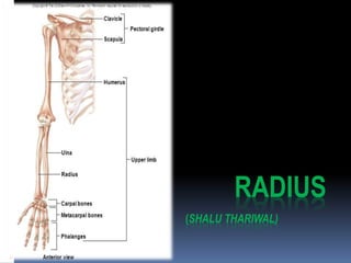
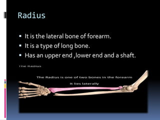
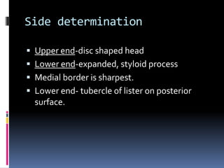
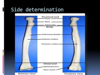
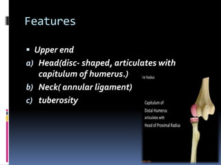
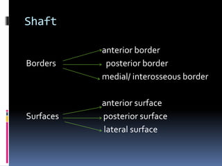
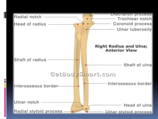
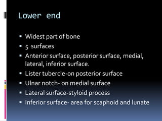
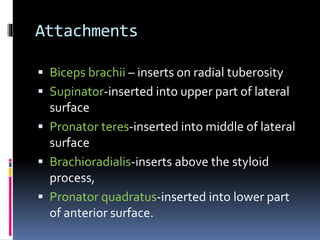
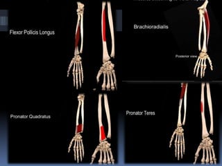
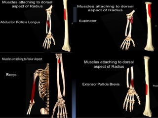
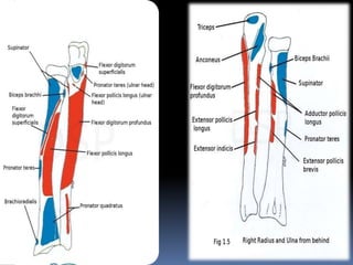
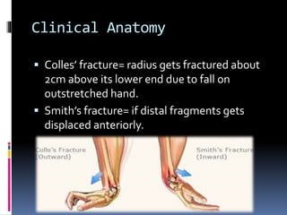
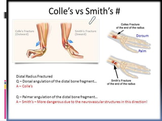
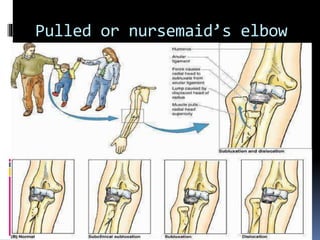
RADIUS
(SHALU THARIWAL)
Radius
 It is the lateral bone of forearm.
 It is a type of long bone.
 Has an upper end ,lower end and a shaft.
Side determination
 Upper end-disc shaped head
 Lower end-expanded, styloid process
 Medial border is sharpest.
 Lower end- tubercle of lister on posterior
surface.
Side determination
Features
 Upper end
a) Head(disc- shaped, articulates with
capitulum of humerus.)
b) Neck( annular ligament)
c) tuberosity
Shaft
anterior border
Borders posterior border
medial/ interosseous border
anterior surface
Surfaces posterior surface
lateral surface
Lower end
 Widest part of bone
 5 surfaces
 Anterior surface, posterior surface, medial,
lateral, inferior surface.
 Lister tubercle-0n posterior surface
 Ulnar notch- on medial surface
 Lateral surface-styloid process
 Inferior surface- area for scaphoid and lunate
Attachments
 Biceps brachii – inserts on radial tuberosity
 Supinator-inserted into upper part of lateral
surface
 Pronator teres-inserted into middle of lateral
surface
 Brachioradialis-inserts above the styloid
process,
 Pronator quadratus-inserted into lower part
of anterior surface.
Attachments
 Flexor digitorum superficialis-origin from
upper part of anterior border
 Flexor pollicis longus- origin form upper 2/3 of
anterior surface
 Abductor pollicis longus-arise form posterior
surface
 Extensor pollicis brevis- posterior surface
 Interosseous membrane-attached to
interosseous border
Clinical Anatomy
 Colles’ fracture= radius gets fractured about
2cm above its lower end due to fall on
outstretched hand.
 Smith’s fracture= if distal fragments gets
displaced anteriorly.
Pulled elbow
 Subluxation of head of radius – due to sudden
powerful jerk on the hand of a child may
dislodge the head of radius from the annular
ligament.
Pulled or nursemaid’s elbow
Radius bone anatomy

Radius bone anatomy

  • 1.
  • 2.
    Radius  It isthe lateral bone of forearm.  It is a type of long bone.  Has an upper end ,lower end and a shaft.
  • 3.
    Side determination  Upperend-disc shaped head  Lower end-expanded, styloid process  Medial border is sharpest.  Lower end- tubercle of lister on posterior surface.
  • 4.
  • 5.
    Features  Upper end a)Head(disc- shaped, articulates with capitulum of humerus.) b) Neck( annular ligament) c) tuberosity
  • 6.
    Shaft anterior border Borders posteriorborder medial/ interosseous border anterior surface Surfaces posterior surface lateral surface
  • 8.
    Lower end  Widestpart of bone  5 surfaces  Anterior surface, posterior surface, medial, lateral, inferior surface.  Lister tubercle-0n posterior surface  Ulnar notch- on medial surface  Lateral surface-styloid process  Inferior surface- area for scaphoid and lunate
  • 9.
    Attachments  Biceps brachii– inserts on radial tuberosity  Supinator-inserted into upper part of lateral surface  Pronator teres-inserted into middle of lateral surface  Brachioradialis-inserts above the styloid process,  Pronator quadratus-inserted into lower part of anterior surface.
  • 10.
    Attachments  Flexor digitorumsuperficialis-origin from upper part of anterior border  Flexor pollicis longus- origin form upper 2/3 of anterior surface  Abductor pollicis longus-arise form posterior surface  Extensor pollicis brevis- posterior surface  Interosseous membrane-attached to interosseous border
  • 14.
    Clinical Anatomy  Colles’fracture= radius gets fractured about 2cm above its lower end due to fall on outstretched hand.  Smith’s fracture= if distal fragments gets displaced anteriorly.
  • 16.
    Pulled elbow  Subluxationof head of radius – due to sudden powerful jerk on the hand of a child may dislodge the head of radius from the annular ligament.
  • 17.