Femur
DR. ATIF RAZA
Femur
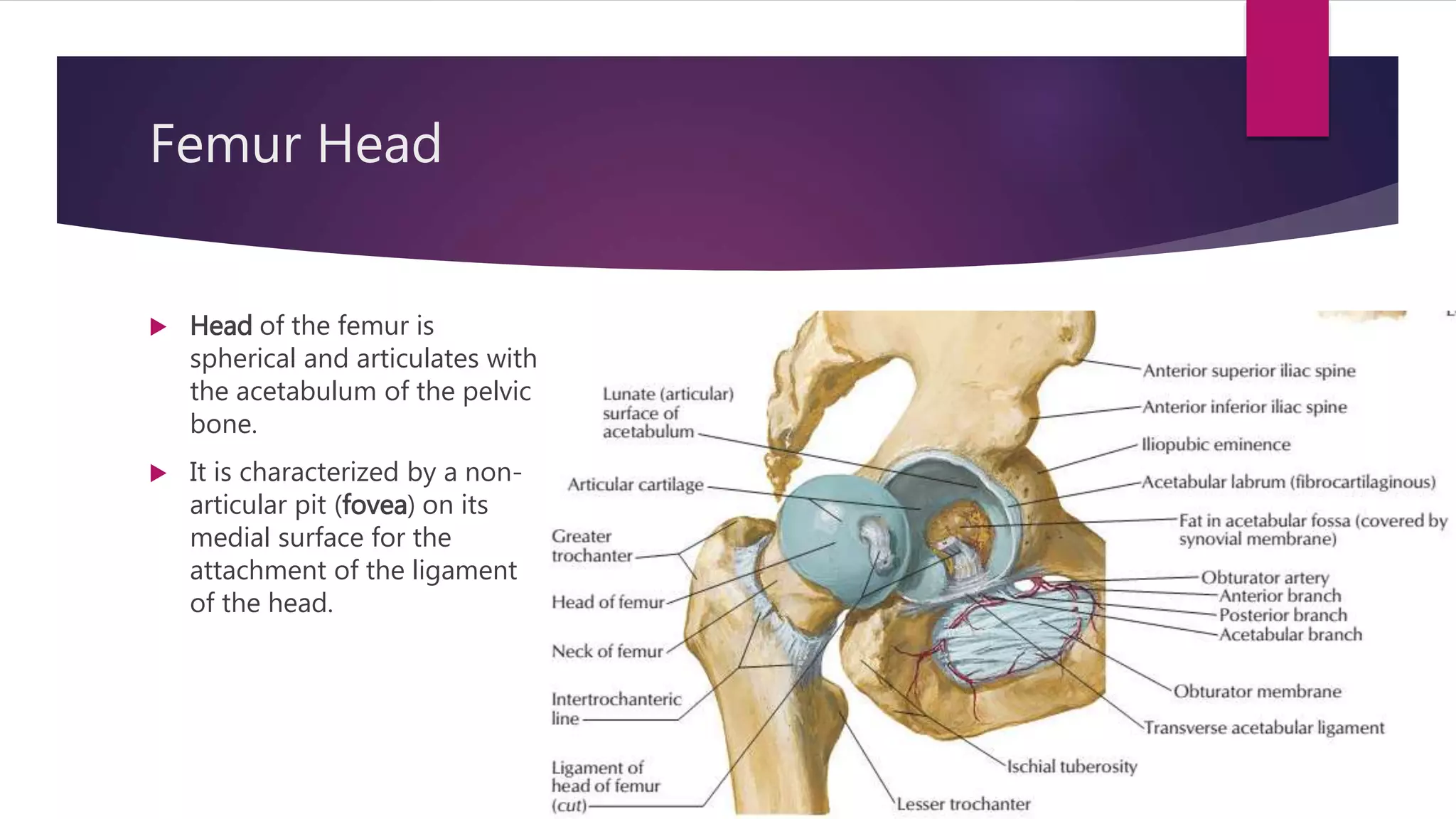
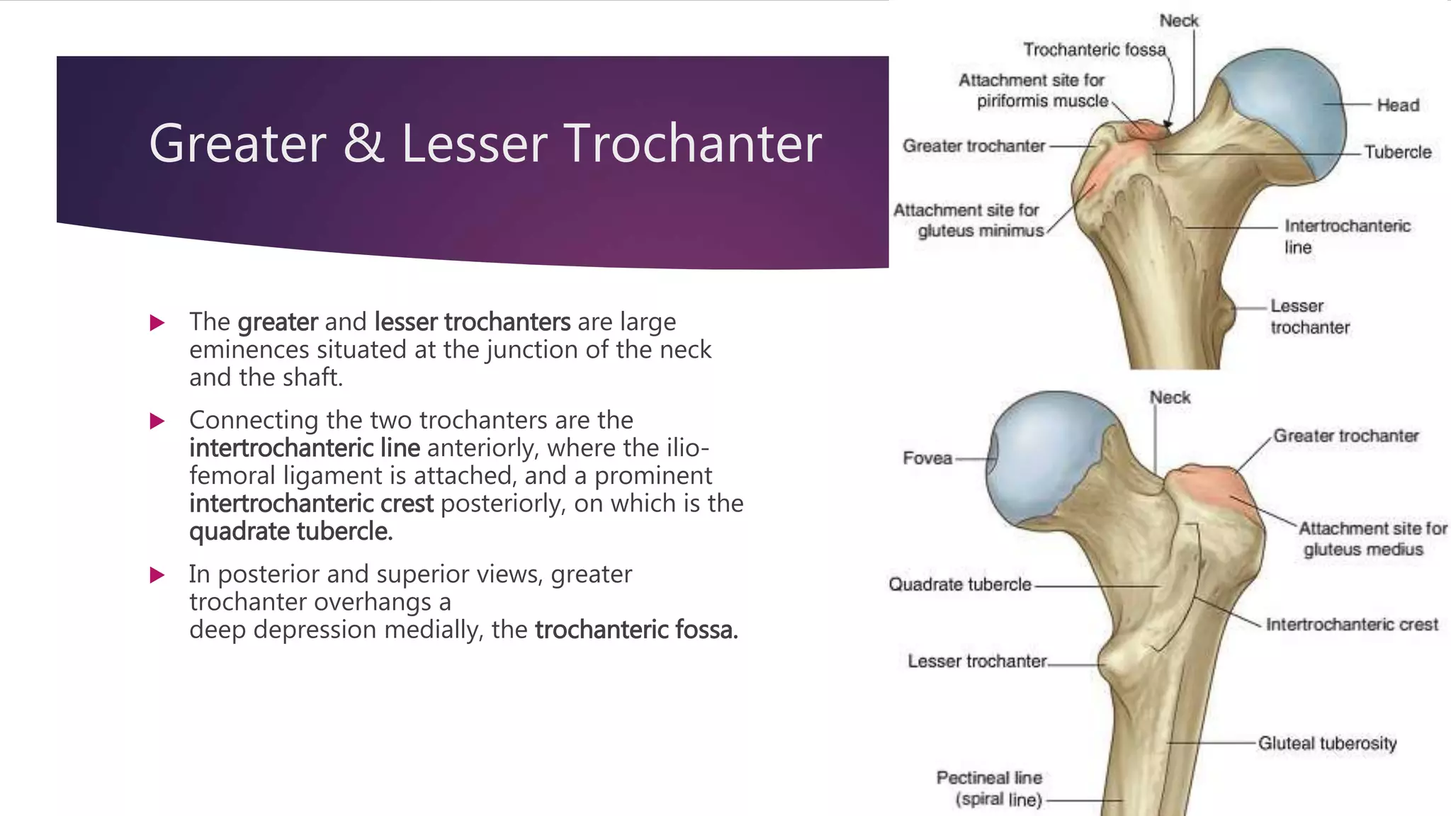
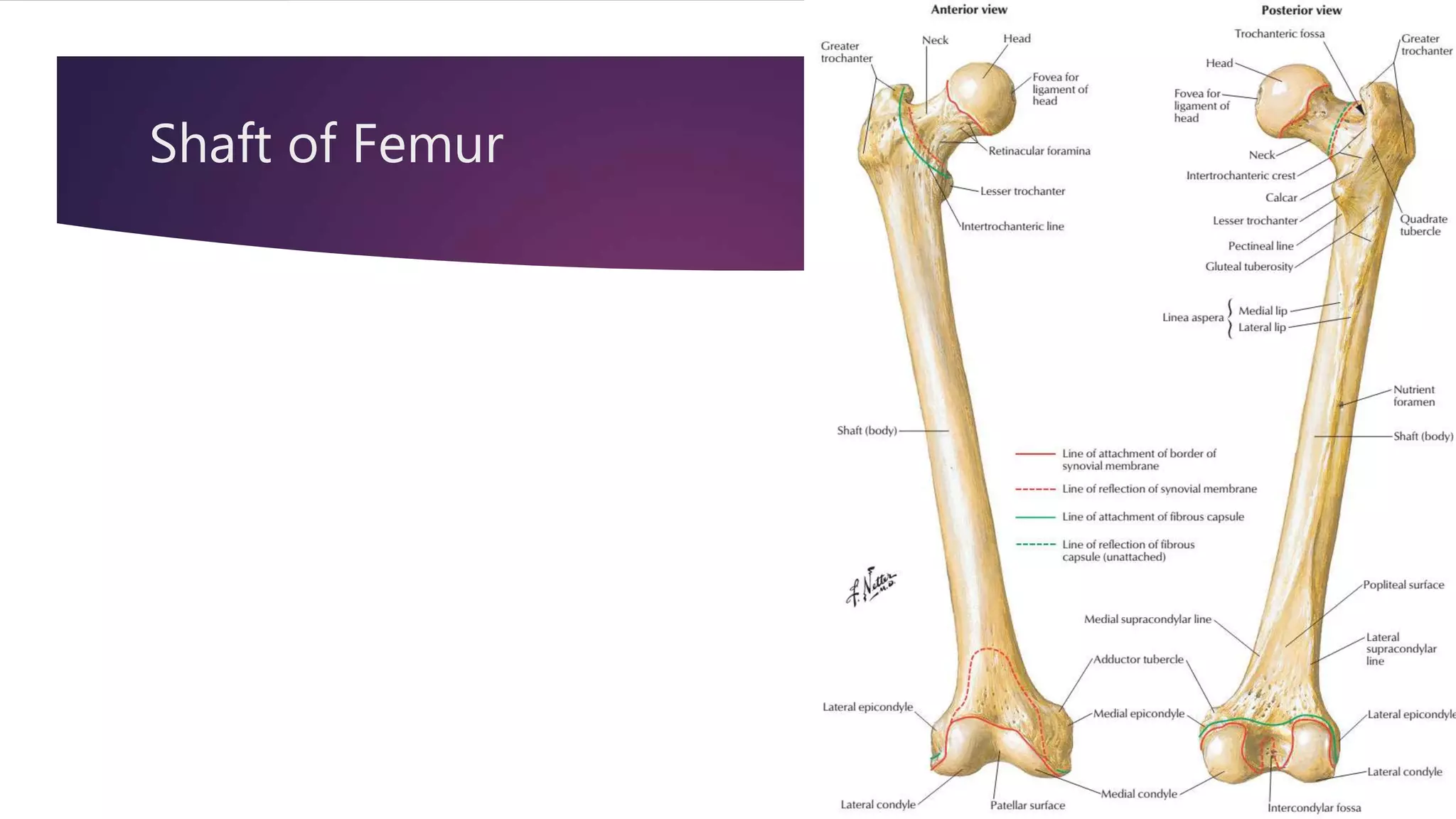
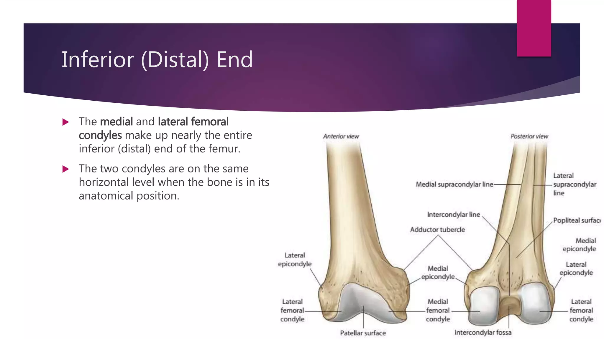
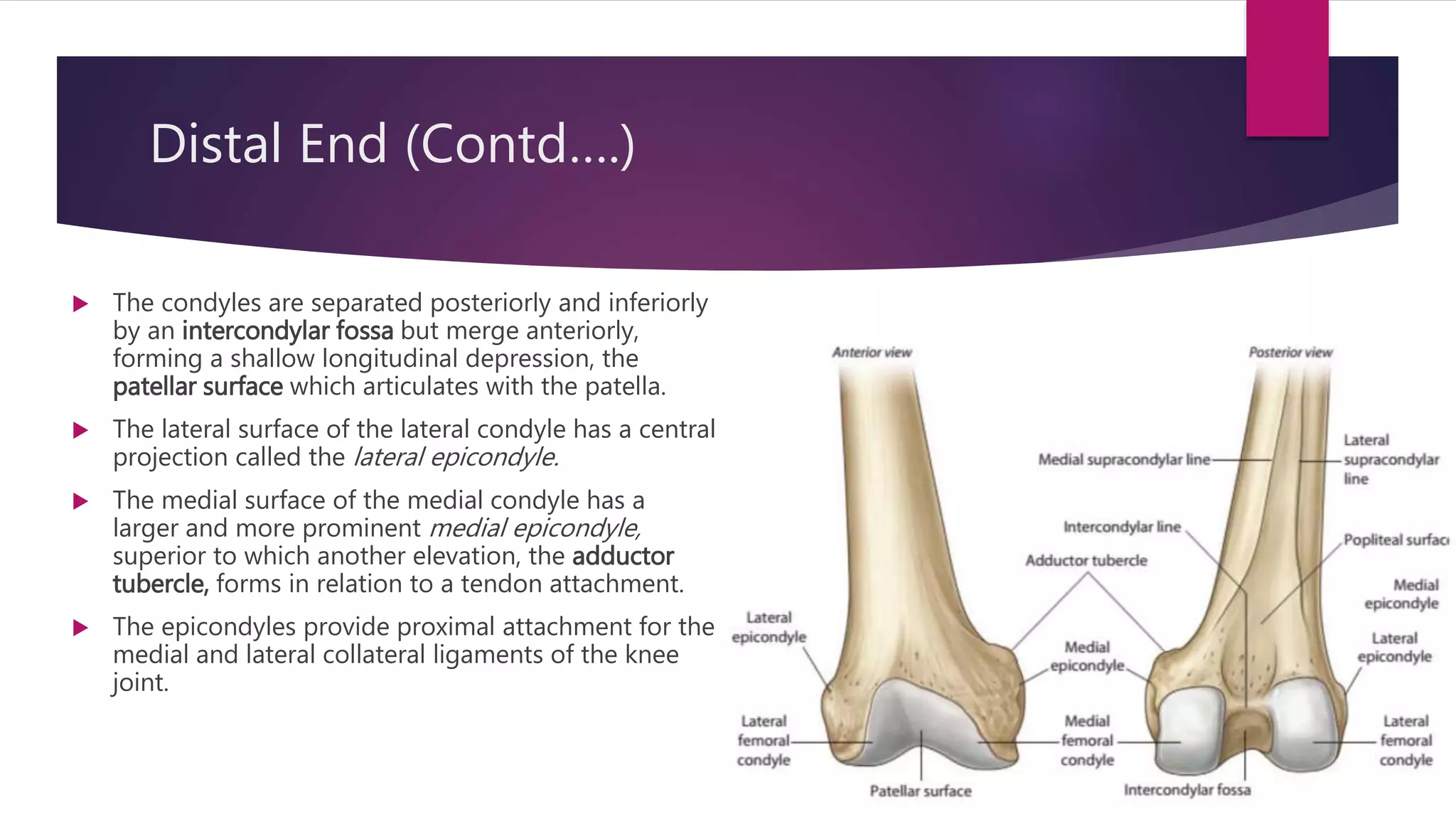
 The femur is the longest and
heaviest bone in the body.
 The femur consists of
 A shaft (body) and
 Superior or proximal and
 Inferior or distal end
Superior (Proximal) End
 Its proximal end is characterized by
 Head
 Neck,
 Two large projections (the greater and lesser
trochanters) on the upper part of the shaft.
Femur Head
 Head of the femur is
spherical and articulates with
the acetabulum of the pelvic
bone.
 It is characterized by a non-
articular pit (fovea) on its
medial surface for the
attachment of the ligament
of the head.
Femur Neck
 Neck passes downward, backward, and laterally and makes an
angle of about 125° (slightly less in the female) with the long
axis of the shaft (Angle of Inclination).
Greater & Lesser Trochanter
 The greater and lesser trochanters are large
eminences situated at the junction of the neck
and the shaft.
 Connecting the two trochanters are the
intertrochanteric line anteriorly, where the ilio-
femoral ligament is attached, and a prominent
intertrochanteric crest posteriorly, on which is the
quadrate tubercle.
 In posterior and superior views, greater
trochanter overhangs a
deep depression medially, the trochanteric fossa.
Shaft of Femur
Shaft of Femur
 The middle third of the shaft of the femur is triangular in shape with smooth lateral and
medial margins between anterior, lateral (posterolateral), and medial (posteromedial)
surfaces. The posterior margin is broad and forms a prominent raised crest (the linea
aspera).
 The linea aspera is a major site of muscle attachment in the thigh.
 In the proximal third of the femur, the medial and lateral margins of the linea aspera
diverge and continue superiorly as the pectineal line and gluteal tuberosity, respectively.
 Inferiorly, the linea aspera divides into medial and lateral supracondylar lines, which lead
to the medial and lateral femoral condyles.
 The popliteal surface, triangular in outline, lies between the medial
and lateral supracondylar lines.
Inferior (Distal) End
 The medial and lateral femoral
condyles make up nearly the entire
inferior (distal) end of the femur.
 The two condyles are on the same
horizontal level when the bone is in its
anatomical position.
Distal End (Contd….)
 The condyles are separated posteriorly and inferiorly
by an intercondylar fossa but merge anteriorly,
forming a shallow longitudinal depression, the
patellar surface which articulates with the patella.
 The lateral surface of the lateral condyle has a central
projection called the lateral epicondyle.
 The medial surface of the medial condyle has a
larger and more prominent medial epicondyle,
superior to which another elevation, the adductor
tubercle, forms in relation to a tendon attachment.
 The epicondyles provide proximal attachment for the
medial and lateral collateral ligaments of the knee
joint.
Fracture Sites:
 The neck of the femur is most frequently fractured because
it is the narrowest and weakest part of the bone and it lies
at a marked angle to the line of weight-bearing.
 Fractures of the femoral neck cause lateral rotation of the
lower limb.
 Fractures of the femoral neck often disrupt the
blood supply to the head of the femur. Most of the blood to
the head and neck of the femur is supplied by the medial
circumflex femoral artery.
Contd….
 Fractures of the greater trochanter and femoral shaft
usually result from direct trauma and are most
common during the more active years.
 Intertrochanteric fractures usually runs from the
greater trochanter through to the lesser trochanter
and does not involve the femoral neck.
Intertrochanteric fractures preserve the femoral neck
blood supply and do not render the femoral head
ischemic.
Thank You

Femur (Gross Anatomy)

  • 2.
  • 3.
    Femur  The femuris the longest and heaviest bone in the body.  The femur consists of  A shaft (body) and  Superior or proximal and  Inferior or distal end
  • 4.
    Superior (Proximal) End Its proximal end is characterized by  Head  Neck,  Two large projections (the greater and lesser trochanters) on the upper part of the shaft.
  • 5.
    Femur Head  Headof the femur is spherical and articulates with the acetabulum of the pelvic bone.  It is characterized by a non- articular pit (fovea) on its medial surface for the attachment of the ligament of the head.
  • 6.
    Femur Neck  Neckpasses downward, backward, and laterally and makes an angle of about 125° (slightly less in the female) with the long axis of the shaft (Angle of Inclination).
  • 7.
    Greater & LesserTrochanter  The greater and lesser trochanters are large eminences situated at the junction of the neck and the shaft.  Connecting the two trochanters are the intertrochanteric line anteriorly, where the ilio- femoral ligament is attached, and a prominent intertrochanteric crest posteriorly, on which is the quadrate tubercle.  In posterior and superior views, greater trochanter overhangs a deep depression medially, the trochanteric fossa.
  • 8.
  • 9.
    Shaft of Femur The middle third of the shaft of the femur is triangular in shape with smooth lateral and medial margins between anterior, lateral (posterolateral), and medial (posteromedial) surfaces. The posterior margin is broad and forms a prominent raised crest (the linea aspera).  The linea aspera is a major site of muscle attachment in the thigh.  In the proximal third of the femur, the medial and lateral margins of the linea aspera diverge and continue superiorly as the pectineal line and gluteal tuberosity, respectively.  Inferiorly, the linea aspera divides into medial and lateral supracondylar lines, which lead to the medial and lateral femoral condyles.  The popliteal surface, triangular in outline, lies between the medial and lateral supracondylar lines.
  • 10.
    Inferior (Distal) End The medial and lateral femoral condyles make up nearly the entire inferior (distal) end of the femur.  The two condyles are on the same horizontal level when the bone is in its anatomical position.
  • 11.
    Distal End (Contd….) The condyles are separated posteriorly and inferiorly by an intercondylar fossa but merge anteriorly, forming a shallow longitudinal depression, the patellar surface which articulates with the patella.  The lateral surface of the lateral condyle has a central projection called the lateral epicondyle.  The medial surface of the medial condyle has a larger and more prominent medial epicondyle, superior to which another elevation, the adductor tubercle, forms in relation to a tendon attachment.  The epicondyles provide proximal attachment for the medial and lateral collateral ligaments of the knee joint.
  • 12.
    Fracture Sites:  Theneck of the femur is most frequently fractured because it is the narrowest and weakest part of the bone and it lies at a marked angle to the line of weight-bearing.  Fractures of the femoral neck cause lateral rotation of the lower limb.  Fractures of the femoral neck often disrupt the blood supply to the head of the femur. Most of the blood to the head and neck of the femur is supplied by the medial circumflex femoral artery.
  • 13.
    Contd….  Fractures ofthe greater trochanter and femoral shaft usually result from direct trauma and are most common during the more active years.  Intertrochanteric fractures usually runs from the greater trochanter through to the lesser trochanter and does not involve the femoral neck. Intertrochanteric fractures preserve the femoral neck blood supply and do not render the femoral head ischemic.
  • 14.