TNM Staging of tumor
•Download as PPTX, PDF•
25 likes•19,373 views

1. The document discusses the TNM classification system for staging tumors, which evaluates the size of the primary tumor (T), whether the cancer has spread to regional lymph nodes (N), and the presence of distant metastasis (M). 2. Staging provides information on cancer prognosis and treatment by assessing how far the cancer has progressed. The TNM system is overseen by organizations like the International Union Against Cancer and the American Joint Committee on Cancer. 3. In addition to staging, tumors are also graded based on their histopathological characteristics like differentiation and growth rate, with higher grades indicating faster growth and worse prognosis. Grading provides additional details beyond tumor staging.

Recommended
More Related Content
What's hot
What's hot (20)
Similar to TNM Staging of tumor
Similar to TNM Staging of tumor (20)
More from Dr. Binu Babu Nursing Lectures Incredibly Easy
More from Dr. Binu Babu Nursing Lectures Incredibly Easy (20)
Recently uploaded
Recently uploaded (20)
TNM Staging of tumor
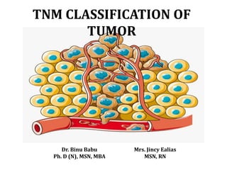
- 1. TNM CLASSIFICATION OF TUMOR Dr. Binu Babu Ph. D (N), MSN, MBA Mrs. Jincy Ealias MSN, RN
- 2. DEFINITION • Tumor: A Tumor is a swelling or mass of tissue that may be benign or malignant. • Benign- A noncancerous growth • Malignant- Dangerous to health; characterized by progressive and uncontrolled growth
- 3. Benign and Malignant Tumor Characteristics Benign Malignant Slow growing Rapid growing Tumor stasis occur It invade surrounding tissue Do not occur adjacent tissue They are anaplastic Death from benign tumor is rare
- 4. STAGING Staging is a way of describing how much a cancer has grown and spread. Purpose of staging To know the amount of cancer and where it is in the body to be able to choose the best possible treatment.
- 5. Aims of cancer staging 1. Selection of primary and adjuvant therapy 2. Estimation of prognosis, 3. Assistance in evaluation of the results of treatment 4. Facilitation of the exchange of information among treatment 5. Contribution to the continuing investigation of human cancers
- 6. Elements of Staging Staging is based on the following factors: • Location of the primary (original) tumor • Tumor size and number of tumors • Lymph node involvement – whether or not the cancer has spread to the nearby lymph nodes • Presence or absence of metastasis – whether or not the cancer has spread to distant areas of the body
- 7. • Gather different types of information about a cancer to determine its stage. The various tests used for staging depend on the type of cancer. Tests include the following: Physical exams provide clues as to the extent of the cancer. Imaging tests Laboratory tests
- 8. Pathology reports may include information about the size of the tumor, the growth into other tissues and organs, the type of cancer cells, and the grade of tumor. Surgical reports tell what is found during surgery.
- 9. Factors of Cancer Staging The staging is based on 3 main factors, T, N, and M: • T: is based on the size of the original (primary) tumor and whether or not it has grown into nearby tissues • N: whether or not the cancer has spread to the nearby lymph nodes • M: whether or not the cancer has spread to distant areas of the body
- 10. • TNM committe on International Union against cancer (UICC) and American Joint Committee on cancer (AJCC) have agreed on the TNM staging system. • In the TNM system, TNM stands for Tumor, Nodes, and Metastases.
- 11. T: Tumor • T Classifies the extent of the primary tumor, and is normally given as T0 through T4. • T0 represents a tumor that has not even started to invade the local tissues. This is called "In Situ". • T4 on the other hand represents a large primary tumor that has probably invaded other organs
- 12. N: Lymph Nodes • N classifies the amount of regional lymph node involvement. N0 means no lymph node involvement while N4 means extensive involvement.
- 13. M: Metastasis • M is either M0 if there are no metastases or M1 if there are metastases.
- 14. TNM classification system PRIMARY TUMOR (T) • Tx Primary tumor cannot be assessed • T0 No evidence of primary tumor • Tis Carcinoma in situ • T1, T2, T3, T4 Increasing size and /or local extent of the primary tumor
- 15. REGIONAL LYMPH NODES (N) • Nx regional lymph nodes cannot be assessed • N0 no regional lymph node metastasis • N1,N2,N3 increasing involvement of regional lymph nodes
- 16. DISTANT METASTASIS (M) • Mx Distant metastasis cannot be assessed • M0:- No distant metastasis • M1 Distant metastasis
- 17. Classification of TNM • Clinical classification, designated cTNM or TNM • Pathologic classification, designated pTNM • Retreatment classification, designated rTNM • Autopsy classification, designated aTNM
- 18. Clinical classification • It is based on evidence acquired before primary treatment. Clinical assessment uses information available prior to first definitive treatment, including but not limited to physical examination, imaging, endoscopy, biopsy, and surgical exploration.
- 19. Pathologic classification Which uses the evidence acquired before treatment, supplemented or modified by the additional evidence acquired during and from surgery, particularly from pathologic examination.
- 20. Retreatment classification • It is assigned when further treatment (such as chemotherapy) is planned for a caner that recurs after a disease-free interval. All information available at the time of retreatment should be used in determining the stage of the recurrent tumor (rTNM).
- 21. Autopsy classification • Occurs when classification of a cancer by postmortem examination is done after the death of a patient (cancer was not evident prior to death). The classification of the stage is identified as aTNM and includes all pathologic information obtained at the time of death.
- 22. AJC (American Joint Committee) system Divides all cancers into stages 0 to IV. • Stage 0: Tumor of microscopic size (carcinoma in situ) • Stage I: tumor confined to the organ of origin (cancers are localized to one part of the body) • Stage II: Local spread, not interfering with surgical removal. • Stage III: Fixation to the surrounding structures. • Stage IV: Distant metastasis.
- 24. GRADING • Grading is a method of classification based on histopathologic characteristics of the tissue. • The grade is a measure of how abnormal the cancer cells look under the microscope, is called differentiation. • Cancers with more abnormal-looking cells tend to grow and spread faster. The grade is usually assigned a number from 1 to 3 or 4.
- 25. • Grading is based on two important histological features. – Degree of anaplasia – Rate of growth
- 26. Two grading systems are commonly seen • One descriptively identifies the tumor as well differentiated, moderately well differentiated, poorly differentiated, or undifferentiated. • The other system numerically grades from 1 to 3 or 4, with 1 being the most differentiated and 3 and 4 being the least well-differentiated ; grade 4 applies to tumors with no specific diffentiation.
- 27. The AJCC recommends the grading classification as: • GX Grade cannot be assessed • G1 well diffentiated (<25% anaplastic cells) • G2 moderatly well-diffentiated (25-50% anaplastic cells) • G3 Poorly diffentiated (>75% anaplastic cells) • G4 Undifferentiated
- 28. Bibliography • Michele Goodman,Cancer Nursing, principle and Practise, 6th edition, 172-177. • Brunner and Sidharth, Text book of Medical Surgical Nursing, 11th edition. • AJCC Cancer Staging Atlas • Understanding Cancer types and Staging • SEER program: comparative staging guide for cancer