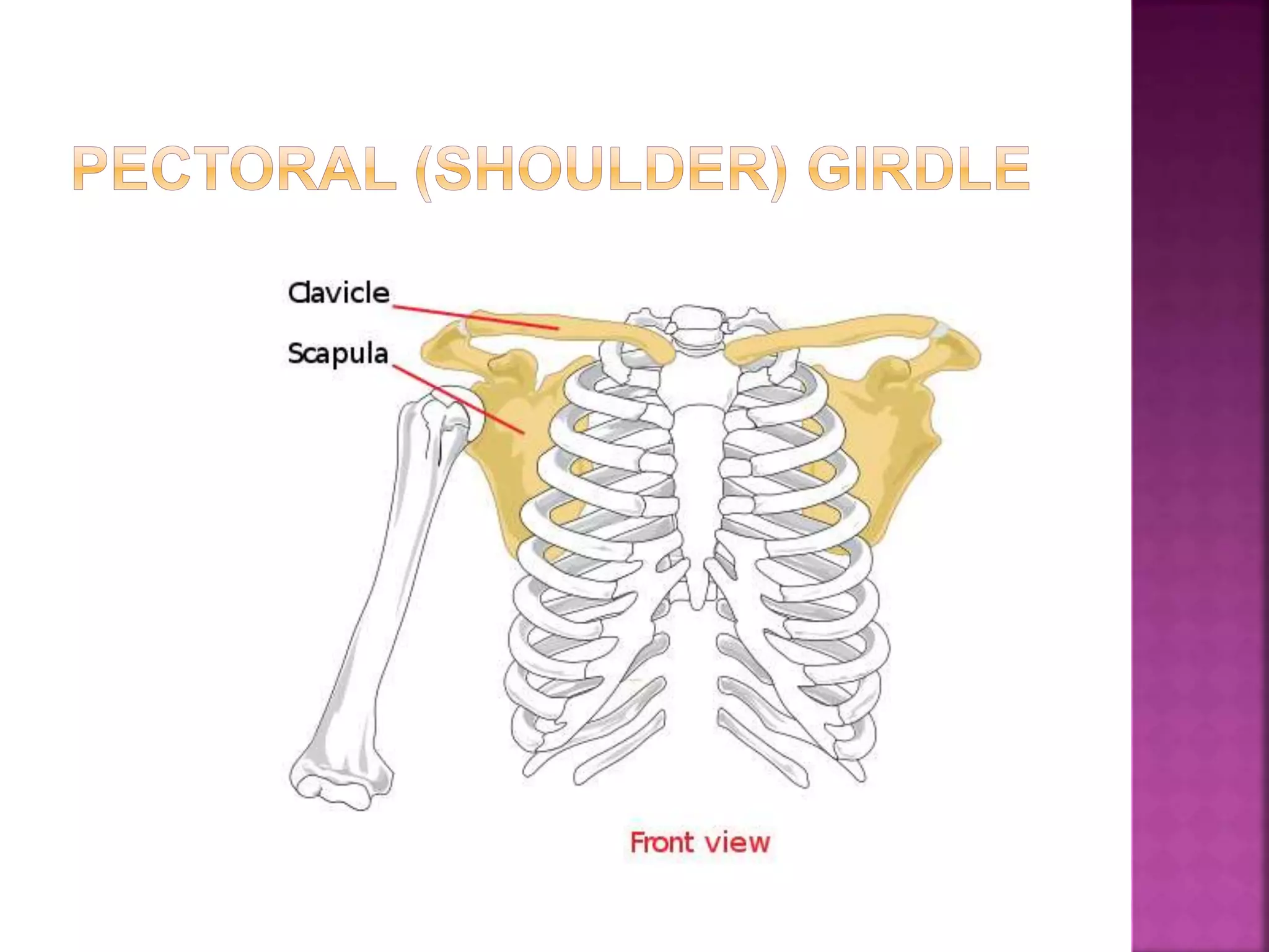
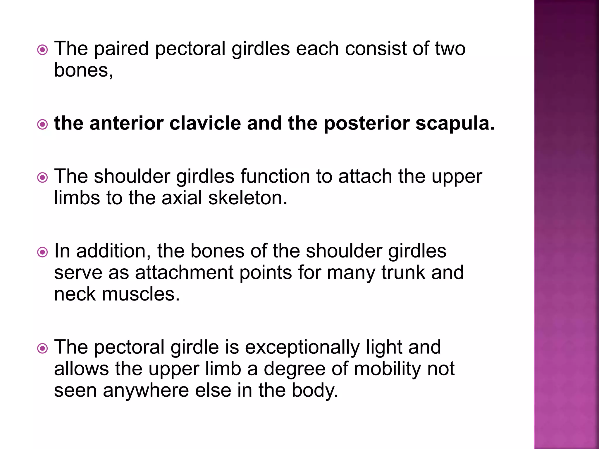
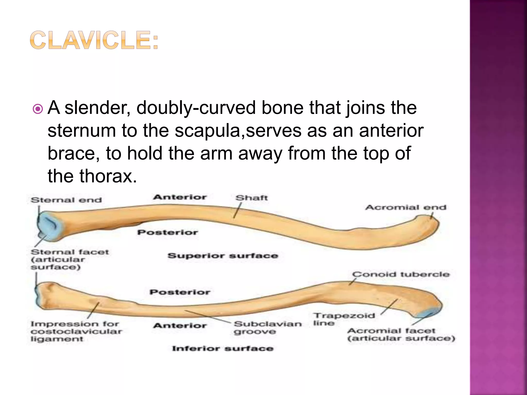
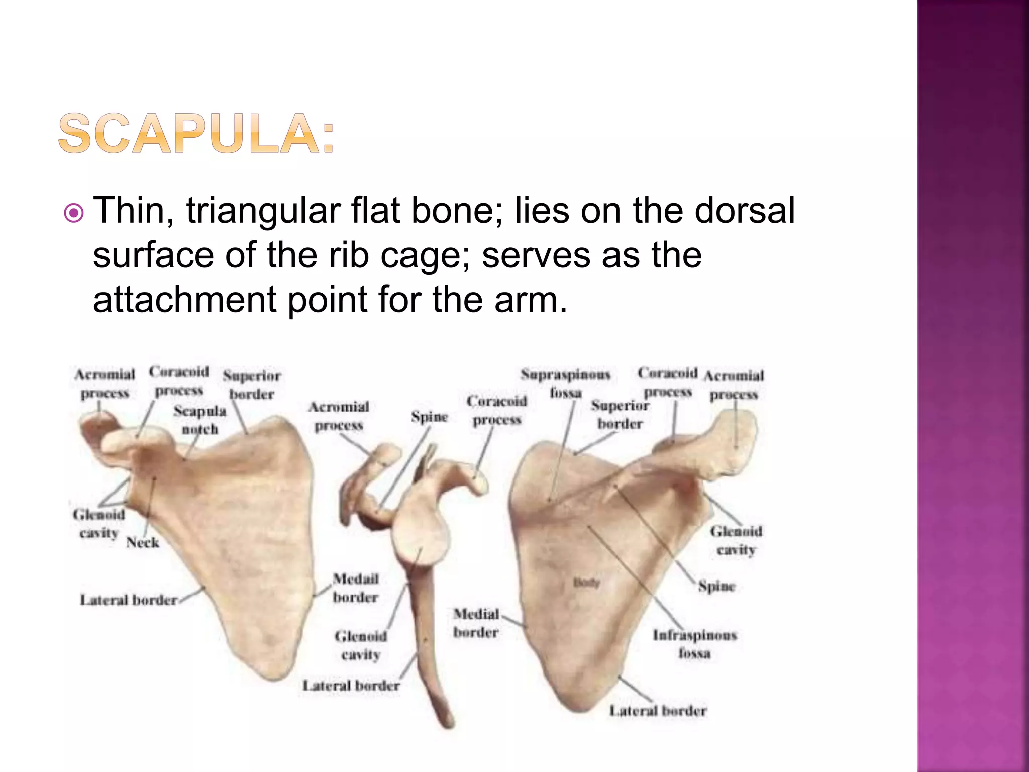
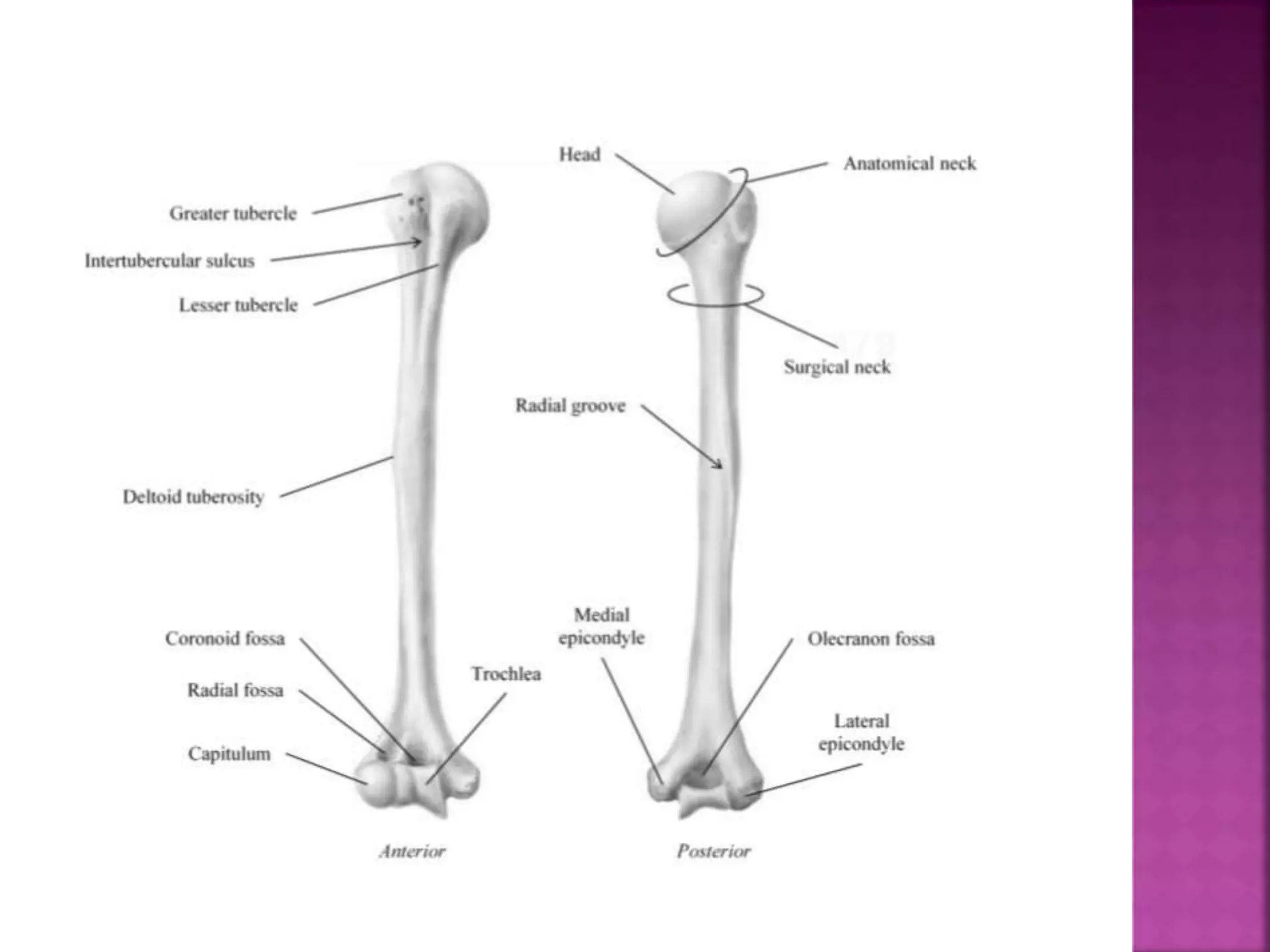
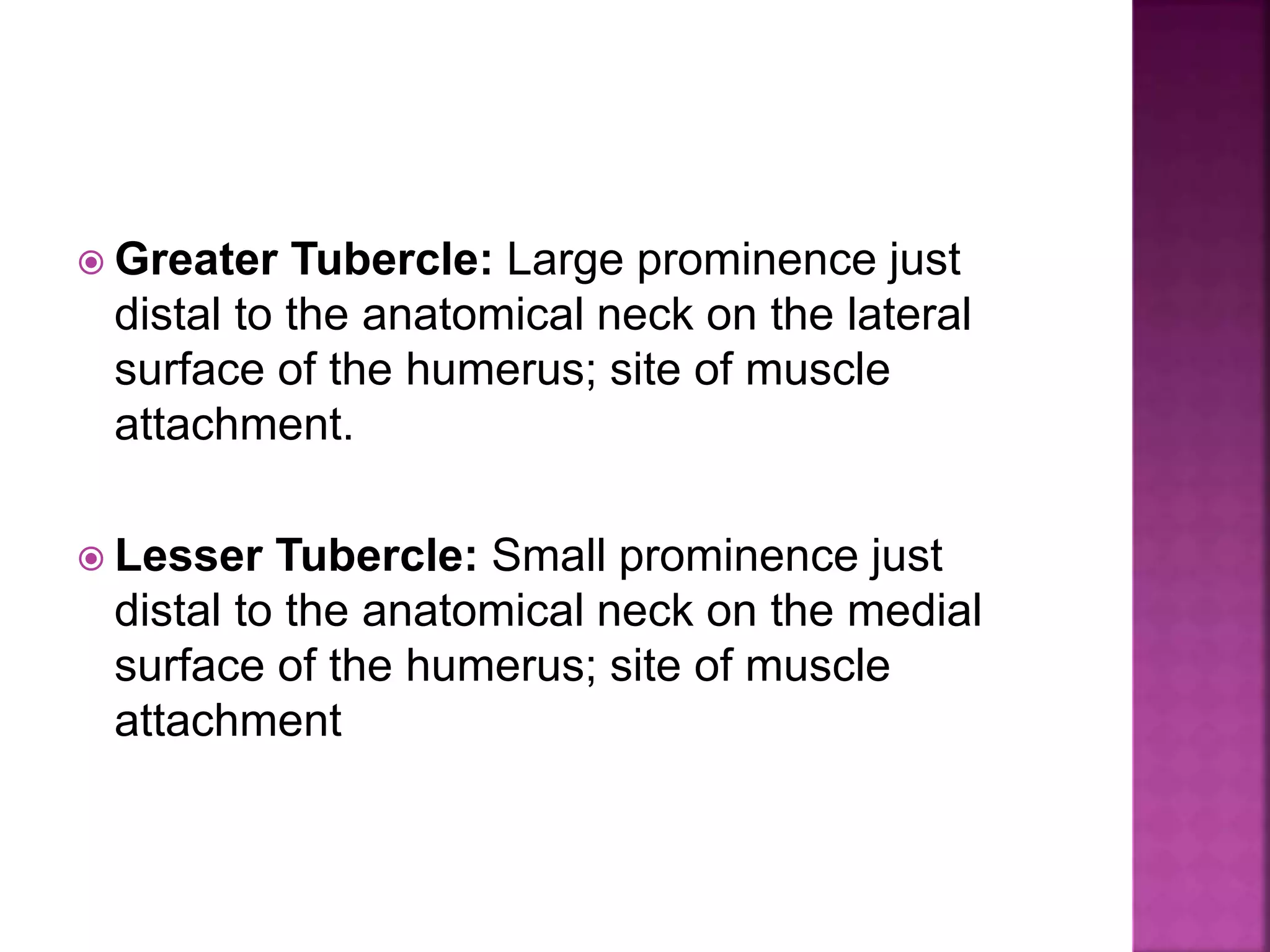
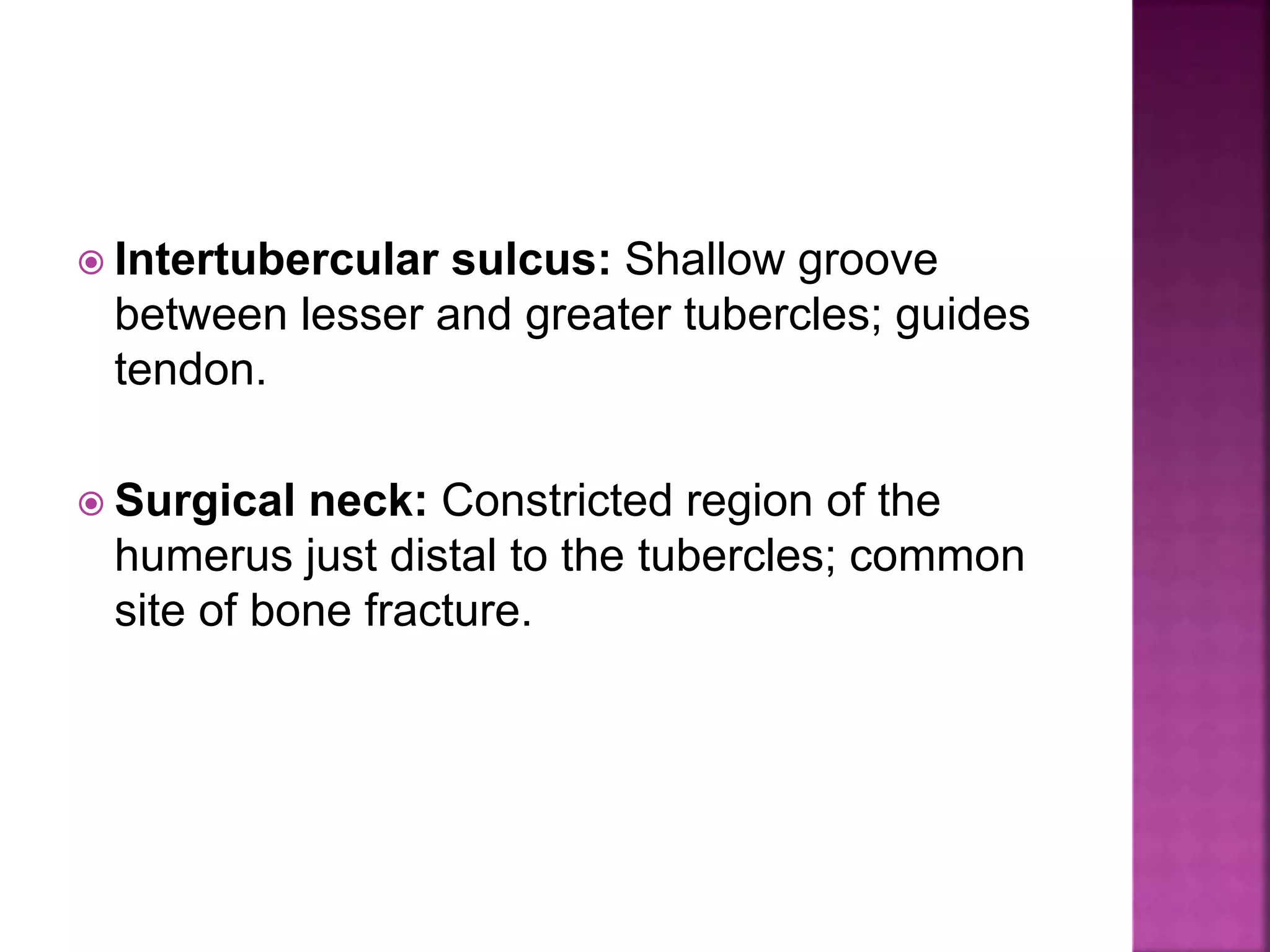
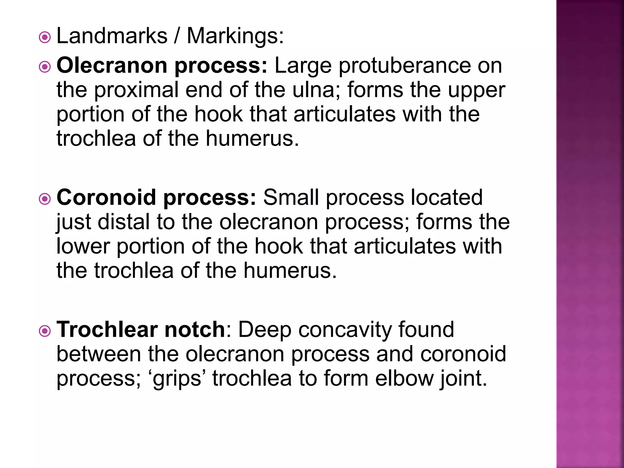
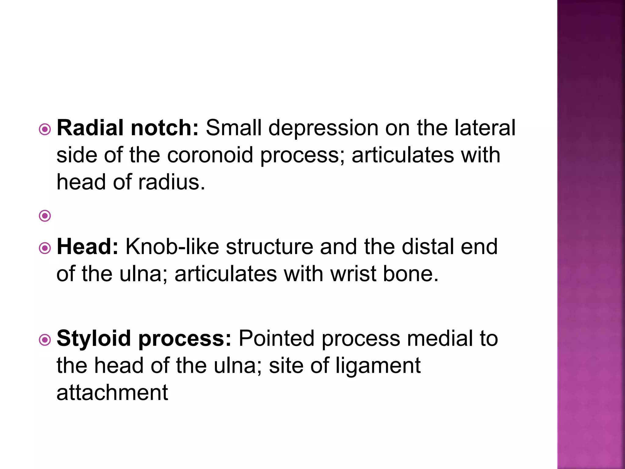
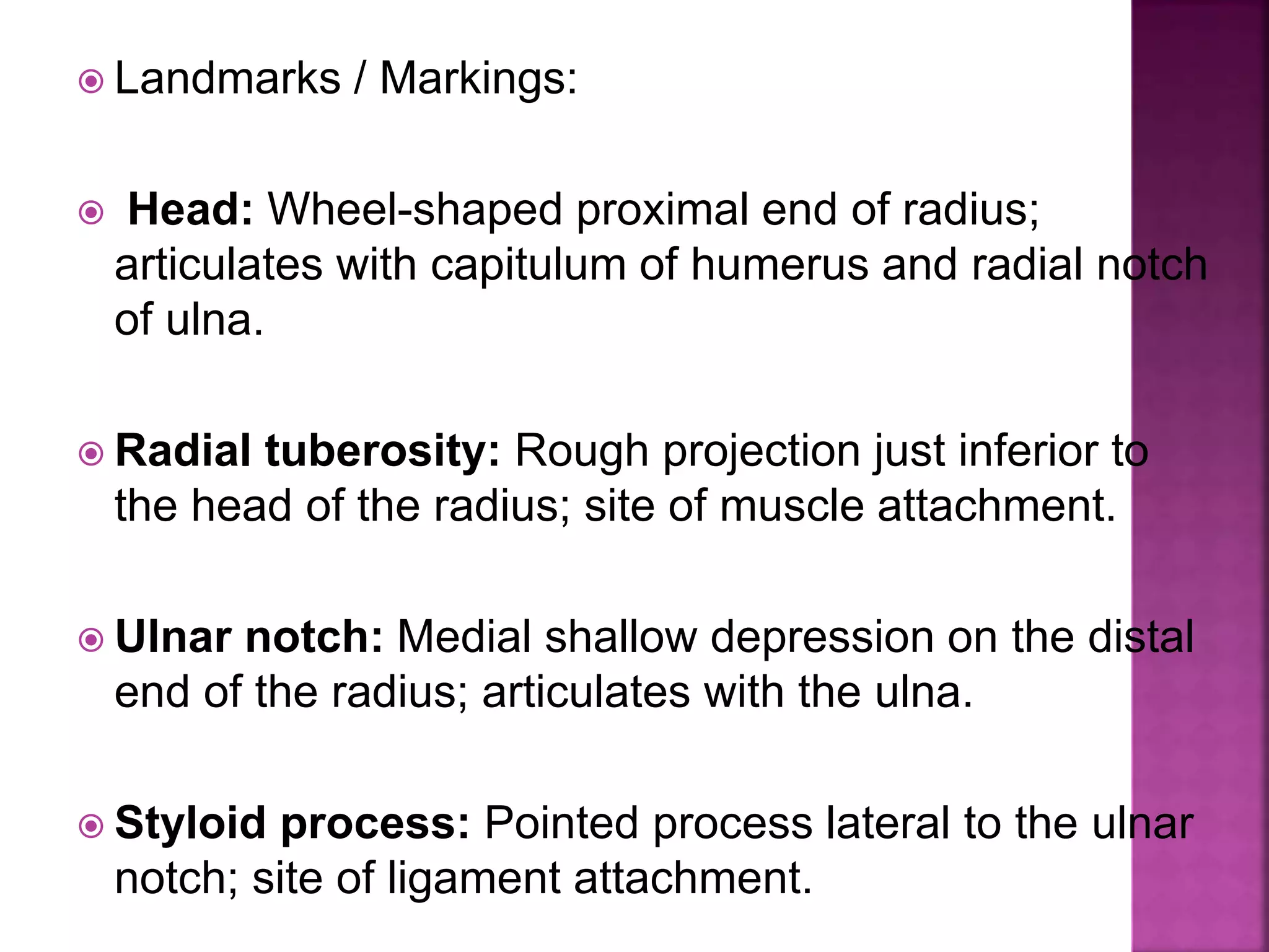
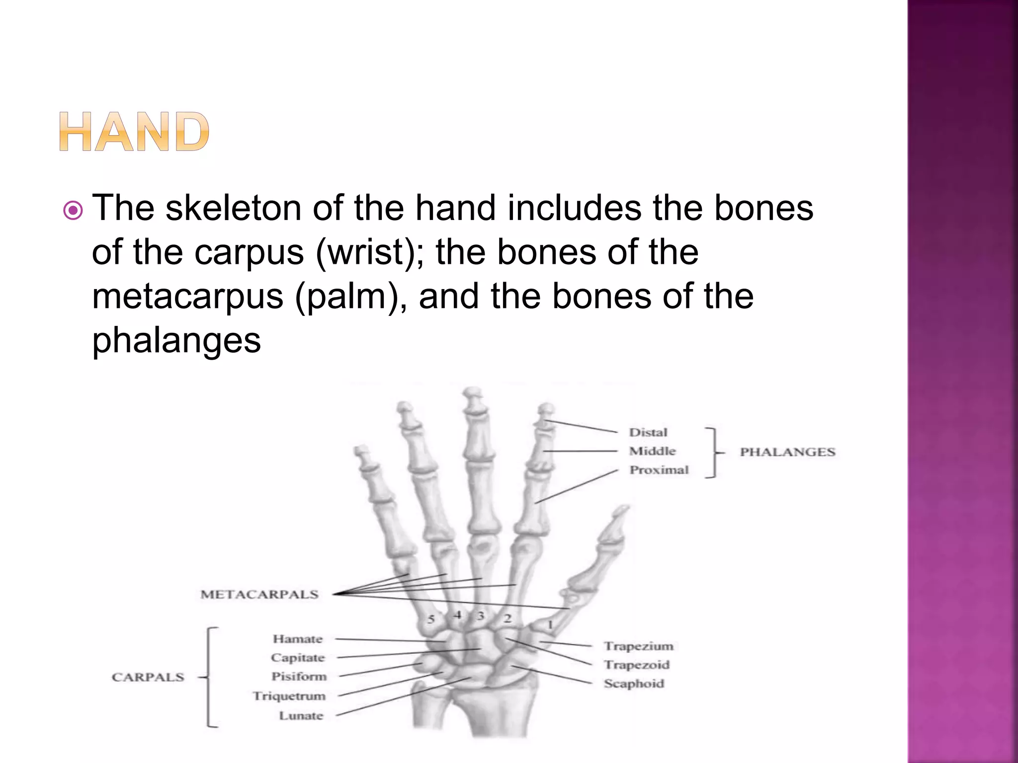
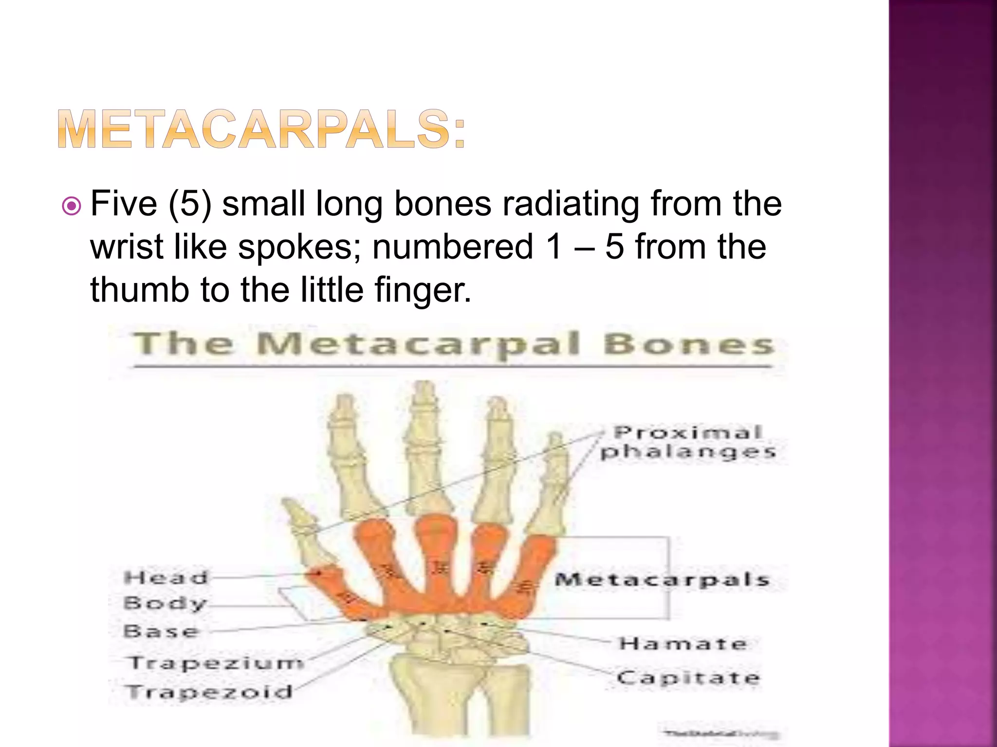
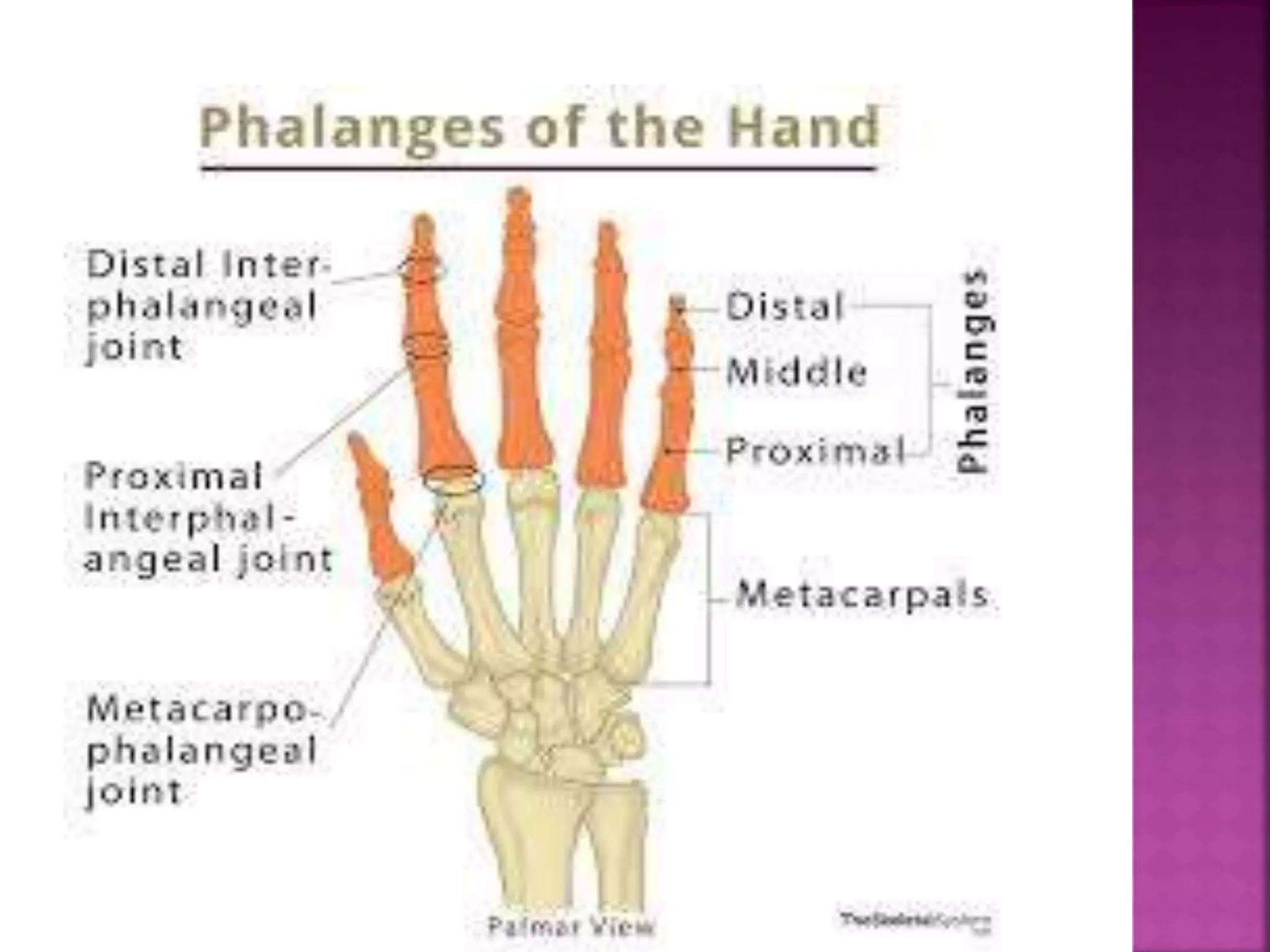
SlideShare a Scribd company logo
 The paired pectoral girdles each consist of two
bones,
 the anterior clavicle and the posterior scapula.
 The shoulder girdles function to attach the upper
limbs to the axial skeleton.
 In addition, the bones of the shoulder girdles
serve as attachment points for many trunk and
neck muscles.
 The pectoral girdle is exceptionally light and
allows the upper limb a degree of mobility not
seen anywhere else in the body.
 A slender, doubly-curved bone that joins the
sternum to the scapula,serves as an anterior
brace, to hold the arm away from the top of
the thorax.
 Landmarks / Markings:
 STERNAL END: Rounded terminus of
clavicle; articulates with the sternal
manubrium.
 ACROMIAL END: Flattened terminus of
clavicle; articulates with the scapula to form
part of the shoulder joint.
 Thin, triangular flat bone; lies on the dorsal
surface of the rib cage; serves as the
attachment point for the arm.
 Borders / angles:
 Superior border: Short, sharp border that forms
the upper margin of the scapula when articulated
with the axial skeleton.
 Medial (vertebral) border: Border which parallels
the vertebral column when articulated with the axial
skeleton; joins with superior border at the superior
angle.
 Lateral (axillary) border: The thick border that
abuts the armpit when articulated with the axial
skeleton; joins with superior border at the lateral
angle and joins with medial border at the inferior
angle.
 Landmarks / Markings:
 Glenoid cavity (fossa): Small, shallow
depression superior to the lateral border, near
the lateral angle; articulates with humerus of
the arm.
 Spine: Prominent ridge rising from the upper
posterior surface of the scapula; site of
muscle attachment.
 Acromion: Enlarged, roughened triangular
structure projecting projection off the lateral
end of the scapular spine; articulates with the
acromial end of the clavicle.
 Coracoid process: Beak-like structure
projecting anteriorly from the superior
scapular border; site of muscle attachment.
 Suprascapular notch: Shallow groove in the
superior border of the scapula at the base of
the coracoid process; passageway for nerves.
 Supraspinous fossa: Deep depression
superior to the spine on the posterior surface
of the scapula; site of muscle attachment.
 Infraspinous fossa: Shallow depression
inferior to the spine on the posterior surface of
the scapula; site of muscle attachment.
 Subscapular fossa: Shallow depression
formed by the entire anterior scapular surface;
site of muscle attachment.
 The upper arm consists of a single bone, the
humerus .
 The largest and longest bone of the upper
limb, it articulates with the scapula at the
shoulder and with the radius and ulna
(forearm bones) at the elbow.
 Landmarks / Markings:
 Head: Smooth, hemispherical projection at
the proximal end of the humerus; articulates
with glenoid cavity of scapula.
 Anatomical neck: Slight constriction just
distal to the head of the humerus.
 Greater Tubercle: Large prominence just
distal to the anatomical neck on the lateral
surface of the humerus; site of muscle
attachment.
 Lesser Tubercle: Small prominence just
distal to the anatomical neck on the medial
surface of the humerus; site of muscle
attachment
 Intertubercular sulcus: Shallow groove
between lesser and greater tubercles; guides
tendon.
 Surgical neck: Constricted region of the
humerus just distal to the tubercles; common
site of bone fracture.
 Deltoid tuberosity: V-shaped rough region
on the lateral aspect of the humerus about
midway down the shaft; site of muscle
attachment.
 Radial groove: A shallow depression running
obliquely down the posterior aspect of the
humerus shaft; nerve passageway
 Trochlea: Medial spool-shaped structure on
the distal end of the humerus; articulates with
ulna of the forearm.
 Capitulum: Lateral ball-like structure on the
distal end of the humerus; articulates with
radius of the forearm.
 Medial epicondyle: Small outward projection
flanking trochlea; site of muscle attachment.
 Lateral epicondyle: Small outward
projection flanking capitulum; site of muscle
attachment
 Coronoid fossa: Large, shallow depression
superior to the trochlea on the anterior
surface of the humerus; receives
corresponding process from ulna when elbow
flexes / extends.
 Radial fossa: Small, shallow depression
lateral to the coronoid fossa; receives head of
radius when elbow flexes.

 Olecranon fossa: Large, deep depression
superior to the trochlea on the posterior
surface of the humerus; anchors
corresponding process from ulna to form
elbow joint.
 Two parallel bones, the radius and the ulna,
form the forearm .
 Their proximal ends articulate with the
humerus and their distal ends articulate with
the wrist bones.
 Long, slender bone with a hook at the
proximal end that forms the elbow joint with
the humerus; lies medially in the forearm
when the body is in anatomical position.
 Landmarks / Markings:
 Olecranon process: Large protuberance on
the proximal end of the ulna; forms the upper
portion of the hook that articulates with the
trochlea of the humerus.
 Coronoid process: Small process located
just distal to the olecranon process; forms the
lower portion of the hook that articulates with
the trochlea of the humerus.
 Trochlear notch: Deep concavity found
between the olecranon process and coronoid
process; ‘grips’ trochlea to form elbow joint.
 Radial notch: Small depression on the lateral
side of the coronoid process; articulates with
head of radius.

 Head: Knob-like structure and the distal end
of the ulna; articulates with wrist bone.
 Styloid process: Pointed process medial to
the head of the ulna; site of ligament
attachment
 Long bone that is thin at its proximal end and
wide at its distal end; lies laterally in the
forearm when the body is in anatomical
position.
 Landmarks / Markings:
 Head: Wheel-shaped proximal end of radius;
articulates with capitulum of humerus and radial notch
of ulna.
 Radial tuberosity: Rough projection just inferior to
the head of the radius; site of muscle attachment.
 Ulnar notch: Medial shallow depression on the distal
end of the radius; articulates with the ulna.
 Styloid process: Pointed process lateral to the ulnar
notch; site of ligament attachment.
 The skeleton of the hand includes the bones
of the carpus (wrist); the bones of the
metacarpus (palm), and the bones of the
phalanges
 Eight (8) marble-size short bones closely
united by ligaments; quite flexible due to
gliding movements between bones.
 1) Scaphoid 5) Trapezium
6) Trapezoid
2)Lunate 7) Capitate
8) Hamate
3) Triquetrum
4) Pisiform
 Five (5) small long bones radiating from the
wrist like spokes; numbered 1 – 5 from the
thumb to the little finger.
 Fourteen (14) miniature long bones that form
the fingers; numbered 1 – 5 from the thumb
(pollex) to the little finger.
 1) Proximal phalange (1 – 5)
 2) Middle phalange (2 – 5)
 3) Distal phalange (1 – 5)
Appendicular Skeleton-Shoulder girdle and upper limb

More Related Content

What's hot

The ribs
The ribsThe ribs
The ribs
Idris Siddiqui
 
Clinical anatomy thorax
Clinical anatomy thoraxClinical anatomy thorax
Clinical anatomy thorax
Idris Siddiqui
 
Anatomy of Upper extremity
Anatomy of Upper extremityAnatomy of Upper extremity
Anatomy of Upper extremity
Sunil Pahari
 
bones of Upper limbs and anatomy of upper limbs
bones of Upper limbs and anatomy of upper limbsbones of Upper limbs and anatomy of upper limbs
bones of Upper limbs and anatomy of upper limbs
sadhamhussain52
 
Anatomy bones of upper limbs
Anatomy bones of upper limbsAnatomy bones of upper limbs
Anatomy bones of upper limbs
Shifa medicine collage
 
Appendicular skeleton
Appendicular skeletonAppendicular skeleton
Appendicular skeleton
PARUL UNIVERSITY
 
Skeleton system- bones and their number with detailed description.
Skeleton system- bones and their number with detailed description.Skeleton system- bones and their number with detailed description.
Skeleton system- bones and their number with detailed description.
bhartisharma175
 
07 Appendicular Skeleton Pelvic Girdle And Lower Limbs
07 Appendicular Skeleton   Pelvic Girdle And Lower Limbs07 Appendicular Skeleton   Pelvic Girdle And Lower Limbs
07 Appendicular Skeleton Pelvic Girdle And Lower Limbs
Kevin Young
 
Joints of thorax
Joints of thoraxJoints of thorax
Joints of thorax
Idris Siddiqui
 
bones and muscles of upper limb
bones and muscles of upper limbbones and muscles of upper limb
bones and muscles of upper limb
Aqsa Mushtaq
 
Anatomy of foot and ankle
Anatomy of foot and ankleAnatomy of foot and ankle
Anatomy of foot and ankle
Junaid Ahmad
 
Osteology upper limb by Dr G Kamau
Osteology upper limb by Dr G KamauOsteology upper limb by Dr G Kamau
Osteology upper limb by Dr G Kamau
MathewJude
 
Femur
FemurFemur
Femur
Aqib Javed
 
Anatomy (anatomy of bone, joint & muscles of upper limb)
Anatomy (anatomy of bone, joint & muscles of upper limb)Anatomy (anatomy of bone, joint & muscles of upper limb)
Anatomy (anatomy of bone, joint & muscles of upper limb)
Osama Al-Zahrani
 
Femur (Gross Anatomy)
Femur (Gross Anatomy)Femur (Gross Anatomy)
Femur (Gross Anatomy)
AtifRaza11
 
Clinical anatomy
Clinical anatomyClinical anatomy
Clinical anatomy
Idris Siddiqui
 

What's hot (20)

The ribs
The ribsThe ribs
The ribs
 
Clinical anatomy thorax
Clinical anatomy thoraxClinical anatomy thorax
Clinical anatomy thorax
 
Anatomy of Upper extremity
Anatomy of Upper extremityAnatomy of Upper extremity
Anatomy of Upper extremity
 
210 lower limb rs
210 lower limb rs210 lower limb rs
210 lower limb rs
 
bones of Upper limbs and anatomy of upper limbs
bones of Upper limbs and anatomy of upper limbsbones of Upper limbs and anatomy of upper limbs
bones of Upper limbs and anatomy of upper limbs
 
Anatomy of the Knee Joint
Anatomy of the Knee JointAnatomy of the Knee Joint
Anatomy of the Knee Joint
 
Anatomy bones of upper limbs
Anatomy bones of upper limbsAnatomy bones of upper limbs
Anatomy bones of upper limbs
 
Femur
FemurFemur
Femur
 
Appendicular skeleton
Appendicular skeletonAppendicular skeleton
Appendicular skeleton
 
Skeleton system- bones and their number with detailed description.
Skeleton system- bones and their number with detailed description.Skeleton system- bones and their number with detailed description.
Skeleton system- bones and their number with detailed description.
 
07 Appendicular Skeleton Pelvic Girdle And Lower Limbs
07 Appendicular Skeleton   Pelvic Girdle And Lower Limbs07 Appendicular Skeleton   Pelvic Girdle And Lower Limbs
07 Appendicular Skeleton Pelvic Girdle And Lower Limbs
 
Joints of thorax
Joints of thoraxJoints of thorax
Joints of thorax
 
bones and muscles of upper limb
bones and muscles of upper limbbones and muscles of upper limb
bones and muscles of upper limb
 
Anatomy of foot and ankle
Anatomy of foot and ankleAnatomy of foot and ankle
Anatomy of foot and ankle
 
Osteology upper limb by Dr G Kamau
Osteology upper limb by Dr G KamauOsteology upper limb by Dr G Kamau
Osteology upper limb by Dr G Kamau
 
Brain
BrainBrain
Brain
 
Femur
FemurFemur
Femur
 
Anatomy (anatomy of bone, joint & muscles of upper limb)
Anatomy (anatomy of bone, joint & muscles of upper limb)Anatomy (anatomy of bone, joint & muscles of upper limb)
Anatomy (anatomy of bone, joint & muscles of upper limb)
 
Femur (Gross Anatomy)
Femur (Gross Anatomy)Femur (Gross Anatomy)
Femur (Gross Anatomy)
 
Clinical anatomy
Clinical anatomyClinical anatomy
Clinical anatomy
 

Similar to Appendicular Skeleton-Shoulder girdle and upper limb

UPPER LIMB 4 ppt of human body..........
UPPER LIMB 4 ppt of human body..........UPPER LIMB 4 ppt of human body..........
UPPER LIMB 4 ppt of human body..........
aahanak787
 
Anatomy of Humerus
Anatomy of HumerusAnatomy of Humerus
Anatomy of Humerus
Dr Adnan Sami
 
Bones of upper limb, By Nida Manzoor.pptx
Bones of upper limb, By Nida Manzoor.pptxBones of upper limb, By Nida Manzoor.pptx
Bones of upper limb, By Nida Manzoor.pptx
ssuser1d41c52
 
Appendicular Skeleton.pptx
Appendicular Skeleton.pptxAppendicular Skeleton.pptx
Appendicular Skeleton.pptx
karthikeyan104187
 
Upper limb bones.pptx
Upper limb bones.pptxUpper limb bones.pptx
Upper limb bones.pptx
KhizerHussain10
 
عملي تشريح سنة اولي ترم اول
عملي تشريح سنة اولي ترم اولعملي تشريح سنة اولي ترم اول
عملي تشريح سنة اولي ترم اول
ebrahim mahmoud
 
تشريح اولي عملي
تشريح اولي عمليتشريح اولي عملي
تشريح اولي عملي
ebrahim mahmoud
 
Skeleton of the upper limb
Skeleton of the upper limbSkeleton of the upper limb
Skeleton of the upper limb
Kamal Deen
 
UL upper limb-1 anatomy,bonemuscle and neurovusculatur
UL upper limb-1 anatomy,bonemuscle and neurovusculaturUL upper limb-1 anatomy,bonemuscle and neurovusculatur
UL upper limb-1 anatomy,bonemuscle and neurovusculatur
DiribaErko
 
UL upper limb-1.pdfassignment presentation 123
UL upper limb-1.pdfassignment presentation 123UL upper limb-1.pdfassignment presentation 123
UL upper limb-1.pdfassignment presentation 123
AmanuelIbrahim
 
UPPERLIMB bs1.pptx
UPPERLIMB bs1.pptxUPPERLIMB bs1.pptx
UPPERLIMB bs1.pptx
THIRUVAZAHAN
 
Myology and ostrology of upper limb
Myology and ostrology of upper limbMyology and ostrology of upper limb
Myology and ostrology of upper limb
zarabano1
 
Presentation1.pptx, radiological anatomy of the upper limb joint.
Presentation1.pptx, radiological anatomy of the upper limb joint.Presentation1.pptx, radiological anatomy of the upper limb joint.
Presentation1.pptx, radiological anatomy of the upper limb joint.Abdellah Nazeer
 
Bio 201 chapter 8 lecture
Bio 201 chapter 8 lectureBio 201 chapter 8 lecture
Bio 201 chapter 8 lectureMatt
 
Skeletal system pharma
Skeletal system pharmaSkeletal system pharma
Skeletal system pharma
Lama K Banna
 
Radius, Ulna, Elbow and Radioulnar Joint
Radius, Ulna, Elbow and Radioulnar JointRadius, Ulna, Elbow and Radioulnar Joint
Radius, Ulna, Elbow and Radioulnar JointSado Anatomist
 
Humerus and Shoulder Joint
Humerus and Shoulder JointHumerus and Shoulder Joint
Humerus and Shoulder JointSado Anatomist
 
01 Bones of upper limb.ppt111111111111111
01 Bones of upper limb.ppt11111111111111101 Bones of upper limb.ppt111111111111111
01 Bones of upper limb.ppt111111111111111
JamesAmaduKamara
 
Anatomy Muskuloskeletal Pectoral Girdle & Upper Limb
Anatomy Muskuloskeletal Pectoral Girdle & Upper LimbAnatomy Muskuloskeletal Pectoral Girdle & Upper Limb
Anatomy Muskuloskeletal Pectoral Girdle & Upper Limb
Sado Anatomist
 

Similar to Appendicular Skeleton-Shoulder girdle and upper limb (20)

UPPER LIMB 4 ppt of human body..........
UPPER LIMB 4 ppt of human body..........UPPER LIMB 4 ppt of human body..........
UPPER LIMB 4 ppt of human body..........
 
Anatomy of Humerus
Anatomy of HumerusAnatomy of Humerus
Anatomy of Humerus
 
Bones of upper limb, By Nida Manzoor.pptx
Bones of upper limb, By Nida Manzoor.pptxBones of upper limb, By Nida Manzoor.pptx
Bones of upper limb, By Nida Manzoor.pptx
 
Appendicular Skeleton.pptx
Appendicular Skeleton.pptxAppendicular Skeleton.pptx
Appendicular Skeleton.pptx
 
Upper limb bones.pptx
Upper limb bones.pptxUpper limb bones.pptx
Upper limb bones.pptx
 
عملي تشريح سنة اولي ترم اول
عملي تشريح سنة اولي ترم اولعملي تشريح سنة اولي ترم اول
عملي تشريح سنة اولي ترم اول
 
upper limb 1.pdf
upper limb 1.pdfupper limb 1.pdf
upper limb 1.pdf
 
تشريح اولي عملي
تشريح اولي عمليتشريح اولي عملي
تشريح اولي عملي
 
Skeleton of the upper limb
Skeleton of the upper limbSkeleton of the upper limb
Skeleton of the upper limb
 
UL upper limb-1 anatomy,bonemuscle and neurovusculatur
UL upper limb-1 anatomy,bonemuscle and neurovusculaturUL upper limb-1 anatomy,bonemuscle and neurovusculatur
UL upper limb-1 anatomy,bonemuscle and neurovusculatur
 
UL upper limb-1.pdfassignment presentation 123
UL upper limb-1.pdfassignment presentation 123UL upper limb-1.pdfassignment presentation 123
UL upper limb-1.pdfassignment presentation 123
 
UPPERLIMB bs1.pptx
UPPERLIMB bs1.pptxUPPERLIMB bs1.pptx
UPPERLIMB bs1.pptx
 
Myology and ostrology of upper limb
Myology and ostrology of upper limbMyology and ostrology of upper limb
Myology and ostrology of upper limb
 
Presentation1.pptx, radiological anatomy of the upper limb joint.
Presentation1.pptx, radiological anatomy of the upper limb joint.Presentation1.pptx, radiological anatomy of the upper limb joint.
Presentation1.pptx, radiological anatomy of the upper limb joint.
 
Bio 201 chapter 8 lecture
Bio 201 chapter 8 lectureBio 201 chapter 8 lecture
Bio 201 chapter 8 lecture
 
Skeletal system pharma
Skeletal system pharmaSkeletal system pharma
Skeletal system pharma
 
Radius, Ulna, Elbow and Radioulnar Joint
Radius, Ulna, Elbow and Radioulnar JointRadius, Ulna, Elbow and Radioulnar Joint
Radius, Ulna, Elbow and Radioulnar Joint
 
Humerus and Shoulder Joint
Humerus and Shoulder JointHumerus and Shoulder Joint
Humerus and Shoulder Joint
 
01 Bones of upper limb.ppt111111111111111
01 Bones of upper limb.ppt11111111111111101 Bones of upper limb.ppt111111111111111
01 Bones of upper limb.ppt111111111111111
 
Anatomy Muskuloskeletal Pectoral Girdle & Upper Limb
Anatomy Muskuloskeletal Pectoral Girdle & Upper LimbAnatomy Muskuloskeletal Pectoral Girdle & Upper Limb
Anatomy Muskuloskeletal Pectoral Girdle & Upper Limb
 

More from ZENITH PARMAR

Skeletal organization
Skeletal organizationSkeletal organization
Skeletal organization
ZENITH PARMAR
 
Introduction of bones, types, structure
Introduction of bones, types, structureIntroduction of bones, types, structure
Introduction of bones, types, structure
ZENITH PARMAR
 
Tissues
TissuesTissues
Tissues
ZENITH PARMAR
 
Transport across cell membrane
Transport across cell membraneTransport across cell membrane
Transport across cell membrane
ZENITH PARMAR
 
Cell Structure
Cell Structure Cell Structure
Cell Structure
ZENITH PARMAR
 
Introduction to Anatomy and Organization of human body
Introduction to Anatomy and Organization of human bodyIntroduction to Anatomy and Organization of human body
Introduction to Anatomy and Organization of human body
ZENITH PARMAR
 

More from ZENITH PARMAR (6)

Skeletal organization
Skeletal organizationSkeletal organization
Skeletal organization
 
Introduction of bones, types, structure
Introduction of bones, types, structureIntroduction of bones, types, structure
Introduction of bones, types, structure
 
Tissues
TissuesTissues
Tissues
 
Transport across cell membrane
Transport across cell membraneTransport across cell membrane
Transport across cell membrane
 
Cell Structure
Cell Structure Cell Structure
Cell Structure
 
Introduction to Anatomy and Organization of human body
Introduction to Anatomy and Organization of human bodyIntroduction to Anatomy and Organization of human body
Introduction to Anatomy and Organization of human body
 

Recently uploaded

Hemodialysis: Chapter 3, Dialysis Water Unit - Dr.Gawad
Hemodialysis: Chapter 3, Dialysis Water Unit - Dr.GawadHemodialysis: Chapter 3, Dialysis Water Unit - Dr.Gawad
Hemodialysis: Chapter 3, Dialysis Water Unit - Dr.Gawad
NephroTube - Dr.Gawad
 
Triangles of Neck and Clinical Correlation by Dr. RIG.pptx
Triangles of Neck and Clinical Correlation by Dr. RIG.pptxTriangles of Neck and Clinical Correlation by Dr. RIG.pptx
Triangles of Neck and Clinical Correlation by Dr. RIG.pptx
Dr. Rabia Inam Gandapore
 
How STIs Influence the Development of Pelvic Inflammatory Disease.pptx
How STIs Influence the Development of Pelvic Inflammatory Disease.pptxHow STIs Influence the Development of Pelvic Inflammatory Disease.pptx
How STIs Influence the Development of Pelvic Inflammatory Disease.pptx
FFragrant
 
Pulmonary Thromboembolism - etilogy, types, medical- Surgical and nursing man...
Pulmonary Thromboembolism - etilogy, types, medical- Surgical and nursing man...Pulmonary Thromboembolism - etilogy, types, medical- Surgical and nursing man...
Pulmonary Thromboembolism - etilogy, types, medical- Surgical and nursing man...
VarunMahajani
 
micro teaching on communication m.sc nursing.pdf
micro teaching on communication m.sc nursing.pdfmicro teaching on communication m.sc nursing.pdf
micro teaching on communication m.sc nursing.pdf
Anurag Sharma
 
BENIGN PROSTATIC HYPERPLASIA.BPH. BPHpdf
BENIGN PROSTATIC HYPERPLASIA.BPH. BPHpdfBENIGN PROSTATIC HYPERPLASIA.BPH. BPHpdf
BENIGN PROSTATIC HYPERPLASIA.BPH. BPHpdf
DR SETH JOTHAM
 
POST OPERATIVE OLIGURIA and its management
POST OPERATIVE OLIGURIA and its managementPOST OPERATIVE OLIGURIA and its management
POST OPERATIVE OLIGURIA and its management
touseefaziz1
 
New Directions in Targeted Therapeutic Approaches for Older Adults With Mantl...
New Directions in Targeted Therapeutic Approaches for Older Adults With Mantl...New Directions in Targeted Therapeutic Approaches for Older Adults With Mantl...
New Directions in Targeted Therapeutic Approaches for Older Adults With Mantl...
i3 Health
 
263778731218 Abortion Clinic /Pills In Harare ,
263778731218 Abortion Clinic /Pills In Harare ,263778731218 Abortion Clinic /Pills In Harare ,
263778731218 Abortion Clinic /Pills In Harare ,
sisternakatoto
 
Physiology of Special Chemical Sensation of Taste
Physiology of Special Chemical Sensation of TastePhysiology of Special Chemical Sensation of Taste
Physiology of Special Chemical Sensation of Taste
MedicoseAcademics
 
heat stroke and heat exhaustion in children
heat stroke and heat exhaustion in childrenheat stroke and heat exhaustion in children
heat stroke and heat exhaustion in children
SumeraAhmad5
 
BRACHYTHERAPY OVERVIEW AND APPLICATORS
BRACHYTHERAPY OVERVIEW  AND  APPLICATORSBRACHYTHERAPY OVERVIEW  AND  APPLICATORS
BRACHYTHERAPY OVERVIEW AND APPLICATORS
Krishan Murari
 
ANATOMY AND PHYSIOLOGY OF URINARY SYSTEM.pptx
ANATOMY AND PHYSIOLOGY OF URINARY SYSTEM.pptxANATOMY AND PHYSIOLOGY OF URINARY SYSTEM.pptx
ANATOMY AND PHYSIOLOGY OF URINARY SYSTEM.pptx
Swetaba Besh
 
Superficial & Deep Fascia of the NECK.pptx
Superficial & Deep Fascia of the NECK.pptxSuperficial & Deep Fascia of the NECK.pptx
Superficial & Deep Fascia of the NECK.pptx
Dr. Rabia Inam Gandapore
 
Prix Galien International 2024 Forum Program
Prix Galien International 2024 Forum ProgramPrix Galien International 2024 Forum Program
Prix Galien International 2024 Forum Program
Levi Shapiro
 
Lung Cancer: Artificial Intelligence, Synergetics, Complex System Analysis, S...
Lung Cancer: Artificial Intelligence, Synergetics, Complex System Analysis, S...Lung Cancer: Artificial Intelligence, Synergetics, Complex System Analysis, S...
Lung Cancer: Artificial Intelligence, Synergetics, Complex System Analysis, S...
Oleg Kshivets
 
basicmodesofventilation2022-220313203758.pdf
basicmodesofventilation2022-220313203758.pdfbasicmodesofventilation2022-220313203758.pdf
basicmodesofventilation2022-220313203758.pdf
aljamhori teaching hospital
 
ARTIFICIAL INTELLIGENCE IN HEALTHCARE.pdf
ARTIFICIAL INTELLIGENCE IN  HEALTHCARE.pdfARTIFICIAL INTELLIGENCE IN  HEALTHCARE.pdf
ARTIFICIAL INTELLIGENCE IN HEALTHCARE.pdf
Anujkumaranit
 
Maxilla, Mandible & Hyoid Bone & Clinical Correlations by Dr. RIG.pptx
Maxilla, Mandible & Hyoid Bone & Clinical Correlations by Dr. RIG.pptxMaxilla, Mandible & Hyoid Bone & Clinical Correlations by Dr. RIG.pptx
Maxilla, Mandible & Hyoid Bone & Clinical Correlations by Dr. RIG.pptx
Dr. Rabia Inam Gandapore
 
Are There Any Natural Remedies To Treat Syphilis.pdf
Are There Any Natural Remedies To Treat Syphilis.pdfAre There Any Natural Remedies To Treat Syphilis.pdf
Are There Any Natural Remedies To Treat Syphilis.pdf
Little Cross Family Clinic
 

Recently uploaded (20)

Hemodialysis: Chapter 3, Dialysis Water Unit - Dr.Gawad
Hemodialysis: Chapter 3, Dialysis Water Unit - Dr.GawadHemodialysis: Chapter 3, Dialysis Water Unit - Dr.Gawad
Hemodialysis: Chapter 3, Dialysis Water Unit - Dr.Gawad
 
Triangles of Neck and Clinical Correlation by Dr. RIG.pptx
Triangles of Neck and Clinical Correlation by Dr. RIG.pptxTriangles of Neck and Clinical Correlation by Dr. RIG.pptx
Triangles of Neck and Clinical Correlation by Dr. RIG.pptx
 
How STIs Influence the Development of Pelvic Inflammatory Disease.pptx
How STIs Influence the Development of Pelvic Inflammatory Disease.pptxHow STIs Influence the Development of Pelvic Inflammatory Disease.pptx
How STIs Influence the Development of Pelvic Inflammatory Disease.pptx
 
Pulmonary Thromboembolism - etilogy, types, medical- Surgical and nursing man...
Pulmonary Thromboembolism - etilogy, types, medical- Surgical and nursing man...Pulmonary Thromboembolism - etilogy, types, medical- Surgical and nursing man...
Pulmonary Thromboembolism - etilogy, types, medical- Surgical and nursing man...
 
micro teaching on communication m.sc nursing.pdf
micro teaching on communication m.sc nursing.pdfmicro teaching on communication m.sc nursing.pdf
micro teaching on communication m.sc nursing.pdf
 
BENIGN PROSTATIC HYPERPLASIA.BPH. BPHpdf
BENIGN PROSTATIC HYPERPLASIA.BPH. BPHpdfBENIGN PROSTATIC HYPERPLASIA.BPH. BPHpdf
BENIGN PROSTATIC HYPERPLASIA.BPH. BPHpdf
 
POST OPERATIVE OLIGURIA and its management
POST OPERATIVE OLIGURIA and its managementPOST OPERATIVE OLIGURIA and its management
POST OPERATIVE OLIGURIA and its management
 
New Directions in Targeted Therapeutic Approaches for Older Adults With Mantl...
New Directions in Targeted Therapeutic Approaches for Older Adults With Mantl...New Directions in Targeted Therapeutic Approaches for Older Adults With Mantl...
New Directions in Targeted Therapeutic Approaches for Older Adults With Mantl...
 
263778731218 Abortion Clinic /Pills In Harare ,
263778731218 Abortion Clinic /Pills In Harare ,263778731218 Abortion Clinic /Pills In Harare ,
263778731218 Abortion Clinic /Pills In Harare ,
 
Physiology of Special Chemical Sensation of Taste
Physiology of Special Chemical Sensation of TastePhysiology of Special Chemical Sensation of Taste
Physiology of Special Chemical Sensation of Taste
 
heat stroke and heat exhaustion in children
heat stroke and heat exhaustion in childrenheat stroke and heat exhaustion in children
heat stroke and heat exhaustion in children
 
BRACHYTHERAPY OVERVIEW AND APPLICATORS
BRACHYTHERAPY OVERVIEW  AND  APPLICATORSBRACHYTHERAPY OVERVIEW  AND  APPLICATORS
BRACHYTHERAPY OVERVIEW AND APPLICATORS
 
ANATOMY AND PHYSIOLOGY OF URINARY SYSTEM.pptx
ANATOMY AND PHYSIOLOGY OF URINARY SYSTEM.pptxANATOMY AND PHYSIOLOGY OF URINARY SYSTEM.pptx
ANATOMY AND PHYSIOLOGY OF URINARY SYSTEM.pptx
 
Superficial & Deep Fascia of the NECK.pptx
Superficial & Deep Fascia of the NECK.pptxSuperficial & Deep Fascia of the NECK.pptx
Superficial & Deep Fascia of the NECK.pptx
 
Prix Galien International 2024 Forum Program
Prix Galien International 2024 Forum ProgramPrix Galien International 2024 Forum Program
Prix Galien International 2024 Forum Program
 
Lung Cancer: Artificial Intelligence, Synergetics, Complex System Analysis, S...
Lung Cancer: Artificial Intelligence, Synergetics, Complex System Analysis, S...Lung Cancer: Artificial Intelligence, Synergetics, Complex System Analysis, S...
Lung Cancer: Artificial Intelligence, Synergetics, Complex System Analysis, S...
 
basicmodesofventilation2022-220313203758.pdf
basicmodesofventilation2022-220313203758.pdfbasicmodesofventilation2022-220313203758.pdf
basicmodesofventilation2022-220313203758.pdf
 
ARTIFICIAL INTELLIGENCE IN HEALTHCARE.pdf
ARTIFICIAL INTELLIGENCE IN  HEALTHCARE.pdfARTIFICIAL INTELLIGENCE IN  HEALTHCARE.pdf
ARTIFICIAL INTELLIGENCE IN HEALTHCARE.pdf
 
Maxilla, Mandible & Hyoid Bone & Clinical Correlations by Dr. RIG.pptx
Maxilla, Mandible & Hyoid Bone & Clinical Correlations by Dr. RIG.pptxMaxilla, Mandible & Hyoid Bone & Clinical Correlations by Dr. RIG.pptx
Maxilla, Mandible & Hyoid Bone & Clinical Correlations by Dr. RIG.pptx
 
Are There Any Natural Remedies To Treat Syphilis.pdf
Are There Any Natural Remedies To Treat Syphilis.pdfAre There Any Natural Remedies To Treat Syphilis.pdf
Are There Any Natural Remedies To Treat Syphilis.pdf
 

Appendicular Skeleton-Shoulder girdle and upper limb

  • 1.
  • 2.
  • 3.  The paired pectoral girdles each consist of two bones,  the anterior clavicle and the posterior scapula.  The shoulder girdles function to attach the upper limbs to the axial skeleton.  In addition, the bones of the shoulder girdles serve as attachment points for many trunk and neck muscles.  The pectoral girdle is exceptionally light and allows the upper limb a degree of mobility not seen anywhere else in the body.
  • 4.  A slender, doubly-curved bone that joins the sternum to the scapula,serves as an anterior brace, to hold the arm away from the top of the thorax.
  • 5.  Landmarks / Markings:  STERNAL END: Rounded terminus of clavicle; articulates with the sternal manubrium.  ACROMIAL END: Flattened terminus of clavicle; articulates with the scapula to form part of the shoulder joint.
  • 6.  Thin, triangular flat bone; lies on the dorsal surface of the rib cage; serves as the attachment point for the arm.
  • 7.  Borders / angles:  Superior border: Short, sharp border that forms the upper margin of the scapula when articulated with the axial skeleton.  Medial (vertebral) border: Border which parallels the vertebral column when articulated with the axial skeleton; joins with superior border at the superior angle.  Lateral (axillary) border: The thick border that abuts the armpit when articulated with the axial skeleton; joins with superior border at the lateral angle and joins with medial border at the inferior angle.
  • 8.  Landmarks / Markings:  Glenoid cavity (fossa): Small, shallow depression superior to the lateral border, near the lateral angle; articulates with humerus of the arm.  Spine: Prominent ridge rising from the upper posterior surface of the scapula; site of muscle attachment.
  • 9.  Acromion: Enlarged, roughened triangular structure projecting projection off the lateral end of the scapular spine; articulates with the acromial end of the clavicle.  Coracoid process: Beak-like structure projecting anteriorly from the superior scapular border; site of muscle attachment.
  • 10.  Suprascapular notch: Shallow groove in the superior border of the scapula at the base of the coracoid process; passageway for nerves.  Supraspinous fossa: Deep depression superior to the spine on the posterior surface of the scapula; site of muscle attachment.
  • 11.  Infraspinous fossa: Shallow depression inferior to the spine on the posterior surface of the scapula; site of muscle attachment.  Subscapular fossa: Shallow depression formed by the entire anterior scapular surface; site of muscle attachment.
  • 12.  The upper arm consists of a single bone, the humerus .  The largest and longest bone of the upper limb, it articulates with the scapula at the shoulder and with the radius and ulna (forearm bones) at the elbow.
  • 13.
  • 14.  Landmarks / Markings:  Head: Smooth, hemispherical projection at the proximal end of the humerus; articulates with glenoid cavity of scapula.  Anatomical neck: Slight constriction just distal to the head of the humerus.
  • 15.  Greater Tubercle: Large prominence just distal to the anatomical neck on the lateral surface of the humerus; site of muscle attachment.  Lesser Tubercle: Small prominence just distal to the anatomical neck on the medial surface of the humerus; site of muscle attachment
  • 16.  Intertubercular sulcus: Shallow groove between lesser and greater tubercles; guides tendon.  Surgical neck: Constricted region of the humerus just distal to the tubercles; common site of bone fracture.
  • 17.  Deltoid tuberosity: V-shaped rough region on the lateral aspect of the humerus about midway down the shaft; site of muscle attachment.  Radial groove: A shallow depression running obliquely down the posterior aspect of the humerus shaft; nerve passageway
  • 18.  Trochlea: Medial spool-shaped structure on the distal end of the humerus; articulates with ulna of the forearm.  Capitulum: Lateral ball-like structure on the distal end of the humerus; articulates with radius of the forearm.  Medial epicondyle: Small outward projection flanking trochlea; site of muscle attachment.  Lateral epicondyle: Small outward projection flanking capitulum; site of muscle attachment
  • 19.  Coronoid fossa: Large, shallow depression superior to the trochlea on the anterior surface of the humerus; receives corresponding process from ulna when elbow flexes / extends.  Radial fossa: Small, shallow depression lateral to the coronoid fossa; receives head of radius when elbow flexes.   Olecranon fossa: Large, deep depression superior to the trochlea on the posterior surface of the humerus; anchors corresponding process from ulna to form elbow joint.
  • 20.  Two parallel bones, the radius and the ulna, form the forearm .  Their proximal ends articulate with the humerus and their distal ends articulate with the wrist bones.
  • 21.
  • 22.  Long, slender bone with a hook at the proximal end that forms the elbow joint with the humerus; lies medially in the forearm when the body is in anatomical position.
  • 23.  Landmarks / Markings:  Olecranon process: Large protuberance on the proximal end of the ulna; forms the upper portion of the hook that articulates with the trochlea of the humerus.  Coronoid process: Small process located just distal to the olecranon process; forms the lower portion of the hook that articulates with the trochlea of the humerus.  Trochlear notch: Deep concavity found between the olecranon process and coronoid process; ‘grips’ trochlea to form elbow joint.
  • 24.  Radial notch: Small depression on the lateral side of the coronoid process; articulates with head of radius.   Head: Knob-like structure and the distal end of the ulna; articulates with wrist bone.  Styloid process: Pointed process medial to the head of the ulna; site of ligament attachment
  • 25.  Long bone that is thin at its proximal end and wide at its distal end; lies laterally in the forearm when the body is in anatomical position.
  • 26.  Landmarks / Markings:  Head: Wheel-shaped proximal end of radius; articulates with capitulum of humerus and radial notch of ulna.  Radial tuberosity: Rough projection just inferior to the head of the radius; site of muscle attachment.  Ulnar notch: Medial shallow depression on the distal end of the radius; articulates with the ulna.  Styloid process: Pointed process lateral to the ulnar notch; site of ligament attachment.
  • 27.  The skeleton of the hand includes the bones of the carpus (wrist); the bones of the metacarpus (palm), and the bones of the phalanges
  • 28.  Eight (8) marble-size short bones closely united by ligaments; quite flexible due to gliding movements between bones.  1) Scaphoid 5) Trapezium 6) Trapezoid 2)Lunate 7) Capitate 8) Hamate 3) Triquetrum 4) Pisiform
  • 29.
  • 30.  Five (5) small long bones radiating from the wrist like spokes; numbered 1 – 5 from the thumb to the little finger.
  • 31.  Fourteen (14) miniature long bones that form the fingers; numbered 1 – 5 from the thumb (pollex) to the little finger.  1) Proximal phalange (1 – 5)  2) Middle phalange (2 – 5)  3) Distal phalange (1 – 5)