The document provides a detailed description of the axillary artery, its continuation from the subclavian artery, and its divisions into three parts around the pectoralis minor muscle. It outlines the various branches of the axillary artery, including their anatomical locations and functions, and presents questions related to its structure and connections. The document serves as a comprehensive overview for anatomical study of the axillary artery and its relationship with surrounding muscles and nerves.
Discusses the axillary artery's pathway, its relation to the brachial plexus, and division by pectoralis minor.
Details the branches of the axillary artery categorized into three parts, including superior thoracic and circumflex arteries.
Examines the superior thoracic artery's role and four terminal branches affecting pectoral and breast supplies.
Describes the larger subscapular artery, its circumflex branches, and their anatomical relationships.
Outlines anatomical relationships of axillary artery, detailing surrounding structures anteriorly, posteriorly, and laterally.
Details lateral and medial structures around the axillary artery, including vein and nerve relationships, and includes clinical notes on artery palpation.
Presents questions regarding the anatomy and branches of the axillary artery, assessing understanding of key concepts.
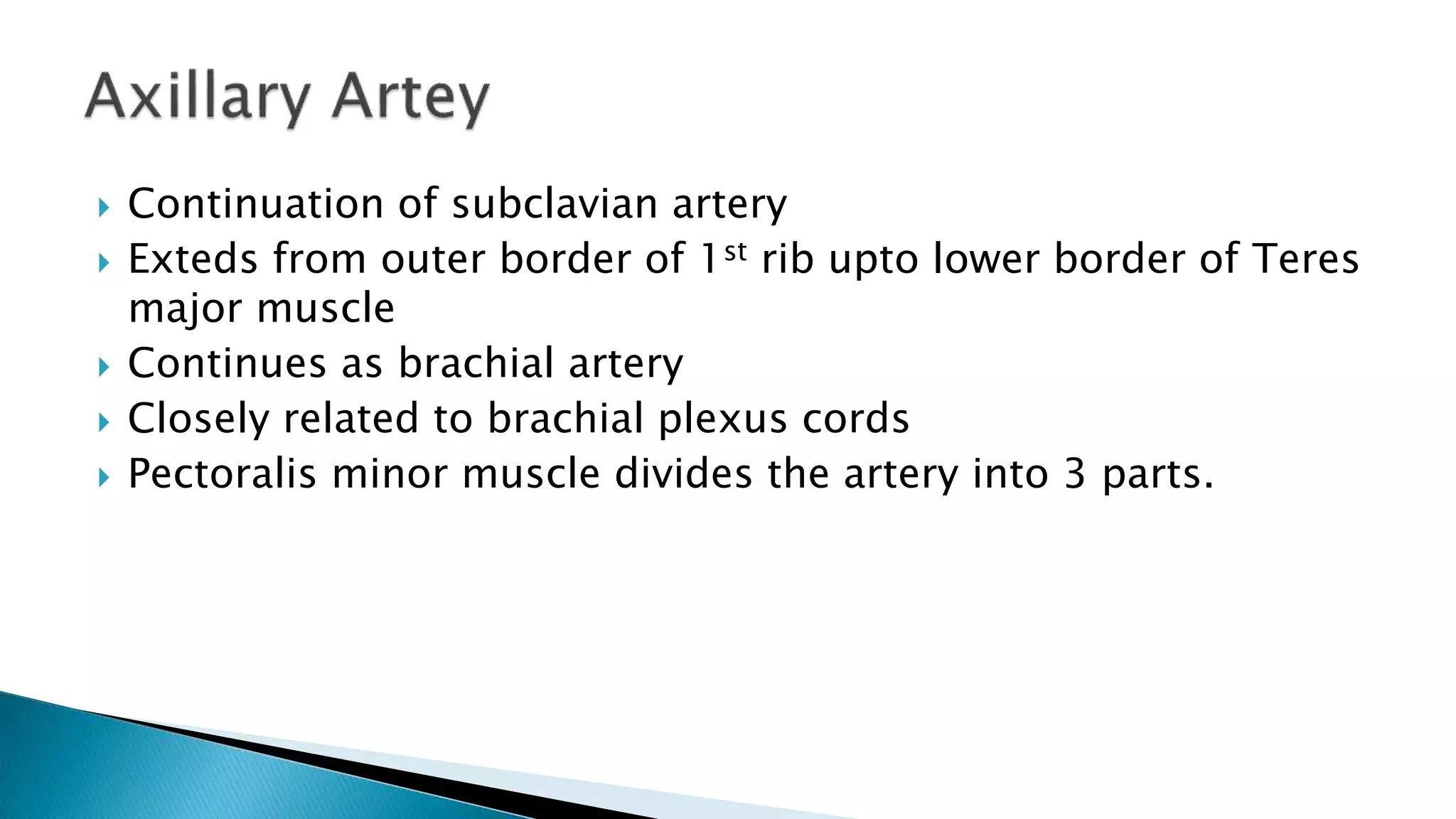
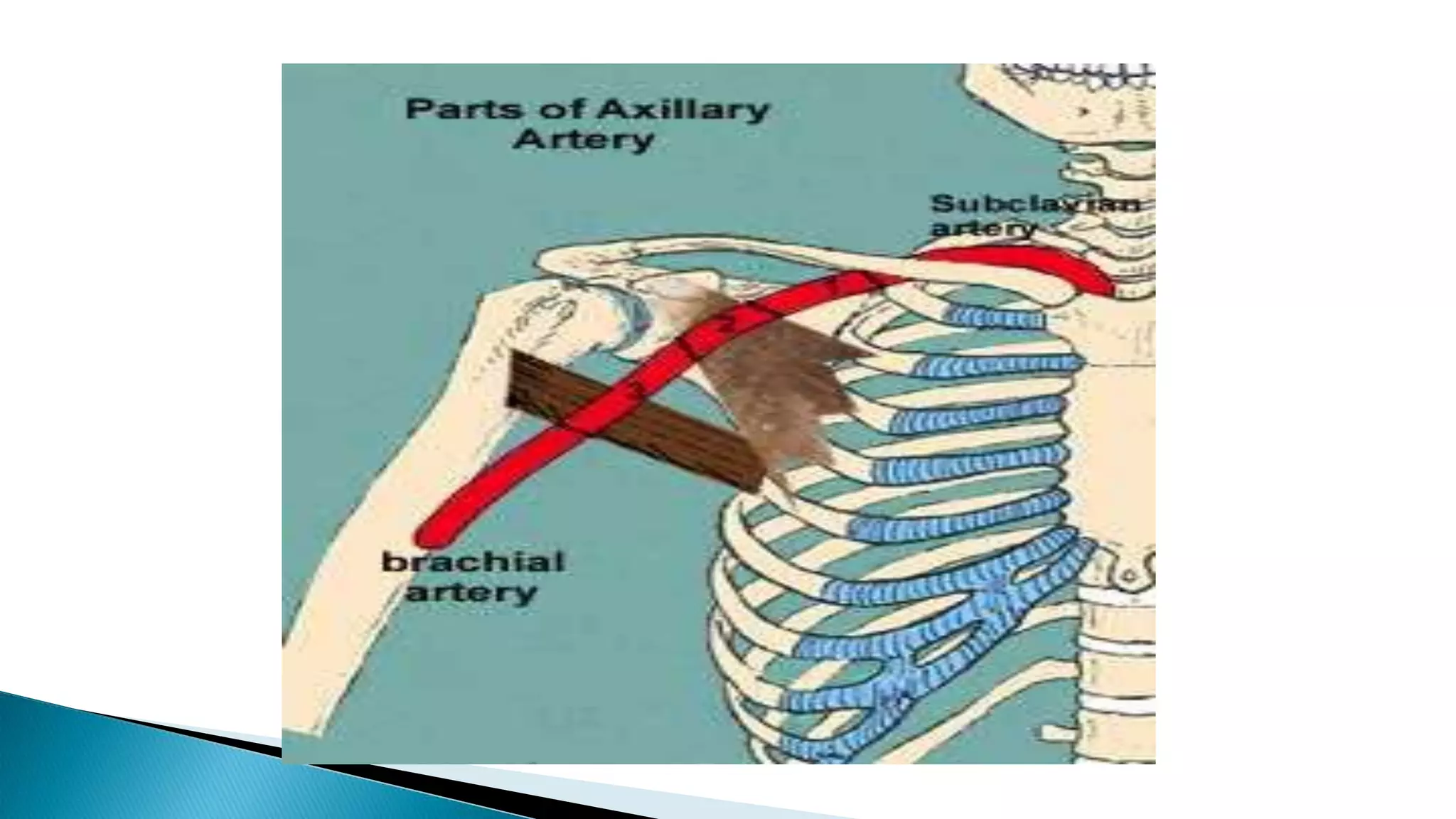
Continuation ofsubclavian artery
Exteds from outer border of 1st rib upto lower border of Teres
major muscle
Continues as brachial artery
Closely related to brachial plexus cords
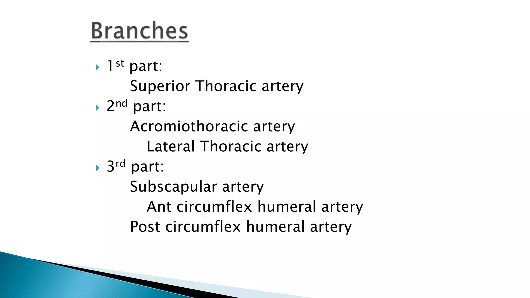
Pectoralis minor muscle divides the artery into 3 parts.
Superior ThoracicA:
o Small branch
o Between Pect. Major & Minor
o Supplies them & thoracic wall
8.
Given atupper border of pect. minor
Four terminal branches:
1. Pectoral – supplies pectoral muscles as well
as breast
2. Deltoid -
3. Acromial - joins the anastomosis over
acromian process
4. Clavicular – supplies sternoclavicular joint
and subclavius
9.
Given atLower border of pect. minor
Ant. axillary LN lies along it
Larger in females, supplies breast tissue
10.
Largest branch
Runs along Lower border of subscapularis
Terminates near inferior angle of scapula
Gives Circumflex Scapular Artery-
Passes through triangular intermuscular
space
Winds around lateral border of scapula,
deep to teres minor
Takes part in anastomosis around
scapula
11.
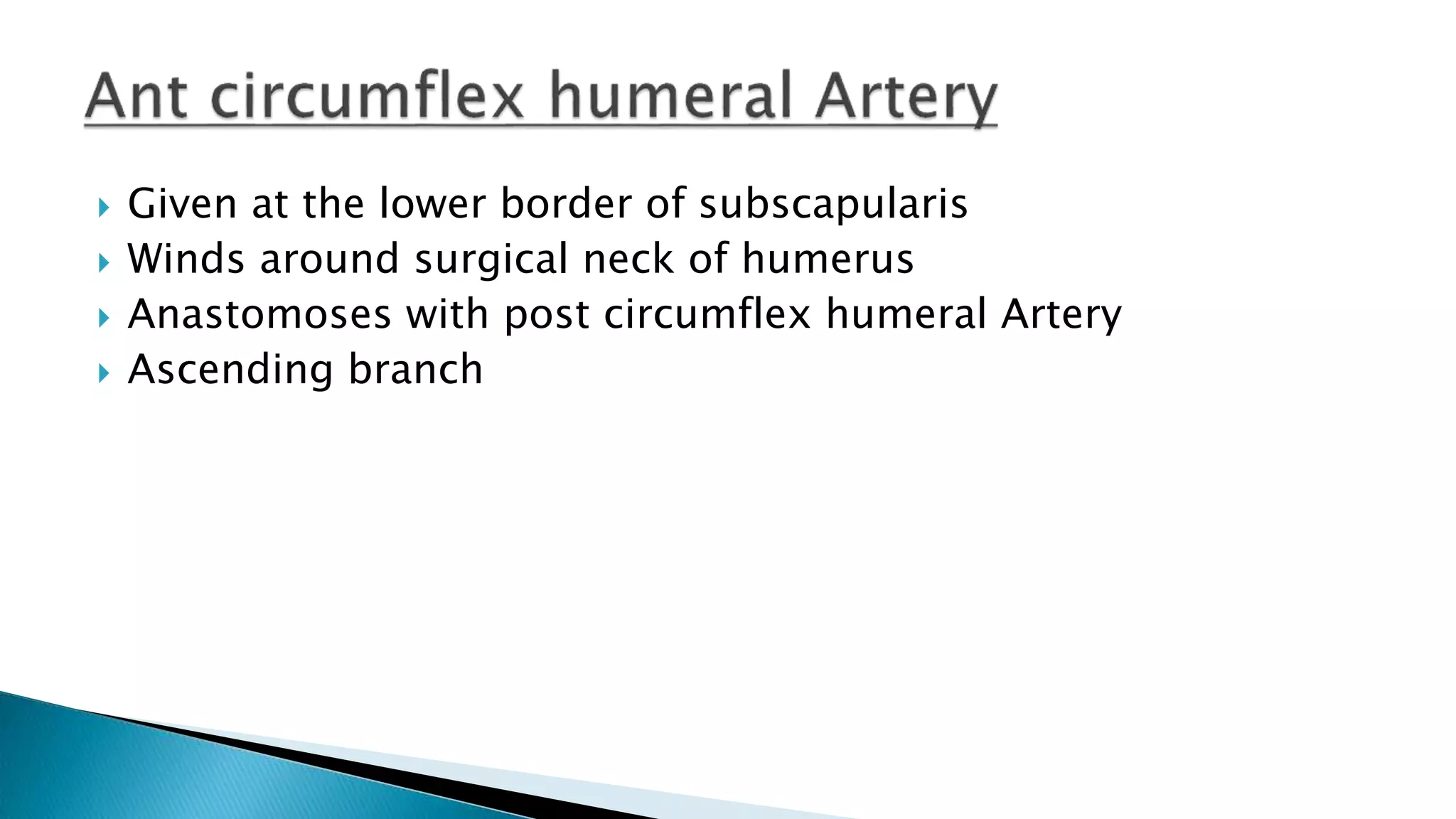
Given atthe lower border of subscapularis
Winds around surgical neck of humerus
Anastomoses with post circumflex humeral Artery
Ascending branch
12.
Larger
Runsbackward, through quadrangular space along with
Axillary N
Supplies shoulder joint, Deltoid mus.
13.
Anteriorly
(i)Skin.
(ii) Superficial fascia
(iii) Deep fascia.
(iv) Clavicular part of the pectoralis major.
(v) Clavipectoral fascia with cephalic vein, lateral pectoral
nerve, and thoracoacromial vessels.
15.
Posteriorly
(i)First intercostal space with the external intercostal muscle.
(ii) First and second digitations of the serratus anterior with
the nerve to serratus anterior.
(iii) Medial cord of brachial plexus with its medial pectoral
branch
16.
laterally
Lateraland posterior cords of the brachial plexus.
Medially
Axillary vein
The first part of the axillary artery is enclosed (together with
the brachial plexus) in the axillary sheath, derived from the
prevertebral layer of deep cervical fascia.
17.
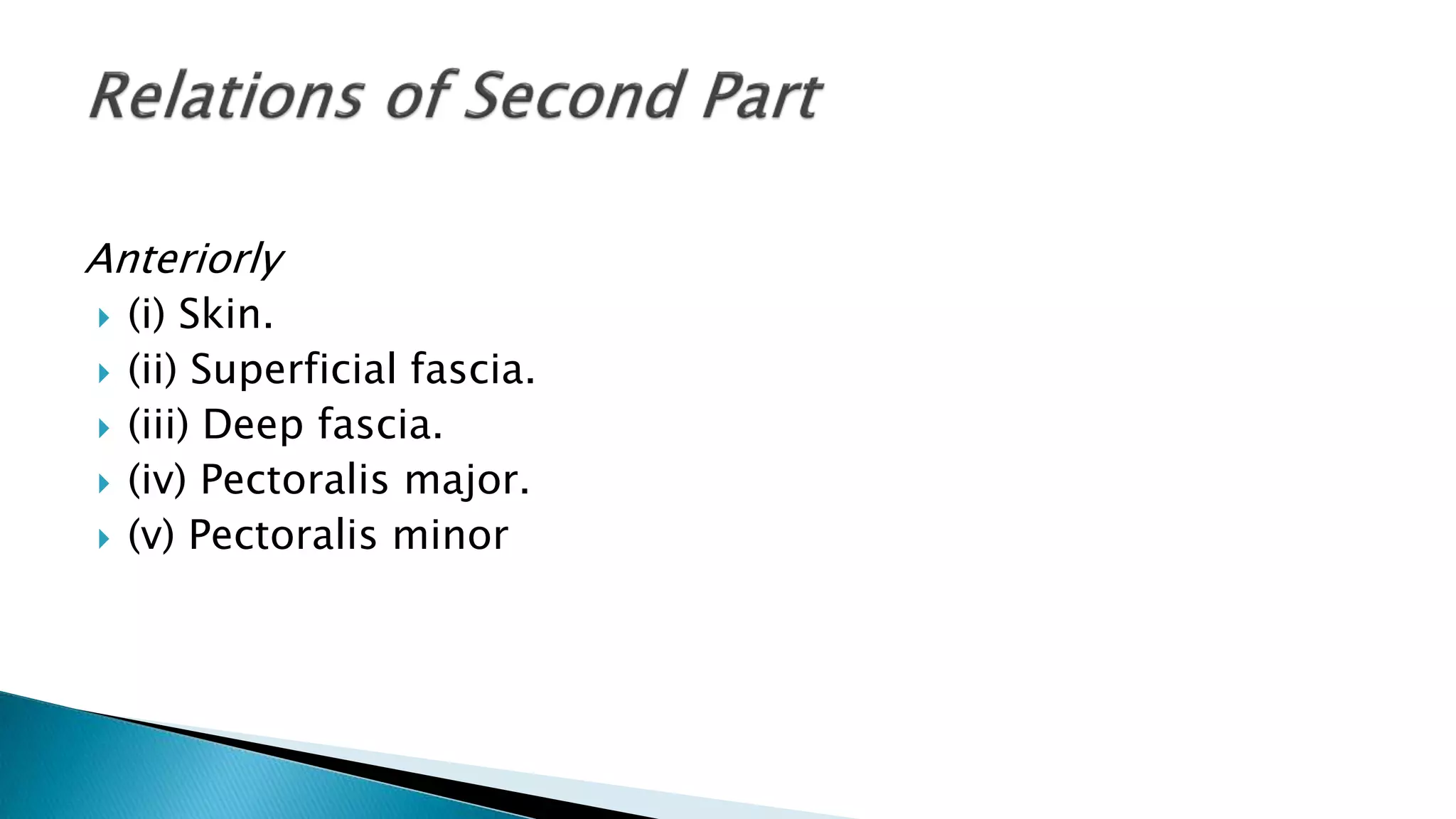
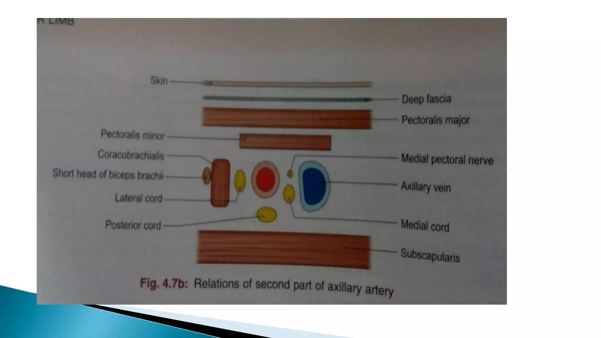
Anteriorly
(i) Skin.
(ii) Superficial fascia.
(iii) Deep fascia.
(iv) Pectoralis major.
(v) Pectoralis minor
19.
Posteriorly
(i) Posteriorcord of brachial plexus.
(ii) subscapularis
Medially
(i) Medial cord of brachial plexus,
(ii) Medial pectoral nerve,
(iii) Axillary vein.
Laterally
Lateral cord of brachial plexus.
20.
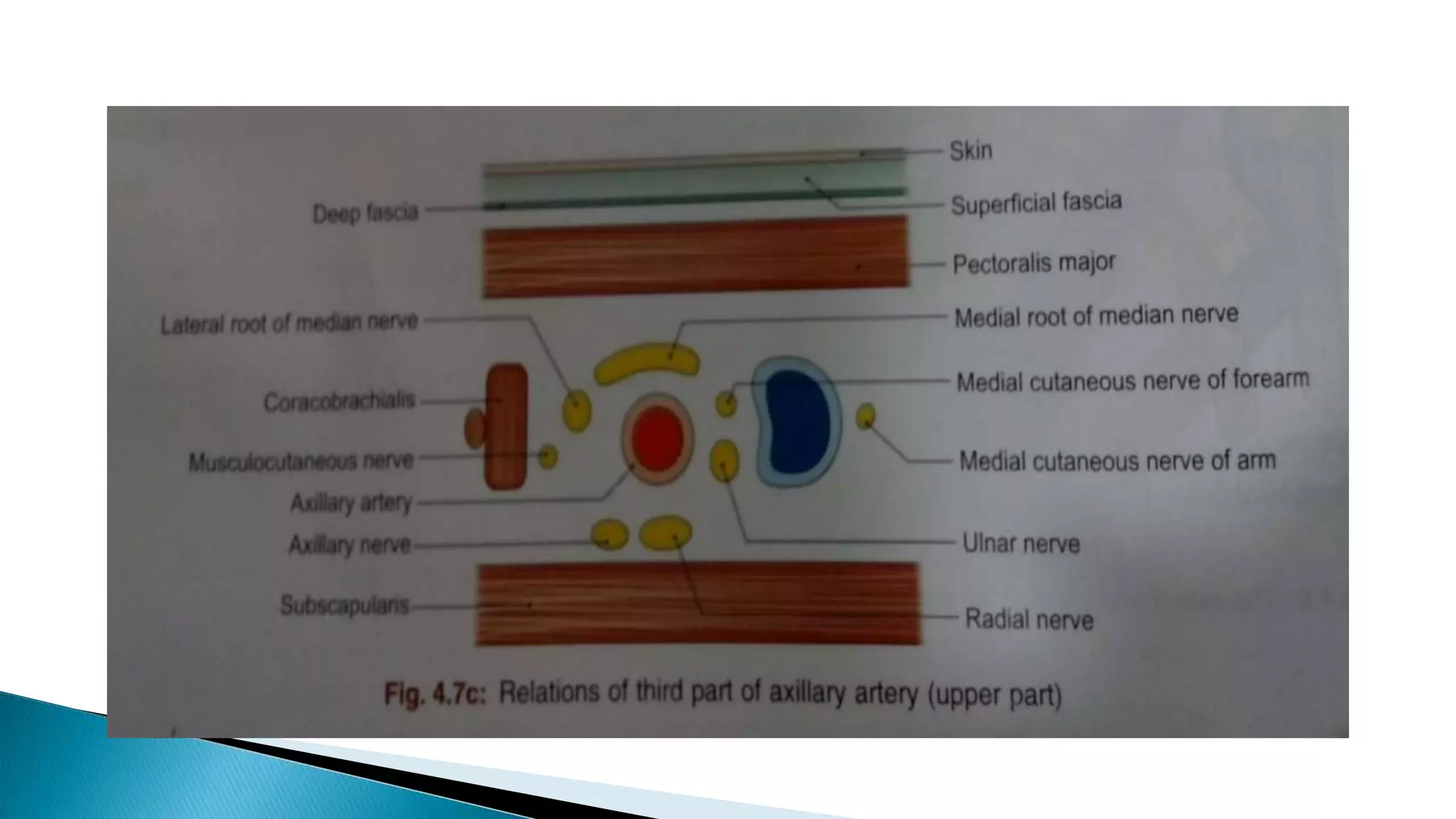
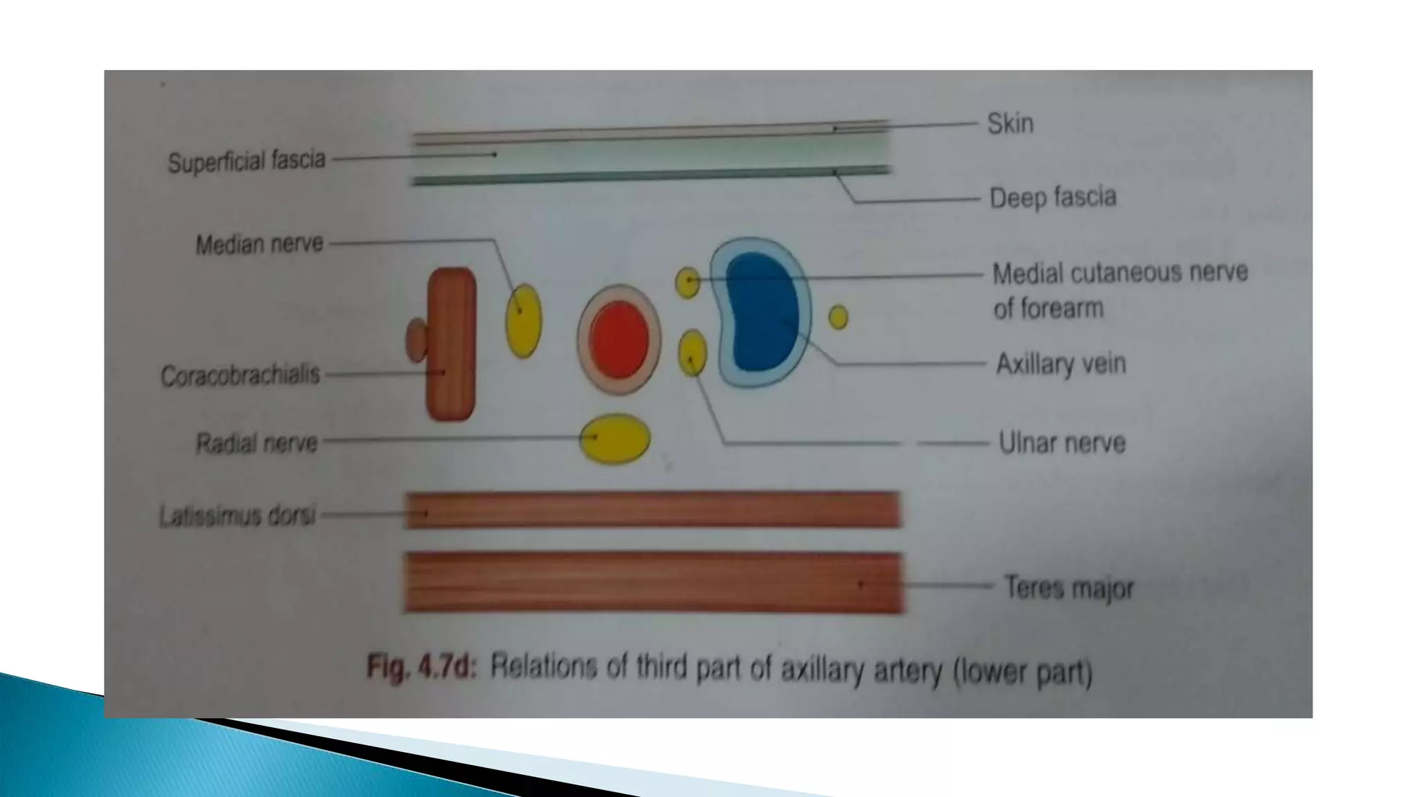
Anteriorly
(i)Skin.
(ii) Superficial fascia, (iii) Deep fascia.
(iv) In the upper part there are the pectoralis major and medial
root of the median nerve.
Posteriorly
(i) Radial nerve.
(ii) Axillary nerve in the upper part, (iii) Subscapularis in the
upper part, (iv) Tendons of the latissimus dorsi and the teres
major in the lower part.
23.
Laterally
(i) Coracobrachialis.
(ii)Musculocutaneous nerve in the upper part,
(iii) Lateral root of median nerve in the upper part,
(iv) Trunk of median nerve in the lower part.
Medially
(i) Axillary vein,
(ii) Medial cutaneous nerve of the forearm and ulnar nerve
(iii) Medial cutaneous nerve of arm
24.
Axial arterypulsastion can be felt against
the lower part of the lateral wall of axilla
In order to check bleeding from the distal part of limb
- The artery can be compressed against the humerus in the
lower part of the lateral wall of axilla
25.
1.Axillary artery isa continuation of which artery?
a) Supraclavicular artery
b) Subclavian artery
c) Subscapular artery
d) Superior thorasic artery
2. Which structure devides the artery into 3 parts
a) Pectoralis major muscle
b) Clavipectoral fascia
c) Pectoralis minor muscle
d) Teres minor muscle
26.
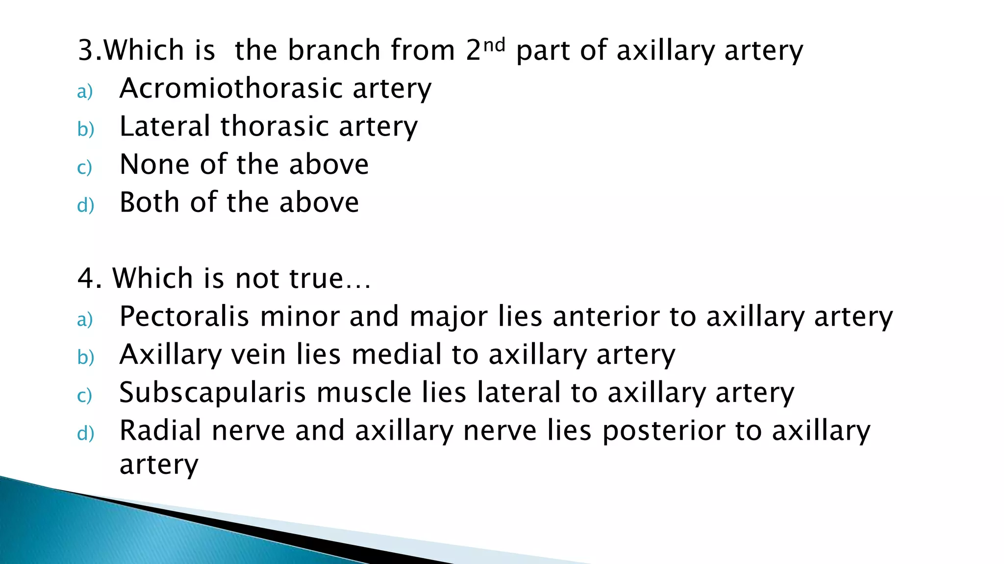
3.Which is thebranch from 2nd part of axillary artery
a) Acromiothorasic artery
b) Lateral thorasic artery
c) None of the above
d) Both of the above
4. Which is not true…
a) Pectoralis minor and major lies anterior to axillary artery
b) Axillary vein lies medial to axillary artery
c) Subscapularis muscle lies lateral to axillary artery
d) Radial nerve and axillary nerve lies posterior to axillary
artery
27.
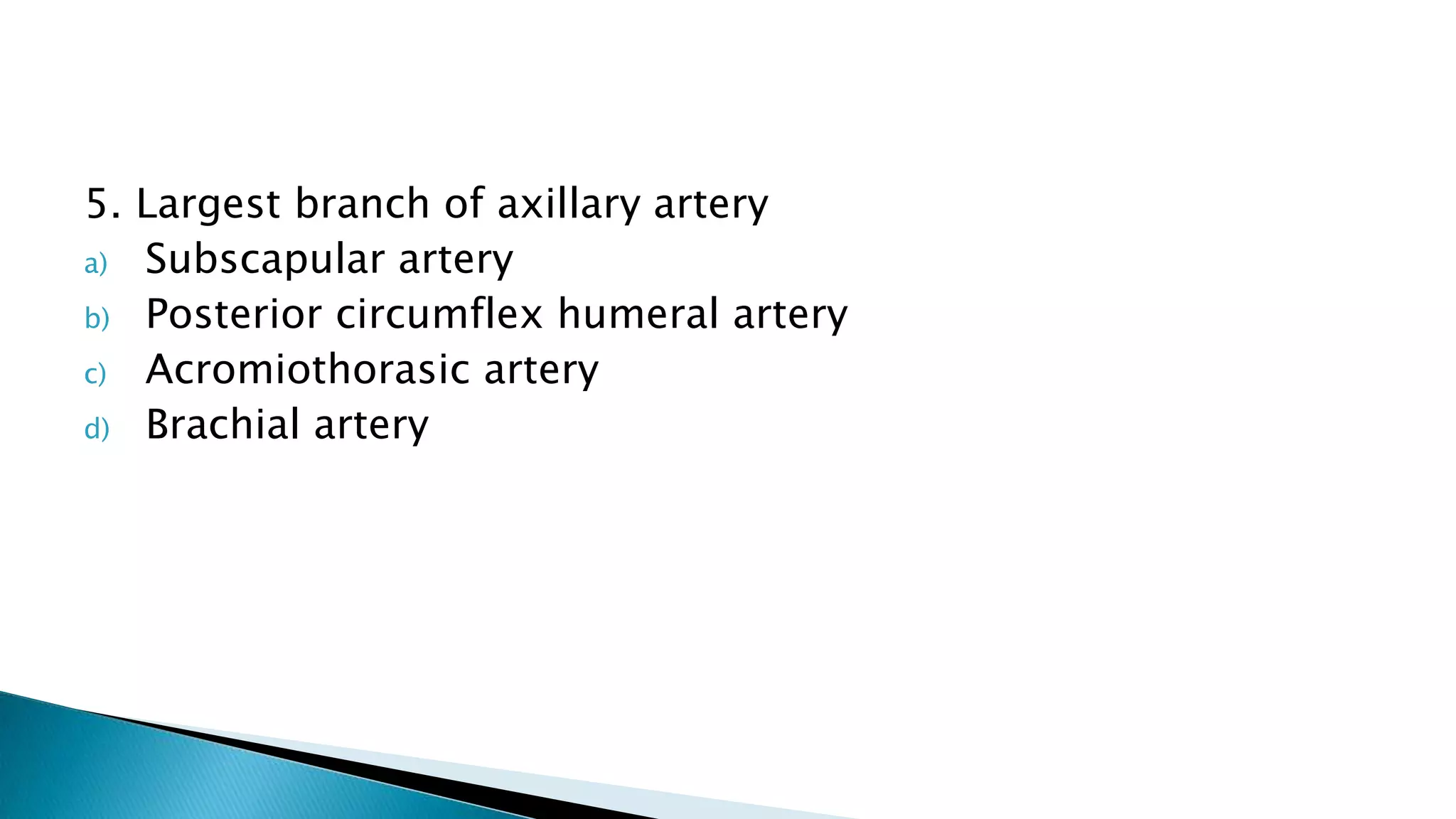
5. Largest branchof axillary artery
a) Subscapular artery
b) Posterior circumflex humeral artery
c) Acromiothorasic artery
d) Brachial artery


![Lecture 25 Intermuscular sapces and axilla [Autosaved].pptx](https://cdn.slidesharecdn.com/ss_thumbnails/lecture25intermuscularsapcesandaxillaautosaved-251110002658-47b36c78-thumbnail.jpg?width=640&height=640&fit=bounds)


