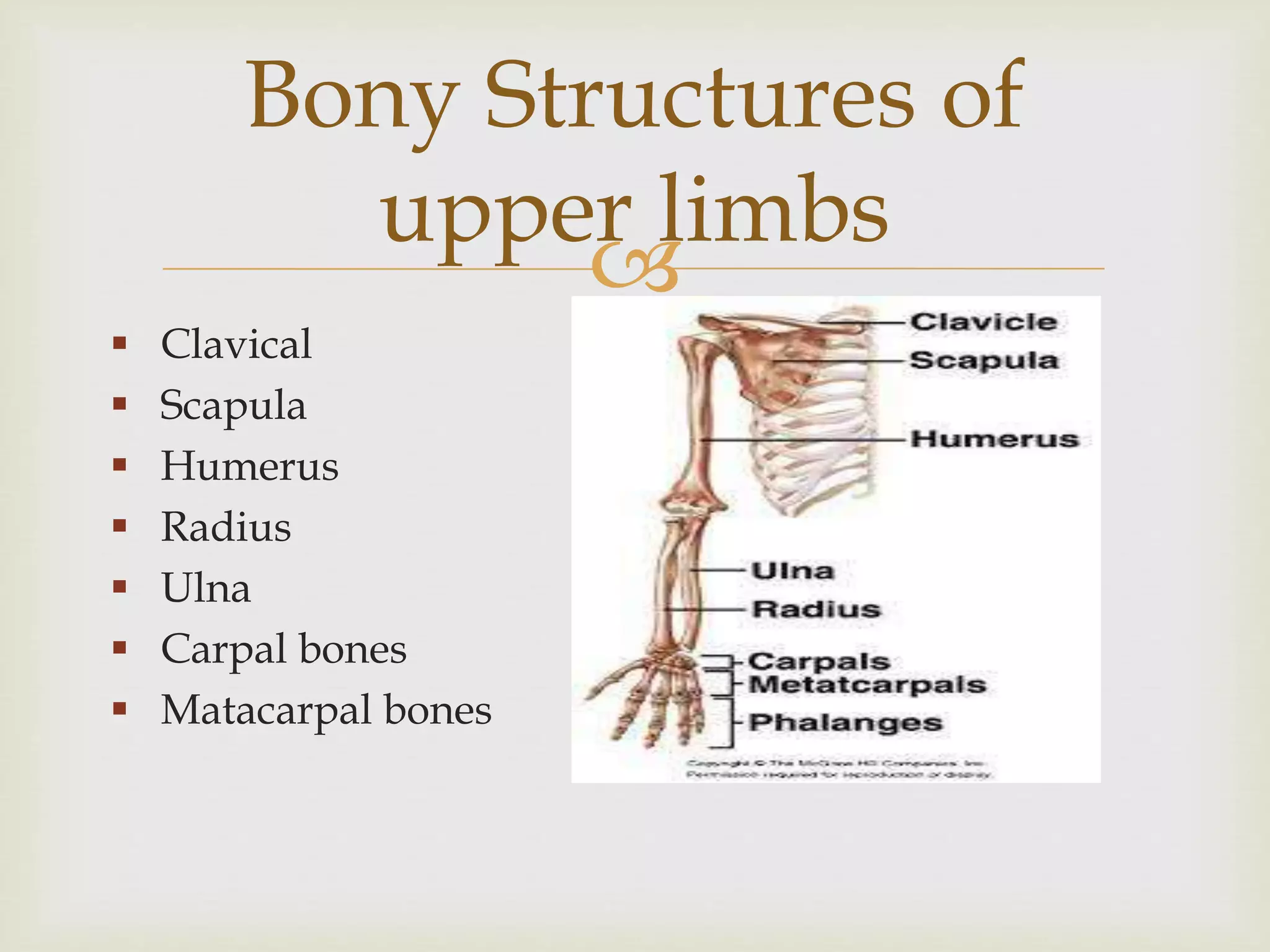
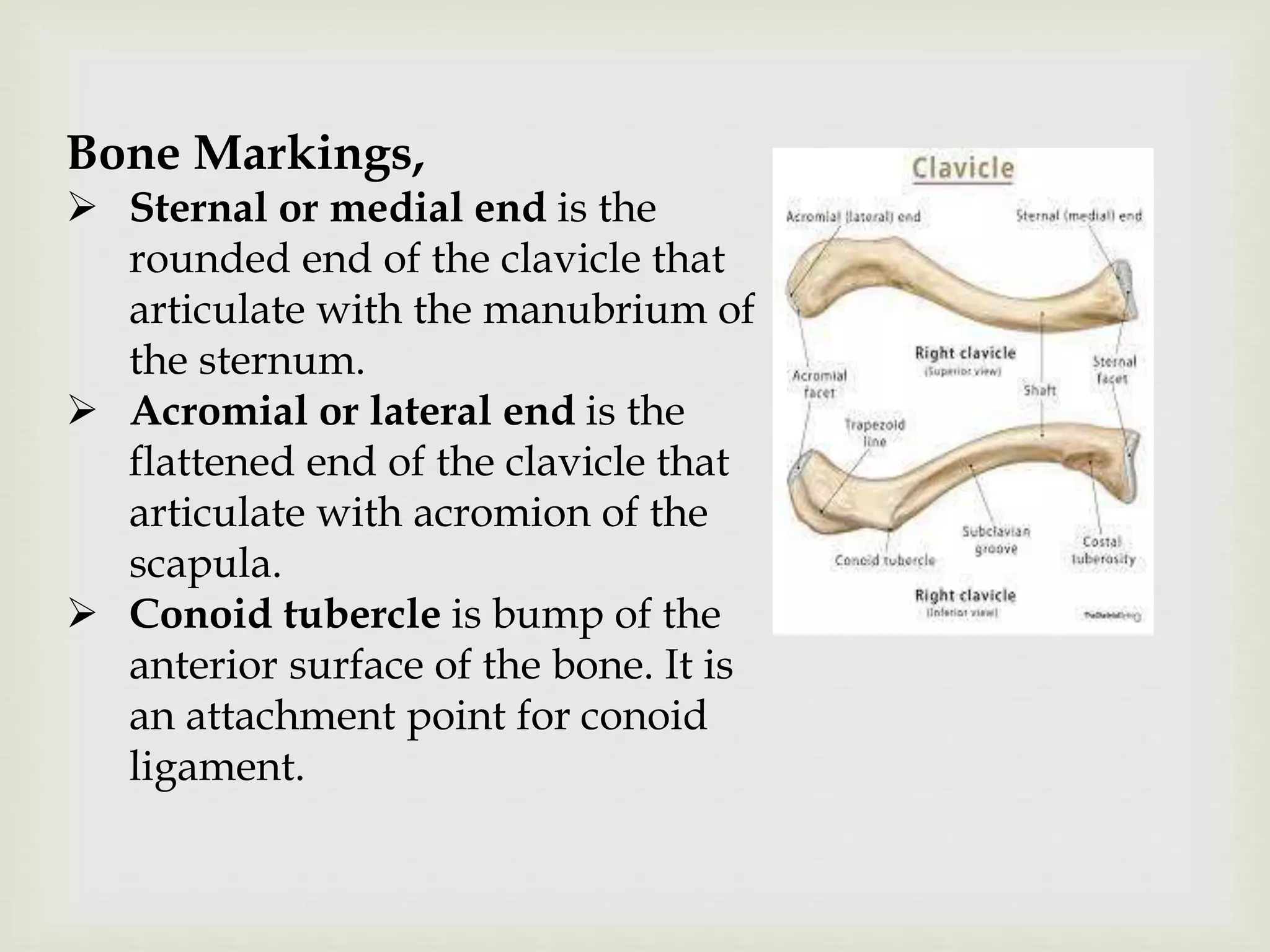
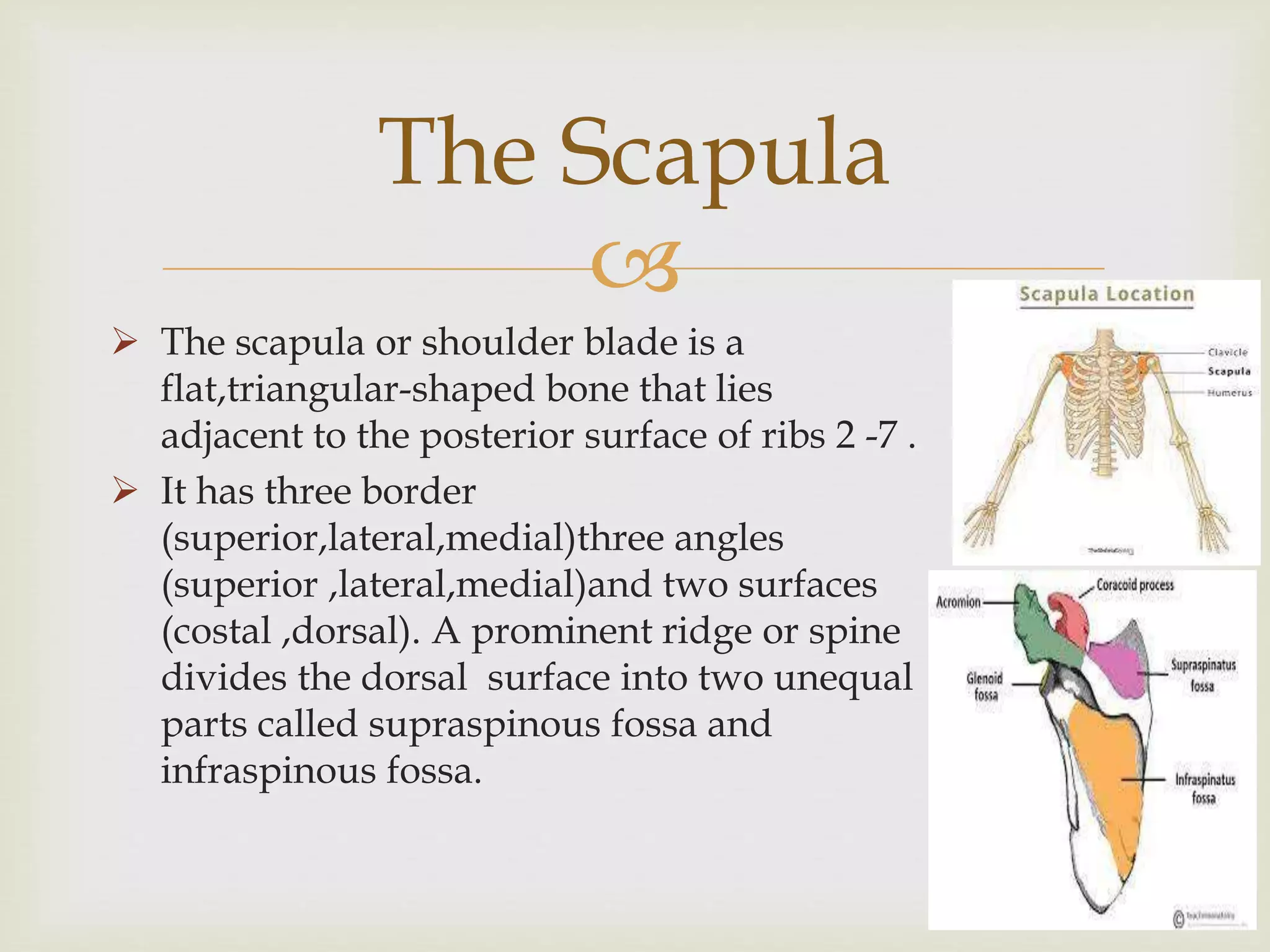
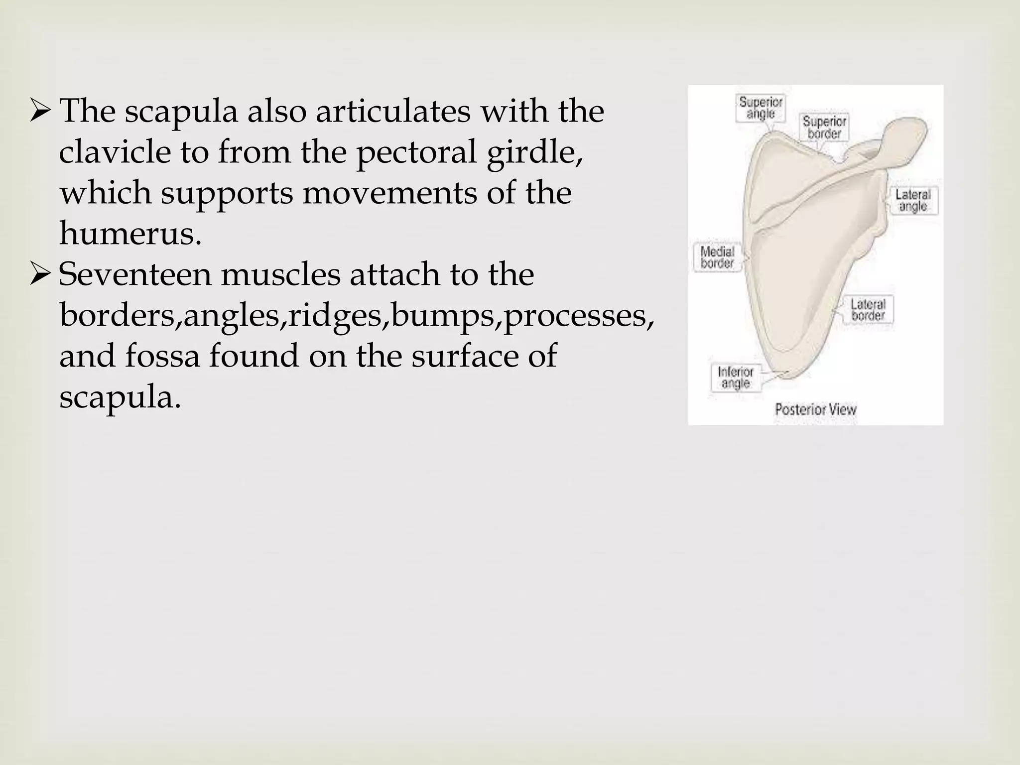
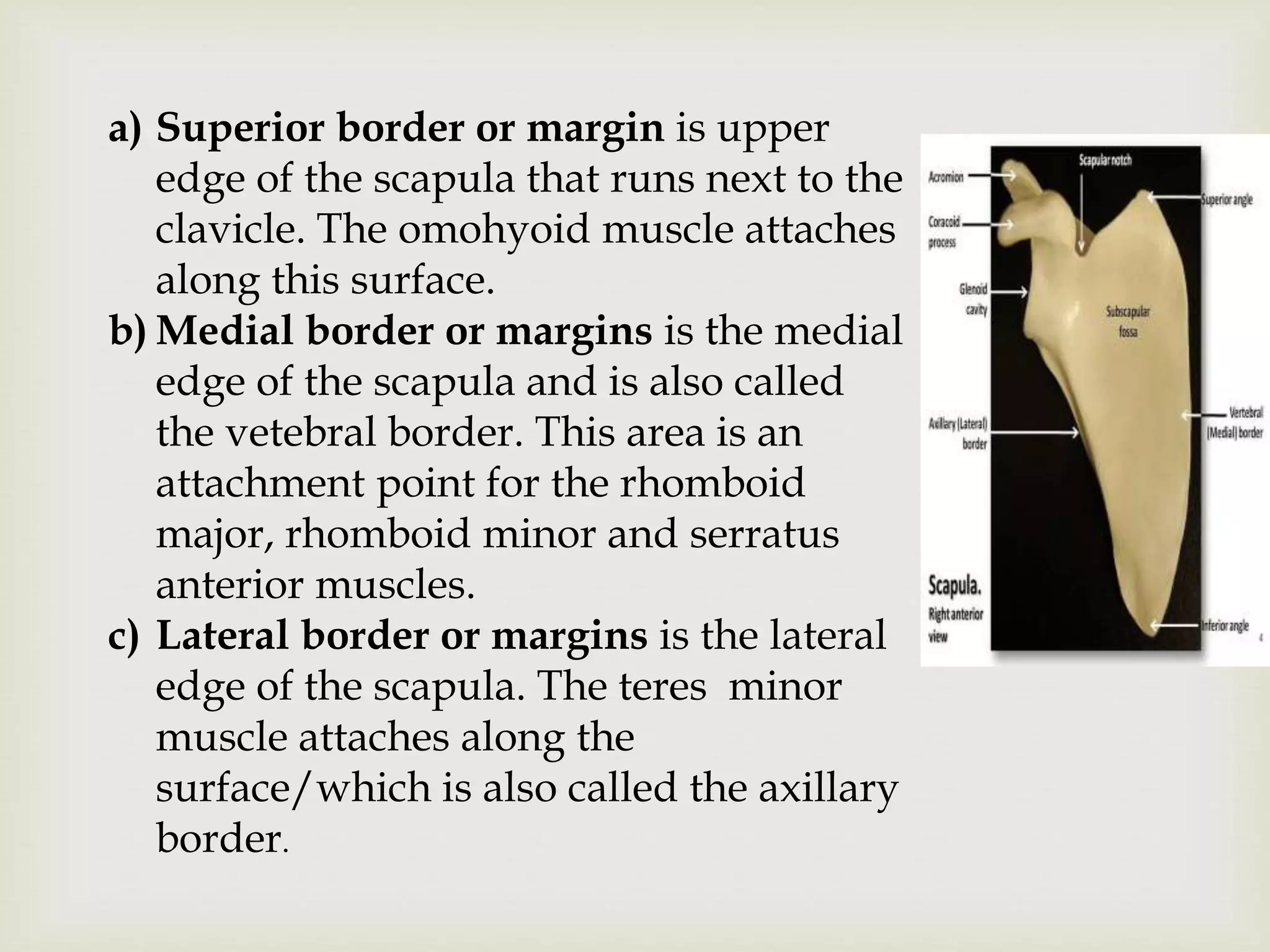
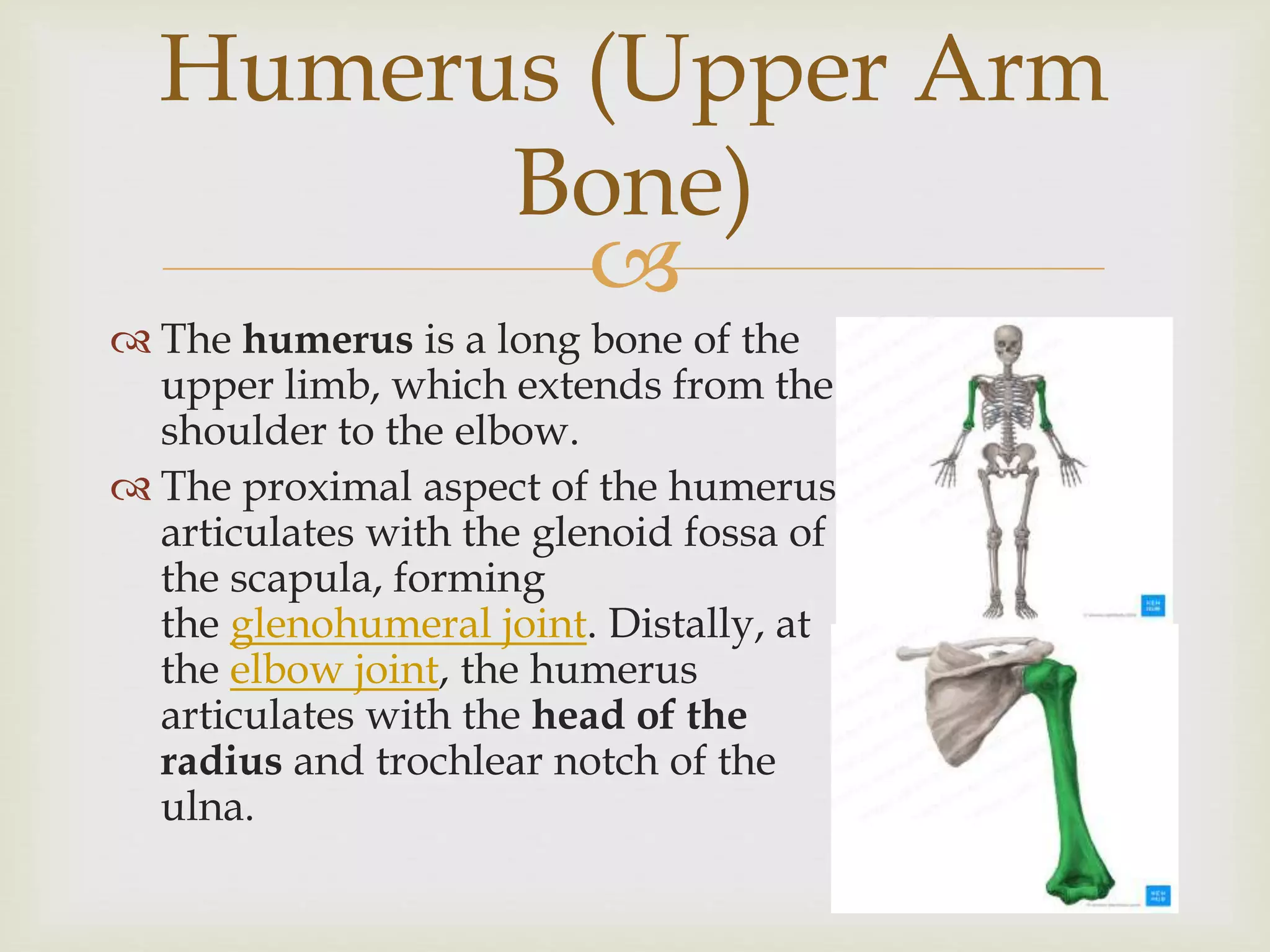
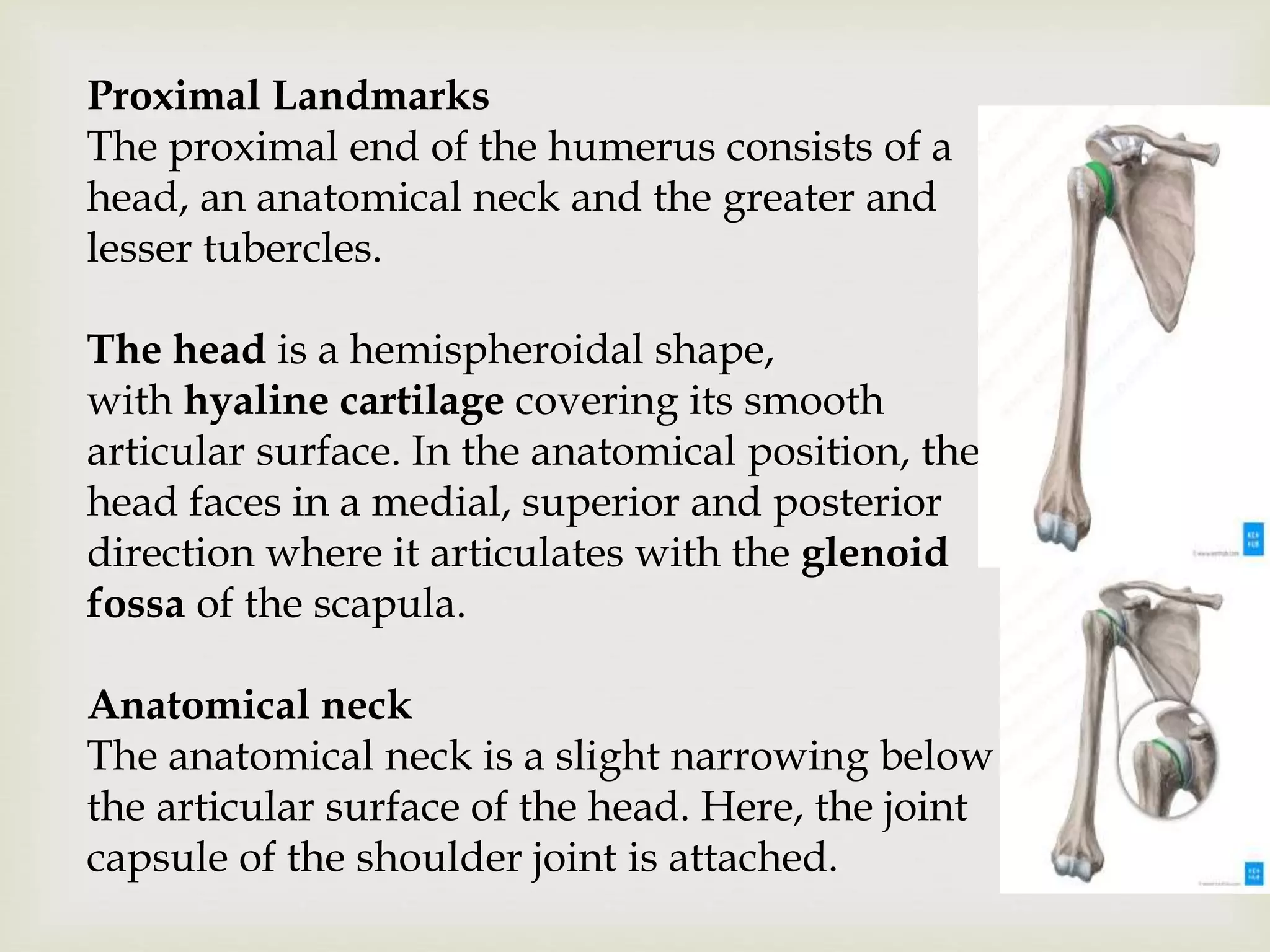
SlideShare a Scribd company logo
1 of 76

UPPER LIMB BONES

 Clavical
 Scapula
 Humerus
 Radius
 Ulna
 Carpal bones
 Matacarpal bones
Bony Structures of
upper limbs

 The clavicle or collarbone is located superior to the
1st rib and runs horizontally from manubrium of the
sternum to the acromion of the scapula.
 A superior view of clavicle shows it is shaped
somewhat like an “S”.The medial end curves
anteriorly and the lateral end curves posteriorly.
The clavicle

 Along with the scapula ,the
clavicle form the shoulder or
pectoral girdle ,which connects
the humerus(arm) to the axial
skeleton.
 The clavicle function like a lever
arm to help support the humerus
and scapula and maintain their
position relative to the rib cage.
Bone Markings,
 Sternal or medial end is the
rounded end of the clavicle that
articulate with the manubrium of
the sternum.
 Acromial or lateral end is the
flattened end of the clavicle that
articulate with acromion of the
scapula.
 Conoid tubercle is bump of the
anterior surface of the bone. It is
an attachment point for conoid
ligament.

 Trapezoid line or ridge is an elevation
that runs obliquely from the conoid
tubercle to the lateral end of the clavicle .it
serves as an attachment point for the
trapezoid ligament.
 Costal tuberosity is a roughened oval
elevation on the inferior surface of the
bone, near the medial(sternal) end.it
serves as an attachment point for
costoclavicular ligament, that attaches the
clavicle to the costal cartilage of the first
rib.

 Subclavian groove or sulcus is an indentation that
runs horizontally along inferior surface of the bone,
from the costal tuberosity to conoid tubercle.it serves
as an attachment point for Subclavian muscle.

 The scapula or shoulder blade is a
flat,triangular-shaped bone that lies
adjacent to the posterior surface of ribs 2 -7 .
 It has three border
(superior,lateral,medial)three angles
(superior ,lateral,medial)and two surfaces
(costal ,dorsal). A prominent ridge or spine
divides the dorsal surface into two unequal
parts called supraspinous fossa and
infraspinous fossa.
The Scapula
The scapula also articulates with the
clavicle to from the pectoral girdle,
which supports movements of the
humerus.
Seventeen muscles attach to the
borders,angles,ridges,bumps,processes,
and fossa found on the surface of
scapula.
a) Superior border or margin is upper
edge of the scapula that runs next to the
clavicle. The omohyoid muscle attaches
along this surface.
b) Medial border or margins is the medial
edge of the scapula and is also called
the vetebral border. This area is an
attachment point for the rhomboid
major, rhomboid minor and serratus
anterior muscles.
c) Lateral border or margins is the lateral
edge of the scapula. The teres minor
muscle attaches along the
surface/which is also called the axillary
border.
d) Superior angle is a sharp curvature at the junction
of the superior and medial borders. The area is an
attachment point for levator scapula muscle.
e) Lateral angle is the junction of the superior and
lateral borders. Its slightly concave lateral edge
articulates with the head of humerus.
f) Inferior angle is a sharp curvature formed at the
junction of the medial and lateral margins. It is an area
of attachment for the teres major muscle.
g) Coracoid process is a curved ,hook-like
anterolateral projection located above the lateral angle.
h) Suprascapular (Scapular) notch is a small groove
located medial to the coracoid process.it forms the
passageway for the Suprascapular nerve.
l)Glenoid cavity or fossa is a slight concavity at the
lateral angle.it forms a shallow socket for the
articulation of the head of the humerus.
i)Infraglenoid tubercle is a small prominence on the
inferior margin of the glenoid fossa.it serves as an
attachment point for the long head of triceps brachii
muscle.
j)Subscapular fossa is a large, slightly depressed
region in the middle of scapula’s anterior surface. The
subscapularis muscle attaches to this region of bone.
a) Superior angle is a sharp curvature at the junction
of the superior and medial borders. This area is an
attachment point for levator scapula muscle
b) Lateral angle is the junction of the superior and
lateral borders, its slightly concave lateral edge
articulates with the head of the humerus.
c) Inferior angle is a Sharpe curvature formed at the
junction of the medial and lateral margins, it is an
area of attachment for the teres major muscle.
d) Scapular spine is a long ridge that runs diagonally
across the upper posterior surface of the bone. The
trapezius and posterior deltoid muscles attach to
the surface of this prominent elevation.
e) Acromion is a lateral, fan-like extension of the spine.it
also is an attachment point for the trapezius and posterior
deltoid muscles.
f)Supraspinous fossa is a slightly depressed area located
superior to the spine. The supraspinous muscle attaches to
this region of scapula
g)Infraspinous fossa is a large, slightly depressed area
located inferior to the spine .it serves as an attachment
point for the infraspinatous muscle.
h) Glenoid cavity or fossa is
a slightly concavity at the
lateral angle.it forms a
shallow socket for the
articulation for the head of
humerus.
i) Infraglenoid tubercle is a
small prominence on the
inferior margin of the glenoid
fossa. The long head of triceps
brachii muscle attaches to the
knob-like ridge.

Humerus (Upper Arm
Bone)
 The humerus is a long bone of the
upper limb, which extends from the
shoulder to the elbow.
 The proximal aspect of the humerus
articulates with the glenoid fossa of
the scapula, forming
the glenohumeral joint. Distally, at
the elbow joint, the humerus
articulates with the head of the
radius and trochlear notch of the
ulna.
Proximal Landmarks
The proximal end of the humerus consists of a
head, an anatomical neck and the greater and
lesser tubercles.
The head is a hemispheroidal shape,
with hyaline cartilage covering its smooth
articular surface. In the anatomical position, the
head faces in a medial, superior and posterior
direction where it articulates with the glenoid
fossa of the scapula.
Anatomical neck
The anatomical neck is a slight narrowing below
the articular surface of the head. Here, the joint
capsule of the shoulder joint is attached.
Greater tubercle
The greater tubercle is the most lateral portion of the
proximal end of the humerus. It consists of three smooth
and flat impressions at the posterosuperior aspect for the
attachment of muscles. From superior to inferior, the
muscles that attach at these impressions are the:
 supraspinatus
 infraspinatus
 teres minor
Remember the muscles attaching to the greater tubercle
of the humerus using the following mnemonic!
Sally and Ingrid Teach Maths
 Supraspinatus
 Infraspinatus
 Teres Minor
The deltoid muscle covers the lateral aspect of the greater
tubercle, resulting in the normal rounded shape of the
shoulder. The lateral aspect also contains multiple vascular
foramina
Lesser tubercle
The lesser tubercle is located anterior to the anatomical neck
and has a smooth, palpable muscular impression. The lateral
part forms the medial margin of the intertubercular sulcus.
The subscapularis muscle attaches at this tubercle and
the transverse ligament of the shoulder also attaches on its
lateral part.
Intertubercular sulcus
The intertubercular sulcus is an indentation located
between the two tubercles. It is sometimes referred to as
the bicipital groove. The long tendon of the biceps
brachii and an ascending branch of the anterior circumflex
humeral artery are located within the sulcus. The sulcus
consists of a lateral lip and a medial lip.
The tendon of the pectoralis major muscle attaches on to
the lateral lip (a.k.a. crest of greater tubercle), while
the teres major tendon attaches on to the medial lip. In
addition, the tendon of lattisimus dorsi attaches to the
posterior aspect.
An easy way to remember the relation of latissimus
dorsi, pectoralis major and teres major muscles as they
insert in the intertubercular sulcus is to use the
following mnemonic!
'Lady between two majors'
 Lady: Latissimus dorsi
 Majors: Teres Major, pectoralis Major
Surgical neck
There is also a
narrowing below the
tubercles referred to as
the surgical neck, which
is a common fracture
site.
It is in close proximity to
the axillary nerve and
the posterior circumflex
humeral artery. This is
where the proximal end
of the humerus joins
with the long shaft.
Shaft
The proximal half of the shaft is of a cylindrical shape,
whereas the distal half is triangular. It consists of three
borders known as the anterior, lateral and medial borders.
The shaft also contains three surfaces referred to as the
anterolateral, anteromedial and posterior surfaces.
Borders
The anterior border begins at the greater tubercle and runs
downward almost to the end of the bone. The proximal
end of the anterior border is continuous with the lateral lip
of the intertubercular sulcus.
The lateral border begins just distal to the greater tubercle
of the humerus. It thickens distally to form the lateral
supracondylar ridge. The middle portion of the lateral
border is adjacent to the rough V shaped area referred to as
the deltoid tubercle/tuberosity.
Anterior border
Lateral
border
The medial border is similar to the lateral border in that it
forms the medial supracondylar ridge distally. The radial
groove is a shallow groove that interrupts the lateral border
in its medial third. The radial nerve and deep
brachial artery are located in this groove.
Surfaces:
The anterolateral surface is an area limited between the
anterior and lateral borders. It has a smooth
proximal surface and is largely covered by the deltoid
muscle. The deltoid inserts into the deltoid
tubercle/tuberosity around the middle of the surface. The
lateral portion of the brachialis muscle originates from the
distal part of this surface, as well as from the proximal two
third of the lateral supracondylar ridge.
Medial border Anterolateral Surface
The anteromedial surface is located between the
anterior and medial borders of the shaft/body,
beginning proximally at the floor of the intertubercular
sulcus. It provides attachment for the coracobrachialis
muscle around its mid-portion, while the distal half of
the surface is largely covered by the medial portion of
the brachialis muscle.
The posterior surface is bounded by the medial and
lateral borders and is covered mostly by the medial
head of the triceps brachii muscle. A ridge on the
proximal third also gives attachment to the lateral
head of the triceps brachii.
Anteromedial surface Posterior surface
Anterior
view
Posterior
view
Distal end
The distal end consists of both articular and non-articular
parts. The articular part of the humerus is a modified
condyle and is wider transversely. It articulates with both
the ulna and radius and consists of a medial trochlea and a
lateral capitulum, which are separated by a faint groove.
The non-articular part consists of the medial and lateral
epicondyles as well as the olecranon fossa, coronoid fossa
and radial fossae
Trochlea
The trochlea has a surface shaped like
a pulley and covers the anterior,
posterior and inferior surfaces of
the medial condyle of the humerus. It
articulates with the ulna at
the trochlear notch.
When the elbow is in the extended
position, the posterior and inferior
aspects of the trochlea are in contact
with the ulna. However, when the
elbow is flexed the posterior part is
no longer in contact, as the trochlear
notch slides towards the anterior
aspect of the humerus.
Capitulum
The capitulum is a convex and rounded projection that
covers the anterior and inferior surfaces of the lateral
condyle of the humerus.
Unlike the trochlea, it doesn’t cover the posterior
surface. It articulates with the head of the radius. In
extension, the inferior surface is in contact with the
radius but in the flexed position the radial head slides
towards the anterior aspect of the humerus.
Differentiate between the trochlea and capitulum at the
elbow joint very easily using this mnemonic!
CRAzy TULips
 Capitulum = RAdius
 Trochlear = ULnar
Medial epicondyle
The medial epicondyle is a blunt projection superomedial to the
medial condyle, which forms at the end of the medial border of the
humerus. The ulnar nerve crosses its smooth posterior surface and
is palpable in this location. The superficial muscles of the anterior
compartment of the forearm originate from the anterior surface of
the medial epicondyle. These muscles are the:
 flexor carpi ulnaris
 palmaris longus
 flexor carpi radialis
 pronator teres
Sometimes it can be difficult to remember if the common flexor
tendon is medial or lateral. Here's a mnemonic that can help you
out.
FM radio
Flexor Medial, so Common Flexor origin is on the medial side
Lateral epicondyle
The lateral border of the humerus ends at the lateral
epicondyle. There is an impression on the lateral and
anterior surfaces where the seven muscles of
the superficial group of the posterior compartment of
the forearm originate. These include the:
 brachioradialis
 extensor carpi radialis longus
 extensor carpi radialis brevis
 extensor digitorium
 extensor digiti minimi
 extensor carpi ulnaris
 anconeus
Olecranon fossa
The olecranon fossa is a deep hollowed area on the
posterior surface, superior to the trochlea. In elbow
extension, the tip of the ulnar olecranon process lodges
into this fossa.
Coronoid fossa
The coronoid fossa is a smaller hollow that is also located
superior to the trochlea, but on the anterior surface.
During flexion of the elbow, the coronoid process of the
ulna lodges into the coronoid fossa. Lateral to the
coronoid fossa and superior to the capitulum is another
depression referred to as the radial fossa. It is so named
as the margin of the head of the radius lodges there in full
flexion.
Olecranon fossa Coronoid Fossa
Radial fossa of humerus
Mnemonic
There are quite a lot of anatomical features at the distal
end of the humerus, don't you think? Remember them
more easily using the following mnemonic!
'CITE two Freaks'
 Capitulum
 Internal (medial) epicondyle
 Trochlea
 External (lateral) epicondyle
 Olecranon and coronoid Fossae
 The radius and ulna are the
bones of the forearm. The
radius bone supports the
lateral( thumb) side of the
forearm and the ulna bone
supports the medial ( little
finger) side.
 At the elbow, the radius
and ulna articulate with
the trochlea and capitulum
of humerus bone.
Radius and Ulna Bones
 At the wrist, the radius articulates with the
proximal row of carpel bones to form an
ellipsoidal joint.
 The forearm bones also articulte with each other.
Proximally, the head of the radius forms a joint
with the radial notch of the ulna, and distally ,
the head of ulna forms a joint with the ulnar
notch of the radius.
 These pivot joints allow the radius to rotate
around the ulna, which turns the palm of the
hand(protation and supination)
Radius Bone Markings
a) Radial Head is a proximal, disc - shaped
prominence. its concave superior surface articulates
with the capitulum of the humerus and its
cylindrical lateral surface articulates with radial
notch of the ulna.
b) Radial Shaft or body is the elongated region of
bone that extends distal to the tuberosity.
e)Interosseous border of the radius is the medial
edge ( margin) of the bone
f)Styloid process of the radius is a thick, pointed,
lateral projection from the distal end of bone.
g)Ulnar notch is a medio- distal concavity that
allows the radius to pivot around tha head of ulna
during pronation and spination of the forearm,
wrist and hand.
a)Olecranon process is a large fan-
shaped superior projection from
proximal end of the ulna.it also is
an attachment site for the triceps
brachii,anconeus and flexor carpi
ulnaris muscles.
b)Coronoid process is a triangular-
shaped,anterior projection from the
proximal end the shaft. it is an
attachment site for the brachialis
muscle.
Ulnar Bone Markings
c)Trochlear notch or semilunar notch is a deep
depression at proximal end of the ulna formed by
the curvatures of the olecranon and coronoid
process.This area articulates with the trochlea of
the humerus at the elbow.
d)Radial notch is a depression along the lateral
edge of the coronoid process that articulates with
the head of the radius.The joint allows the radial
head to rotate against the proximal end of the ulna
during pronation and supination.
e)Ulnar tuberosity is a roughnend eminence
located just inferior to the anterior tip of the
coronoid process. This area serves as the distal
attachment(Insertion) point for the brachialis
muscle.
f)Ulnar shaft is the body of the bone that extends
distal to the ulnae tuberosity.
g)Interosseous border of the ulna is the portion of
the lateral margin of the bone.
h)Ulnar head is a small rounded eminence at the distal
end of the ulna.its inferior surface articulates with the
articular disc cartillage of thr wrist and its lateral
surface articulates with the ulnar notch of the radius
during pronation and supination of foreram ,wrist and
hand.
i)Styloid process of the ulna is a small projection that
descends from the posterior portion of the ulnar
hand.it is an attachment point for the ulnar collateral
ligament, which connects the ulna to the triquetral and
pisiform carpel bones at the wrist.
Radius Bone Markings.
a) Radial Head is a proximal disc-
shaped prominence.its concave
superior surface articulates with the
capitulum of thr humerus and its
cylindrical lateral surface articulates
with the radial notch of the ulna.
b)Radial Neck is the region of bone
between the head and tuberosity.
Posterior
Anatomy
c)Radial Shaft or body is the elongated region of
bone that extends distal to the neck.
d)Interosseous border of the radius is the medial
edge( margin) of the bone where the interosseous
membrane attaches.
e)Styloid process of the radius is a thick , pointed
lateral projection from the distal end of the bone.it
forms the lateral portion of the wrist joint and
serves as an attachment site for the
branchioradialis muscle and radial( lateral)
collateral ligament.
f)Ulnar notch is a medio distal concavity that
allows the radius to pivot around the head of the
ulna during pronation and supination of the
forearm , wrist and hand.
a)Olecranon process
b)Ulnar shaft is the body of the
bone that extends distal to the
olecranon.
c)Interosseous border
d) Ulnar head is a small rounded
eminence at the distal end of the
ulna.
e)Styloid Process of the ulna.
Ulna Bone Markings
 Distal to the radius ana ulna are twenty-seven
bones that forms the hand.
 Eight small carpal bones support the wrist(
carpus).
 The proximal row of carpal bones is convex and
two of the bones articulates with the concave
distal surface of the radius.This junction
permits abduction and adduction ( side to side)
and flexion and extension( front to back) of the
wrist joint.
General Anatomy of the Hand and the
Wrist Bones
 Distal to the metacarpals are fourteen phalanges
which support the fingers or digits of the hand.
 Each finger is supported by three phalanges,
with the exception of the thumb.
a) Scaphoid bone is a boat-shaped bone located on
the lateral ( thumb) side of the carpus( Wrist).it
articulates with the distal end of the radius bone.
b)Lunate bone is cresent- shaped bone located
medial to the scaphoid bone.its concave proximal
surface also articulated with the distal end of the
radius bone.
c) Triquetral bone is a pyramid-shaped bone
located medial to the lunate bone on the ulnar side
of the carpus.
Carpel Bones: Proximal Row
d)Pisiform bone is a small , pea -shaped bone that
articulates with the triquetral bone on the ulnar side
of the carpus.it is embedded in the tendon of the
flexor carpi ulnaris muscle and increases tension
during muscle contraction.
a)Trapezium bone is an irregular-shaped bone
located distal to the scaphoid bone on the lateral (
thumb) side of the carpus.it is the radial( lateral)
border of the carpal tunnel,and its distal surface
articulates with the proximal surface of the first
metacarpal( thumb) bone.
b) Trapezoid bone is a small , four- sided bone that
is wedged between the trapezium and capitate
bones.it articulates with the proximal end of the
second metacarpal bone.
Carpel Bone : Distal Row
c) Capitate bone is located medial to the trapezium in
the center of the distal row of the carpus.The name of
the bone is based on its rounded proximal head
which fits into the concavities of scaphoid and lunate
bones.Distally , the capitate bone articulates with the
proximal end of the third metacarpal bone.
d)Hamate bone is located on the ulnar side of the
carpus. Projecting from its palmar surface is a hook-
like process that forms the ulnar( medial) border of
the carpal tunnel.The hamate bone articulates
distally with the fourth and fifth metacarpal bones.
 Metacarpals 1-5 (ossametacarpi) are the bones
that support the palm of the hand.Each consists
of a elongated body ( shaft),cubital base, and
convex head.The bases articulate with the distal
row of the carpus and the heads articulate with
proximal phalanges.Each bone is assigned a
number from 1 to 5 , starting on the lateral side(
thumb side) of the hand.
Metacarpal Bones
The phalanges ( singular: phalanx) are the fourteen
bones that support the fingers or digits of the
hand.Each finger is supported by three phalanges ,
exception of the thumb, which is supported by
two.
a)Proximal: 1-5 are the phalanges located directly
anterior to metatarsal bones.The proximal
phalanges articulates anteriorly with the middle
phalanges, except the first proximal phalanx which
articulates with the first distal phalanx.
Phalanges
b)Middle : 2-5 are the phalanges located between
the proximal and distal phalnges.
c)Distal: 1-5 are the phalanges that support the tips
of the fingers.All articulate posteriorly with a
middle phalnax,except the first distal
phalanx,which articulates with the first proximal
phalanx.
Upper limb bones.pptx

More Related Content

What's hot

Posterior abdominal wall
Posterior abdominal wallPosterior abdominal wall
Posterior abdominal walldhina raddy
 
Muscles of Lower Limb
Muscles of Lower LimbMuscles of Lower Limb
Muscles of Lower LimbQamar Iqbal
 
Anatomy of the Leg
Anatomy of the Leg Anatomy of the Leg
Anatomy of the Leg Mohamed Amin
 
General anatomy
General anatomyGeneral anatomy
General anatomyKemUnited
 
Ch 10 and 11 part 5
Ch 10 and 11 part 5Ch 10 and 11 part 5
Ch 10 and 11 part 5schwartzcm
 
5.posterior compartment of the thigh
5.posterior compartment of the thigh5.posterior compartment of the thigh
5.posterior compartment of the thighDr. Mohammad Mahmoud
 
Hip bone (Gross Anatomy)
Hip bone (Gross Anatomy)Hip bone (Gross Anatomy)
Hip bone (Gross Anatomy)AtifRaza11
 
Heart and pericardium cvs anatomy
Heart and pericardium cvs anatomyHeart and pericardium cvs anatomy
Heart and pericardium cvs anatomyRobbinsHobbin
 
1-Sternum-Osteology- 1 & 2.pdf
1-Sternum-Osteology- 1 & 2.pdf1-Sternum-Osteology- 1 & 2.pdf
1-Sternum-Osteology- 1 & 2.pdfRohitSah62
 
Vessels & nerves of lower limb
Vessels & nerves of lower limbVessels & nerves of lower limb
Vessels & nerves of lower limbOmar Moatamed
 
Anatomy (anatomy of bone, joint & muscles of upper limb)
Anatomy (anatomy of bone, joint & muscles of upper limb)Anatomy (anatomy of bone, joint & muscles of upper limb)
Anatomy (anatomy of bone, joint & muscles of upper limb)Osama Al-Zahrani
 

What's hot (20)

Posterior abdominal wall
Posterior abdominal wallPosterior abdominal wall
Posterior abdominal wall
 
Slideshow: The Foot
Slideshow: The FootSlideshow: The Foot
Slideshow: The Foot
 
Muscles of thorax
Muscles of thoraxMuscles of thorax
Muscles of thorax
 
Slideshow: Anterior Thigh
Slideshow: Anterior ThighSlideshow: Anterior Thigh
Slideshow: Anterior Thigh
 
Femoral triangle
Femoral triangleFemoral triangle
Femoral triangle
 
Muscles of Lower Limb
Muscles of Lower LimbMuscles of Lower Limb
Muscles of Lower Limb
 
Slideshow: Ulna
Slideshow: UlnaSlideshow: Ulna
Slideshow: Ulna
 
Anatomy of the Leg
Anatomy of the Leg Anatomy of the Leg
Anatomy of the Leg
 
Heart
HeartHeart
Heart
 
Conduction system
Conduction systemConduction system
Conduction system
 
General anatomy
General anatomyGeneral anatomy
General anatomy
 
Ch 10 and 11 part 5
Ch 10 and 11 part 5Ch 10 and 11 part 5
Ch 10 and 11 part 5
 
5.posterior compartment of the thigh
5.posterior compartment of the thigh5.posterior compartment of the thigh
5.posterior compartment of the thigh
 
Hip bone (Gross Anatomy)
Hip bone (Gross Anatomy)Hip bone (Gross Anatomy)
Hip bone (Gross Anatomy)
 
Heart and pericardium cvs anatomy
Heart and pericardium cvs anatomyHeart and pericardium cvs anatomy
Heart and pericardium cvs anatomy
 
1-Sternum-Osteology- 1 & 2.pdf
1-Sternum-Osteology- 1 & 2.pdf1-Sternum-Osteology- 1 & 2.pdf
1-Sternum-Osteology- 1 & 2.pdf
 
Vessels & nerves of lower limb
Vessels & nerves of lower limbVessels & nerves of lower limb
Vessels & nerves of lower limb
 
Anatomy (anatomy of bone, joint & muscles of upper limb)
Anatomy (anatomy of bone, joint & muscles of upper limb)Anatomy (anatomy of bone, joint & muscles of upper limb)
Anatomy (anatomy of bone, joint & muscles of upper limb)
 
Lymphatic system
Lymphatic systemLymphatic system
Lymphatic system
 
Interior of right atrium
Interior of right atriumInterior of right atrium
Interior of right atrium
 

Similar to Upper limb bones.pptx

Femur bone lec3
Femur bone lec3Femur bone lec3
Femur bone lec3Eimaan Ktk
 
UL upper limb-1.pdfassignment presentation 123
UL upper limb-1.pdfassignment presentation 123UL upper limb-1.pdfassignment presentation 123
UL upper limb-1.pdfassignment presentation 123AmanuelIbrahim
 
UL upper limb-1 anatomy,bonemuscle and neurovusculatur
UL upper limb-1 anatomy,bonemuscle and neurovusculaturUL upper limb-1 anatomy,bonemuscle and neurovusculatur
UL upper limb-1 anatomy,bonemuscle and neurovusculaturDiribaErko
 
UPPERLIMB bs1.pptx
UPPERLIMB bs1.pptxUPPERLIMB bs1.pptx
UPPERLIMB bs1.pptxTHIRUVAZAHAN
 
Femur (Gross Anatomy)
Femur (Gross Anatomy)Femur (Gross Anatomy)
Femur (Gross Anatomy)AtifRaza11
 
Anatomy of the Radius Bone
Anatomy of the Radius BoneAnatomy of the Radius Bone
Anatomy of the Radius BoneDr Adnan Sami
 
The skeleton of the lower limb
The skeleton of the lower limbThe skeleton of the lower limb
The skeleton of the lower limbKamal Deen
 
UPPER LIMB 4 ppt of human body..........
UPPER LIMB 4 ppt of human body..........UPPER LIMB 4 ppt of human body..........
UPPER LIMB 4 ppt of human body..........aahanak787
 
Skeletal System 2
Skeletal System 2Skeletal System 2
Skeletal System 2Marc Potter
 
Anatomy of the Lower Limb.pdf
Anatomy of the Lower Limb.pdfAnatomy of the Lower Limb.pdf
Anatomy of the Lower Limb.pdfWalubitaWalubita1
 
Appendicular Skeleton-Shoulder girdle and upper limb
Appendicular Skeleton-Shoulder girdle and upper limbAppendicular Skeleton-Shoulder girdle and upper limb
Appendicular Skeleton-Shoulder girdle and upper limbZENITH PARMAR
 
free lower limb.pptx
free lower limb.pptxfree lower limb.pptx
free lower limb.pptxFkFo
 
Dr. anurag applied anatomy of knee
Dr. anurag   applied anatomy of kneeDr. anurag   applied anatomy of knee
Dr. anurag applied anatomy of kneemrinal joshi
 

Similar to Upper limb bones.pptx (20)

Upper limb bones
Upper limb bonesUpper limb bones
Upper limb bones
 
Anatomy of Humerus
Anatomy of HumerusAnatomy of Humerus
Anatomy of Humerus
 
Femur bone lec3
Femur bone lec3Femur bone lec3
Femur bone lec3
 
UL upper limb-1.pdfassignment presentation 123
UL upper limb-1.pdfassignment presentation 123UL upper limb-1.pdfassignment presentation 123
UL upper limb-1.pdfassignment presentation 123
 
UL upper limb-1 anatomy,bonemuscle and neurovusculatur
UL upper limb-1 anatomy,bonemuscle and neurovusculaturUL upper limb-1 anatomy,bonemuscle and neurovusculatur
UL upper limb-1 anatomy,bonemuscle and neurovusculatur
 
UPPERLIMB bs1.pptx
UPPERLIMB bs1.pptxUPPERLIMB bs1.pptx
UPPERLIMB bs1.pptx
 
Femur (Gross Anatomy)
Femur (Gross Anatomy)Femur (Gross Anatomy)
Femur (Gross Anatomy)
 
Femur
FemurFemur
Femur
 
Anatomy of the Radius Bone
Anatomy of the Radius BoneAnatomy of the Radius Bone
Anatomy of the Radius Bone
 
The skeleton of the lower limb
The skeleton of the lower limbThe skeleton of the lower limb
The skeleton of the lower limb
 
UPPER LIMB 4 ppt of human body..........
UPPER LIMB 4 ppt of human body..........UPPER LIMB 4 ppt of human body..........
UPPER LIMB 4 ppt of human body..........
 
Appendicular Skeleton.pptx
Appendicular Skeleton.pptxAppendicular Skeleton.pptx
Appendicular Skeleton.pptx
 
Skeletal System 2
Skeletal System 2Skeletal System 2
Skeletal System 2
 
Anatomy lec1.pptx
Anatomy lec1.pptxAnatomy lec1.pptx
Anatomy lec1.pptx
 
Anatomy of the Lower Limb.pdf
Anatomy of the Lower Limb.pdfAnatomy of the Lower Limb.pdf
Anatomy of the Lower Limb.pdf
 
Appendicular Skeleton-Shoulder girdle and upper limb
Appendicular Skeleton-Shoulder girdle and upper limbAppendicular Skeleton-Shoulder girdle and upper limb
Appendicular Skeleton-Shoulder girdle and upper limb
 
knee complex.pptx
knee complex.pptxknee complex.pptx
knee complex.pptx
 
free lower limb.pptx
free lower limb.pptxfree lower limb.pptx
free lower limb.pptx
 
Dr. anurag applied anatomy of knee
Dr. anurag   applied anatomy of kneeDr. anurag   applied anatomy of knee
Dr. anurag applied anatomy of knee
 
Lower limb bones
Lower limb bonesLower limb bones
Lower limb bones
 

Recently uploaded

Paris 2024 Olympic Geographies - an activity
Paris 2024 Olympic Geographies - an activityParis 2024 Olympic Geographies - an activity
Paris 2024 Olympic Geographies - an activityGeoBlogs
 
Separation of Lanthanides/ Lanthanides and Actinides
Separation of Lanthanides/ Lanthanides and ActinidesSeparation of Lanthanides/ Lanthanides and Actinides
Separation of Lanthanides/ Lanthanides and ActinidesFatimaKhan178732
 
CARE OF CHILD IN INCUBATOR..........pptx
CARE OF CHILD IN INCUBATOR..........pptxCARE OF CHILD IN INCUBATOR..........pptx
CARE OF CHILD IN INCUBATOR..........pptxGaneshChakor2
 
Interactive Powerpoint_How to Master effective communication
Interactive Powerpoint_How to Master effective communicationInteractive Powerpoint_How to Master effective communication
Interactive Powerpoint_How to Master effective communicationnomboosow
 
Introduction to ArtificiaI Intelligence in Higher Education
Introduction to ArtificiaI Intelligence in Higher EducationIntroduction to ArtificiaI Intelligence in Higher Education
Introduction to ArtificiaI Intelligence in Higher Educationpboyjonauth
 
POINT- BIOCHEMISTRY SEM 2 ENZYMES UNIT 5.pptx
POINT- BIOCHEMISTRY SEM 2 ENZYMES UNIT 5.pptxPOINT- BIOCHEMISTRY SEM 2 ENZYMES UNIT 5.pptx
POINT- BIOCHEMISTRY SEM 2 ENZYMES UNIT 5.pptxSayali Powar
 
Accessible design: Minimum effort, maximum impact
Accessible design: Minimum effort, maximum impactAccessible design: Minimum effort, maximum impact
Accessible design: Minimum effort, maximum impactdawncurless
 
SOCIAL AND HISTORICAL CONTEXT - LFTVD.pptx
SOCIAL AND HISTORICAL CONTEXT - LFTVD.pptxSOCIAL AND HISTORICAL CONTEXT - LFTVD.pptx
SOCIAL AND HISTORICAL CONTEXT - LFTVD.pptxiammrhaywood
 
Organic Name Reactions for the students and aspirants of Chemistry12th.pptx
Organic Name Reactions  for the students and aspirants of Chemistry12th.pptxOrganic Name Reactions  for the students and aspirants of Chemistry12th.pptx
Organic Name Reactions for the students and aspirants of Chemistry12th.pptxVS Mahajan Coaching Centre
 
KSHARA STURA .pptx---KSHARA KARMA THERAPY (CAUSTIC THERAPY)————IMP.OF KSHARA ...
KSHARA STURA .pptx---KSHARA KARMA THERAPY (CAUSTIC THERAPY)————IMP.OF KSHARA ...KSHARA STURA .pptx---KSHARA KARMA THERAPY (CAUSTIC THERAPY)————IMP.OF KSHARA ...
KSHARA STURA .pptx---KSHARA KARMA THERAPY (CAUSTIC THERAPY)————IMP.OF KSHARA ...M56BOOKSTORE PRODUCT/SERVICE
 
microwave assisted reaction. General introduction
microwave assisted reaction. General introductionmicrowave assisted reaction. General introduction
microwave assisted reaction. General introductionMaksud Ahmed
 
URLs and Routing in the Odoo 17 Website App
URLs and Routing in the Odoo 17 Website AppURLs and Routing in the Odoo 17 Website App
URLs and Routing in the Odoo 17 Website AppCeline George
 
How to Configure Email Server in Odoo 17
How to Configure Email Server in Odoo 17How to Configure Email Server in Odoo 17
How to Configure Email Server in Odoo 17Celine George
 
Alper Gobel In Media Res Media Component
Alper Gobel In Media Res Media ComponentAlper Gobel In Media Res Media Component
Alper Gobel In Media Res Media ComponentInMediaRes1
 
Contemporary philippine arts from the regions_PPT_Module_12 [Autosaved] (1).pptx
Contemporary philippine arts from the regions_PPT_Module_12 [Autosaved] (1).pptxContemporary philippine arts from the regions_PPT_Module_12 [Autosaved] (1).pptx
Contemporary philippine arts from the regions_PPT_Module_12 [Autosaved] (1).pptxRoyAbrique
 
“Oh GOSH! Reflecting on Hackteria's Collaborative Practices in a Global Do-It...
“Oh GOSH! Reflecting on Hackteria's Collaborative Practices in a Global Do-It...“Oh GOSH! Reflecting on Hackteria's Collaborative Practices in a Global Do-It...
“Oh GOSH! Reflecting on Hackteria's Collaborative Practices in a Global Do-It...Marc Dusseiller Dusjagr
 
Sanyam Choudhary Chemistry practical.pdf
Sanyam Choudhary Chemistry practical.pdfSanyam Choudhary Chemistry practical.pdf
Sanyam Choudhary Chemistry practical.pdfsanyamsingh5019
 
ECONOMIC CONTEXT - LONG FORM TV DRAMA - PPT
ECONOMIC CONTEXT - LONG FORM TV DRAMA - PPTECONOMIC CONTEXT - LONG FORM TV DRAMA - PPT
ECONOMIC CONTEXT - LONG FORM TV DRAMA - PPTiammrhaywood
 
Class 11 Legal Studies Ch-1 Concept of State .pdf
Class 11 Legal Studies Ch-1 Concept of State .pdfClass 11 Legal Studies Ch-1 Concept of State .pdf
Class 11 Legal Studies Ch-1 Concept of State .pdfakmcokerachita
 

Recently uploaded (20)

Paris 2024 Olympic Geographies - an activity
Paris 2024 Olympic Geographies - an activityParis 2024 Olympic Geographies - an activity
Paris 2024 Olympic Geographies - an activity
 
Separation of Lanthanides/ Lanthanides and Actinides
Separation of Lanthanides/ Lanthanides and ActinidesSeparation of Lanthanides/ Lanthanides and Actinides
Separation of Lanthanides/ Lanthanides and Actinides
 
CARE OF CHILD IN INCUBATOR..........pptx
CARE OF CHILD IN INCUBATOR..........pptxCARE OF CHILD IN INCUBATOR..........pptx
CARE OF CHILD IN INCUBATOR..........pptx
 
Interactive Powerpoint_How to Master effective communication
Interactive Powerpoint_How to Master effective communicationInteractive Powerpoint_How to Master effective communication
Interactive Powerpoint_How to Master effective communication
 
Introduction to ArtificiaI Intelligence in Higher Education
Introduction to ArtificiaI Intelligence in Higher EducationIntroduction to ArtificiaI Intelligence in Higher Education
Introduction to ArtificiaI Intelligence in Higher Education
 
POINT- BIOCHEMISTRY SEM 2 ENZYMES UNIT 5.pptx
POINT- BIOCHEMISTRY SEM 2 ENZYMES UNIT 5.pptxPOINT- BIOCHEMISTRY SEM 2 ENZYMES UNIT 5.pptx
POINT- BIOCHEMISTRY SEM 2 ENZYMES UNIT 5.pptx
 
Accessible design: Minimum effort, maximum impact
Accessible design: Minimum effort, maximum impactAccessible design: Minimum effort, maximum impact
Accessible design: Minimum effort, maximum impact
 
SOCIAL AND HISTORICAL CONTEXT - LFTVD.pptx
SOCIAL AND HISTORICAL CONTEXT - LFTVD.pptxSOCIAL AND HISTORICAL CONTEXT - LFTVD.pptx
SOCIAL AND HISTORICAL CONTEXT - LFTVD.pptx
 
Organic Name Reactions for the students and aspirants of Chemistry12th.pptx
Organic Name Reactions  for the students and aspirants of Chemistry12th.pptxOrganic Name Reactions  for the students and aspirants of Chemistry12th.pptx
Organic Name Reactions for the students and aspirants of Chemistry12th.pptx
 
Staff of Color (SOC) Retention Efforts DDSD
Staff of Color (SOC) Retention Efforts DDSDStaff of Color (SOC) Retention Efforts DDSD
Staff of Color (SOC) Retention Efforts DDSD
 
KSHARA STURA .pptx---KSHARA KARMA THERAPY (CAUSTIC THERAPY)————IMP.OF KSHARA ...
KSHARA STURA .pptx---KSHARA KARMA THERAPY (CAUSTIC THERAPY)————IMP.OF KSHARA ...KSHARA STURA .pptx---KSHARA KARMA THERAPY (CAUSTIC THERAPY)————IMP.OF KSHARA ...
KSHARA STURA .pptx---KSHARA KARMA THERAPY (CAUSTIC THERAPY)————IMP.OF KSHARA ...
 
microwave assisted reaction. General introduction
microwave assisted reaction. General introductionmicrowave assisted reaction. General introduction
microwave assisted reaction. General introduction
 
URLs and Routing in the Odoo 17 Website App
URLs and Routing in the Odoo 17 Website AppURLs and Routing in the Odoo 17 Website App
URLs and Routing in the Odoo 17 Website App
 
How to Configure Email Server in Odoo 17
How to Configure Email Server in Odoo 17How to Configure Email Server in Odoo 17
How to Configure Email Server in Odoo 17
 
Alper Gobel In Media Res Media Component
Alper Gobel In Media Res Media ComponentAlper Gobel In Media Res Media Component
Alper Gobel In Media Res Media Component
 
Contemporary philippine arts from the regions_PPT_Module_12 [Autosaved] (1).pptx
Contemporary philippine arts from the regions_PPT_Module_12 [Autosaved] (1).pptxContemporary philippine arts from the regions_PPT_Module_12 [Autosaved] (1).pptx
Contemporary philippine arts from the regions_PPT_Module_12 [Autosaved] (1).pptx
 
“Oh GOSH! Reflecting on Hackteria's Collaborative Practices in a Global Do-It...
“Oh GOSH! Reflecting on Hackteria's Collaborative Practices in a Global Do-It...“Oh GOSH! Reflecting on Hackteria's Collaborative Practices in a Global Do-It...
“Oh GOSH! Reflecting on Hackteria's Collaborative Practices in a Global Do-It...
 
Sanyam Choudhary Chemistry practical.pdf
Sanyam Choudhary Chemistry practical.pdfSanyam Choudhary Chemistry practical.pdf
Sanyam Choudhary Chemistry practical.pdf
 
ECONOMIC CONTEXT - LONG FORM TV DRAMA - PPT
ECONOMIC CONTEXT - LONG FORM TV DRAMA - PPTECONOMIC CONTEXT - LONG FORM TV DRAMA - PPT
ECONOMIC CONTEXT - LONG FORM TV DRAMA - PPT
 
Class 11 Legal Studies Ch-1 Concept of State .pdf
Class 11 Legal Studies Ch-1 Concept of State .pdfClass 11 Legal Studies Ch-1 Concept of State .pdf
Class 11 Legal Studies Ch-1 Concept of State .pdf
 

Upper limb bones.pptx

  • 2.   Clavical  Scapula  Humerus  Radius  Ulna  Carpal bones  Matacarpal bones Bony Structures of upper limbs
  • 3.   The clavicle or collarbone is located superior to the 1st rib and runs horizontally from manubrium of the sternum to the acromion of the scapula.  A superior view of clavicle shows it is shaped somewhat like an “S”.The medial end curves anteriorly and the lateral end curves posteriorly. The clavicle
  • 4.   Along with the scapula ,the clavicle form the shoulder or pectoral girdle ,which connects the humerus(arm) to the axial skeleton.  The clavicle function like a lever arm to help support the humerus and scapula and maintain their position relative to the rib cage.
  • 5. Bone Markings,  Sternal or medial end is the rounded end of the clavicle that articulate with the manubrium of the sternum.  Acromial or lateral end is the flattened end of the clavicle that articulate with acromion of the scapula.  Conoid tubercle is bump of the anterior surface of the bone. It is an attachment point for conoid ligament.
  • 6.   Trapezoid line or ridge is an elevation that runs obliquely from the conoid tubercle to the lateral end of the clavicle .it serves as an attachment point for the trapezoid ligament.  Costal tuberosity is a roughened oval elevation on the inferior surface of the bone, near the medial(sternal) end.it serves as an attachment point for costoclavicular ligament, that attaches the clavicle to the costal cartilage of the first rib.
  • 7.   Subclavian groove or sulcus is an indentation that runs horizontally along inferior surface of the bone, from the costal tuberosity to conoid tubercle.it serves as an attachment point for Subclavian muscle.
  • 8.   The scapula or shoulder blade is a flat,triangular-shaped bone that lies adjacent to the posterior surface of ribs 2 -7 .  It has three border (superior,lateral,medial)three angles (superior ,lateral,medial)and two surfaces (costal ,dorsal). A prominent ridge or spine divides the dorsal surface into two unequal parts called supraspinous fossa and infraspinous fossa. The Scapula
  • 9. The scapula also articulates with the clavicle to from the pectoral girdle, which supports movements of the humerus. Seventeen muscles attach to the borders,angles,ridges,bumps,processes, and fossa found on the surface of scapula.
  • 10. a) Superior border or margin is upper edge of the scapula that runs next to the clavicle. The omohyoid muscle attaches along this surface. b) Medial border or margins is the medial edge of the scapula and is also called the vetebral border. This area is an attachment point for the rhomboid major, rhomboid minor and serratus anterior muscles. c) Lateral border or margins is the lateral edge of the scapula. The teres minor muscle attaches along the surface/which is also called the axillary border.
  • 11. d) Superior angle is a sharp curvature at the junction of the superior and medial borders. The area is an attachment point for levator scapula muscle. e) Lateral angle is the junction of the superior and lateral borders. Its slightly concave lateral edge articulates with the head of humerus. f) Inferior angle is a sharp curvature formed at the junction of the medial and lateral margins. It is an area of attachment for the teres major muscle. g) Coracoid process is a curved ,hook-like anterolateral projection located above the lateral angle.
  • 12. h) Suprascapular (Scapular) notch is a small groove located medial to the coracoid process.it forms the passageway for the Suprascapular nerve. l)Glenoid cavity or fossa is a slight concavity at the lateral angle.it forms a shallow socket for the articulation of the head of the humerus. i)Infraglenoid tubercle is a small prominence on the inferior margin of the glenoid fossa.it serves as an attachment point for the long head of triceps brachii muscle. j)Subscapular fossa is a large, slightly depressed region in the middle of scapula’s anterior surface. The subscapularis muscle attaches to this region of bone.
  • 13.
  • 14. a) Superior angle is a sharp curvature at the junction of the superior and medial borders. This area is an attachment point for levator scapula muscle b) Lateral angle is the junction of the superior and lateral borders, its slightly concave lateral edge articulates with the head of the humerus. c) Inferior angle is a Sharpe curvature formed at the junction of the medial and lateral margins, it is an area of attachment for the teres major muscle. d) Scapular spine is a long ridge that runs diagonally across the upper posterior surface of the bone. The trapezius and posterior deltoid muscles attach to the surface of this prominent elevation.
  • 15.
  • 16. e) Acromion is a lateral, fan-like extension of the spine.it also is an attachment point for the trapezius and posterior deltoid muscles. f)Supraspinous fossa is a slightly depressed area located superior to the spine. The supraspinous muscle attaches to this region of scapula g)Infraspinous fossa is a large, slightly depressed area located inferior to the spine .it serves as an attachment point for the infraspinatous muscle.
  • 17. h) Glenoid cavity or fossa is a slightly concavity at the lateral angle.it forms a shallow socket for the articulation for the head of humerus. i) Infraglenoid tubercle is a small prominence on the inferior margin of the glenoid fossa. The long head of triceps brachii muscle attaches to the knob-like ridge.
  • 18.  Humerus (Upper Arm Bone)  The humerus is a long bone of the upper limb, which extends from the shoulder to the elbow.  The proximal aspect of the humerus articulates with the glenoid fossa of the scapula, forming the glenohumeral joint. Distally, at the elbow joint, the humerus articulates with the head of the radius and trochlear notch of the ulna.
  • 19. Proximal Landmarks The proximal end of the humerus consists of a head, an anatomical neck and the greater and lesser tubercles. The head is a hemispheroidal shape, with hyaline cartilage covering its smooth articular surface. In the anatomical position, the head faces in a medial, superior and posterior direction where it articulates with the glenoid fossa of the scapula. Anatomical neck The anatomical neck is a slight narrowing below the articular surface of the head. Here, the joint capsule of the shoulder joint is attached.
  • 20. Greater tubercle The greater tubercle is the most lateral portion of the proximal end of the humerus. It consists of three smooth and flat impressions at the posterosuperior aspect for the attachment of muscles. From superior to inferior, the muscles that attach at these impressions are the:  supraspinatus  infraspinatus  teres minor Remember the muscles attaching to the greater tubercle of the humerus using the following mnemonic! Sally and Ingrid Teach Maths  Supraspinatus  Infraspinatus  Teres Minor
  • 21.
  • 22. The deltoid muscle covers the lateral aspect of the greater tubercle, resulting in the normal rounded shape of the shoulder. The lateral aspect also contains multiple vascular foramina Lesser tubercle The lesser tubercle is located anterior to the anatomical neck and has a smooth, palpable muscular impression. The lateral part forms the medial margin of the intertubercular sulcus. The subscapularis muscle attaches at this tubercle and the transverse ligament of the shoulder also attaches on its lateral part.
  • 23.
  • 24. Intertubercular sulcus The intertubercular sulcus is an indentation located between the two tubercles. It is sometimes referred to as the bicipital groove. The long tendon of the biceps brachii and an ascending branch of the anterior circumflex humeral artery are located within the sulcus. The sulcus consists of a lateral lip and a medial lip.
  • 25. The tendon of the pectoralis major muscle attaches on to the lateral lip (a.k.a. crest of greater tubercle), while the teres major tendon attaches on to the medial lip. In addition, the tendon of lattisimus dorsi attaches to the posterior aspect. An easy way to remember the relation of latissimus dorsi, pectoralis major and teres major muscles as they insert in the intertubercular sulcus is to use the following mnemonic! 'Lady between two majors'  Lady: Latissimus dorsi  Majors: Teres Major, pectoralis Major
  • 26. Surgical neck There is also a narrowing below the tubercles referred to as the surgical neck, which is a common fracture site. It is in close proximity to the axillary nerve and the posterior circumflex humeral artery. This is where the proximal end of the humerus joins with the long shaft.
  • 27. Shaft The proximal half of the shaft is of a cylindrical shape, whereas the distal half is triangular. It consists of three borders known as the anterior, lateral and medial borders. The shaft also contains three surfaces referred to as the anterolateral, anteromedial and posterior surfaces. Borders The anterior border begins at the greater tubercle and runs downward almost to the end of the bone. The proximal end of the anterior border is continuous with the lateral lip of the intertubercular sulcus. The lateral border begins just distal to the greater tubercle of the humerus. It thickens distally to form the lateral supracondylar ridge. The middle portion of the lateral border is adjacent to the rough V shaped area referred to as the deltoid tubercle/tuberosity.
  • 29. The medial border is similar to the lateral border in that it forms the medial supracondylar ridge distally. The radial groove is a shallow groove that interrupts the lateral border in its medial third. The radial nerve and deep brachial artery are located in this groove. Surfaces: The anterolateral surface is an area limited between the anterior and lateral borders. It has a smooth proximal surface and is largely covered by the deltoid muscle. The deltoid inserts into the deltoid tubercle/tuberosity around the middle of the surface. The lateral portion of the brachialis muscle originates from the distal part of this surface, as well as from the proximal two third of the lateral supracondylar ridge.
  • 31. The anteromedial surface is located between the anterior and medial borders of the shaft/body, beginning proximally at the floor of the intertubercular sulcus. It provides attachment for the coracobrachialis muscle around its mid-portion, while the distal half of the surface is largely covered by the medial portion of the brachialis muscle. The posterior surface is bounded by the medial and lateral borders and is covered mostly by the medial head of the triceps brachii muscle. A ridge on the proximal third also gives attachment to the lateral head of the triceps brachii.
  • 35. Distal end The distal end consists of both articular and non-articular parts. The articular part of the humerus is a modified condyle and is wider transversely. It articulates with both the ulna and radius and consists of a medial trochlea and a lateral capitulum, which are separated by a faint groove. The non-articular part consists of the medial and lateral epicondyles as well as the olecranon fossa, coronoid fossa and radial fossae
  • 36. Trochlea The trochlea has a surface shaped like a pulley and covers the anterior, posterior and inferior surfaces of the medial condyle of the humerus. It articulates with the ulna at the trochlear notch. When the elbow is in the extended position, the posterior and inferior aspects of the trochlea are in contact with the ulna. However, when the elbow is flexed the posterior part is no longer in contact, as the trochlear notch slides towards the anterior aspect of the humerus.
  • 37. Capitulum The capitulum is a convex and rounded projection that covers the anterior and inferior surfaces of the lateral condyle of the humerus. Unlike the trochlea, it doesn’t cover the posterior surface. It articulates with the head of the radius. In extension, the inferior surface is in contact with the radius but in the flexed position the radial head slides towards the anterior aspect of the humerus. Differentiate between the trochlea and capitulum at the elbow joint very easily using this mnemonic! CRAzy TULips  Capitulum = RAdius  Trochlear = ULnar
  • 38.
  • 39. Medial epicondyle The medial epicondyle is a blunt projection superomedial to the medial condyle, which forms at the end of the medial border of the humerus. The ulnar nerve crosses its smooth posterior surface and is palpable in this location. The superficial muscles of the anterior compartment of the forearm originate from the anterior surface of the medial epicondyle. These muscles are the:  flexor carpi ulnaris  palmaris longus  flexor carpi radialis  pronator teres Sometimes it can be difficult to remember if the common flexor tendon is medial or lateral. Here's a mnemonic that can help you out. FM radio Flexor Medial, so Common Flexor origin is on the medial side
  • 40.
  • 41. Lateral epicondyle The lateral border of the humerus ends at the lateral epicondyle. There is an impression on the lateral and anterior surfaces where the seven muscles of the superficial group of the posterior compartment of the forearm originate. These include the:  brachioradialis  extensor carpi radialis longus  extensor carpi radialis brevis  extensor digitorium  extensor digiti minimi  extensor carpi ulnaris  anconeus
  • 42.
  • 43. Olecranon fossa The olecranon fossa is a deep hollowed area on the posterior surface, superior to the trochlea. In elbow extension, the tip of the ulnar olecranon process lodges into this fossa. Coronoid fossa The coronoid fossa is a smaller hollow that is also located superior to the trochlea, but on the anterior surface. During flexion of the elbow, the coronoid process of the ulna lodges into the coronoid fossa. Lateral to the coronoid fossa and superior to the capitulum is another depression referred to as the radial fossa. It is so named as the margin of the head of the radius lodges there in full flexion.
  • 44.
  • 46. Radial fossa of humerus
  • 47. Mnemonic There are quite a lot of anatomical features at the distal end of the humerus, don't you think? Remember them more easily using the following mnemonic! 'CITE two Freaks'  Capitulum  Internal (medial) epicondyle  Trochlea  External (lateral) epicondyle  Olecranon and coronoid Fossae
  • 48.  The radius and ulna are the bones of the forearm. The radius bone supports the lateral( thumb) side of the forearm and the ulna bone supports the medial ( little finger) side.  At the elbow, the radius and ulna articulate with the trochlea and capitulum of humerus bone. Radius and Ulna Bones
  • 49.  At the wrist, the radius articulates with the proximal row of carpel bones to form an ellipsoidal joint.  The forearm bones also articulte with each other. Proximally, the head of the radius forms a joint with the radial notch of the ulna, and distally , the head of ulna forms a joint with the ulnar notch of the radius.  These pivot joints allow the radius to rotate around the ulna, which turns the palm of the hand(protation and supination)
  • 50. Radius Bone Markings a) Radial Head is a proximal, disc - shaped prominence. its concave superior surface articulates with the capitulum of the humerus and its cylindrical lateral surface articulates with radial notch of the ulna. b) Radial Shaft or body is the elongated region of bone that extends distal to the tuberosity.
  • 51.
  • 52. e)Interosseous border of the radius is the medial edge ( margin) of the bone f)Styloid process of the radius is a thick, pointed, lateral projection from the distal end of bone. g)Ulnar notch is a medio- distal concavity that allows the radius to pivot around tha head of ulna during pronation and spination of the forearm, wrist and hand.
  • 53.
  • 54. a)Olecranon process is a large fan- shaped superior projection from proximal end of the ulna.it also is an attachment site for the triceps brachii,anconeus and flexor carpi ulnaris muscles. b)Coronoid process is a triangular- shaped,anterior projection from the proximal end the shaft. it is an attachment site for the brachialis muscle. Ulnar Bone Markings
  • 55. c)Trochlear notch or semilunar notch is a deep depression at proximal end of the ulna formed by the curvatures of the olecranon and coronoid process.This area articulates with the trochlea of the humerus at the elbow. d)Radial notch is a depression along the lateral edge of the coronoid process that articulates with the head of the radius.The joint allows the radial head to rotate against the proximal end of the ulna during pronation and supination.
  • 56. e)Ulnar tuberosity is a roughnend eminence located just inferior to the anterior tip of the coronoid process. This area serves as the distal attachment(Insertion) point for the brachialis muscle. f)Ulnar shaft is the body of the bone that extends distal to the ulnae tuberosity. g)Interosseous border of the ulna is the portion of the lateral margin of the bone.
  • 57. h)Ulnar head is a small rounded eminence at the distal end of the ulna.its inferior surface articulates with the articular disc cartillage of thr wrist and its lateral surface articulates with the ulnar notch of the radius during pronation and supination of foreram ,wrist and hand. i)Styloid process of the ulna is a small projection that descends from the posterior portion of the ulnar hand.it is an attachment point for the ulnar collateral ligament, which connects the ulna to the triquetral and pisiform carpel bones at the wrist.
  • 58. Radius Bone Markings. a) Radial Head is a proximal disc- shaped prominence.its concave superior surface articulates with the capitulum of thr humerus and its cylindrical lateral surface articulates with the radial notch of the ulna. b)Radial Neck is the region of bone between the head and tuberosity. Posterior Anatomy
  • 59. c)Radial Shaft or body is the elongated region of bone that extends distal to the neck. d)Interosseous border of the radius is the medial edge( margin) of the bone where the interosseous membrane attaches. e)Styloid process of the radius is a thick , pointed lateral projection from the distal end of the bone.it forms the lateral portion of the wrist joint and serves as an attachment site for the branchioradialis muscle and radial( lateral) collateral ligament.
  • 60. f)Ulnar notch is a medio distal concavity that allows the radius to pivot around the head of the ulna during pronation and supination of the forearm , wrist and hand.
  • 61. a)Olecranon process b)Ulnar shaft is the body of the bone that extends distal to the olecranon. c)Interosseous border d) Ulnar head is a small rounded eminence at the distal end of the ulna. e)Styloid Process of the ulna. Ulna Bone Markings
  • 62.  Distal to the radius ana ulna are twenty-seven bones that forms the hand.  Eight small carpal bones support the wrist( carpus).  The proximal row of carpal bones is convex and two of the bones articulates with the concave distal surface of the radius.This junction permits abduction and adduction ( side to side) and flexion and extension( front to back) of the wrist joint. General Anatomy of the Hand and the Wrist Bones
  • 63.
  • 64.  Distal to the metacarpals are fourteen phalanges which support the fingers or digits of the hand.  Each finger is supported by three phalanges, with the exception of the thumb.
  • 65. a) Scaphoid bone is a boat-shaped bone located on the lateral ( thumb) side of the carpus( Wrist).it articulates with the distal end of the radius bone. b)Lunate bone is cresent- shaped bone located medial to the scaphoid bone.its concave proximal surface also articulated with the distal end of the radius bone. c) Triquetral bone is a pyramid-shaped bone located medial to the lunate bone on the ulnar side of the carpus. Carpel Bones: Proximal Row
  • 66.
  • 67. d)Pisiform bone is a small , pea -shaped bone that articulates with the triquetral bone on the ulnar side of the carpus.it is embedded in the tendon of the flexor carpi ulnaris muscle and increases tension during muscle contraction.
  • 68. a)Trapezium bone is an irregular-shaped bone located distal to the scaphoid bone on the lateral ( thumb) side of the carpus.it is the radial( lateral) border of the carpal tunnel,and its distal surface articulates with the proximal surface of the first metacarpal( thumb) bone. b) Trapezoid bone is a small , four- sided bone that is wedged between the trapezium and capitate bones.it articulates with the proximal end of the second metacarpal bone. Carpel Bone : Distal Row
  • 69. c) Capitate bone is located medial to the trapezium in the center of the distal row of the carpus.The name of the bone is based on its rounded proximal head which fits into the concavities of scaphoid and lunate bones.Distally , the capitate bone articulates with the proximal end of the third metacarpal bone. d)Hamate bone is located on the ulnar side of the carpus. Projecting from its palmar surface is a hook- like process that forms the ulnar( medial) border of the carpal tunnel.The hamate bone articulates distally with the fourth and fifth metacarpal bones.
  • 70.
  • 71.  Metacarpals 1-5 (ossametacarpi) are the bones that support the palm of the hand.Each consists of a elongated body ( shaft),cubital base, and convex head.The bases articulate with the distal row of the carpus and the heads articulate with proximal phalanges.Each bone is assigned a number from 1 to 5 , starting on the lateral side( thumb side) of the hand. Metacarpal Bones
  • 72.
  • 73. The phalanges ( singular: phalanx) are the fourteen bones that support the fingers or digits of the hand.Each finger is supported by three phalanges , exception of the thumb, which is supported by two. a)Proximal: 1-5 are the phalanges located directly anterior to metatarsal bones.The proximal phalanges articulates anteriorly with the middle phalanges, except the first proximal phalanx which articulates with the first distal phalanx. Phalanges
  • 74. b)Middle : 2-5 are the phalanges located between the proximal and distal phalnges. c)Distal: 1-5 are the phalanges that support the tips of the fingers.All articulate posteriorly with a middle phalnax,except the first distal phalanx,which articulates with the first proximal phalanx.