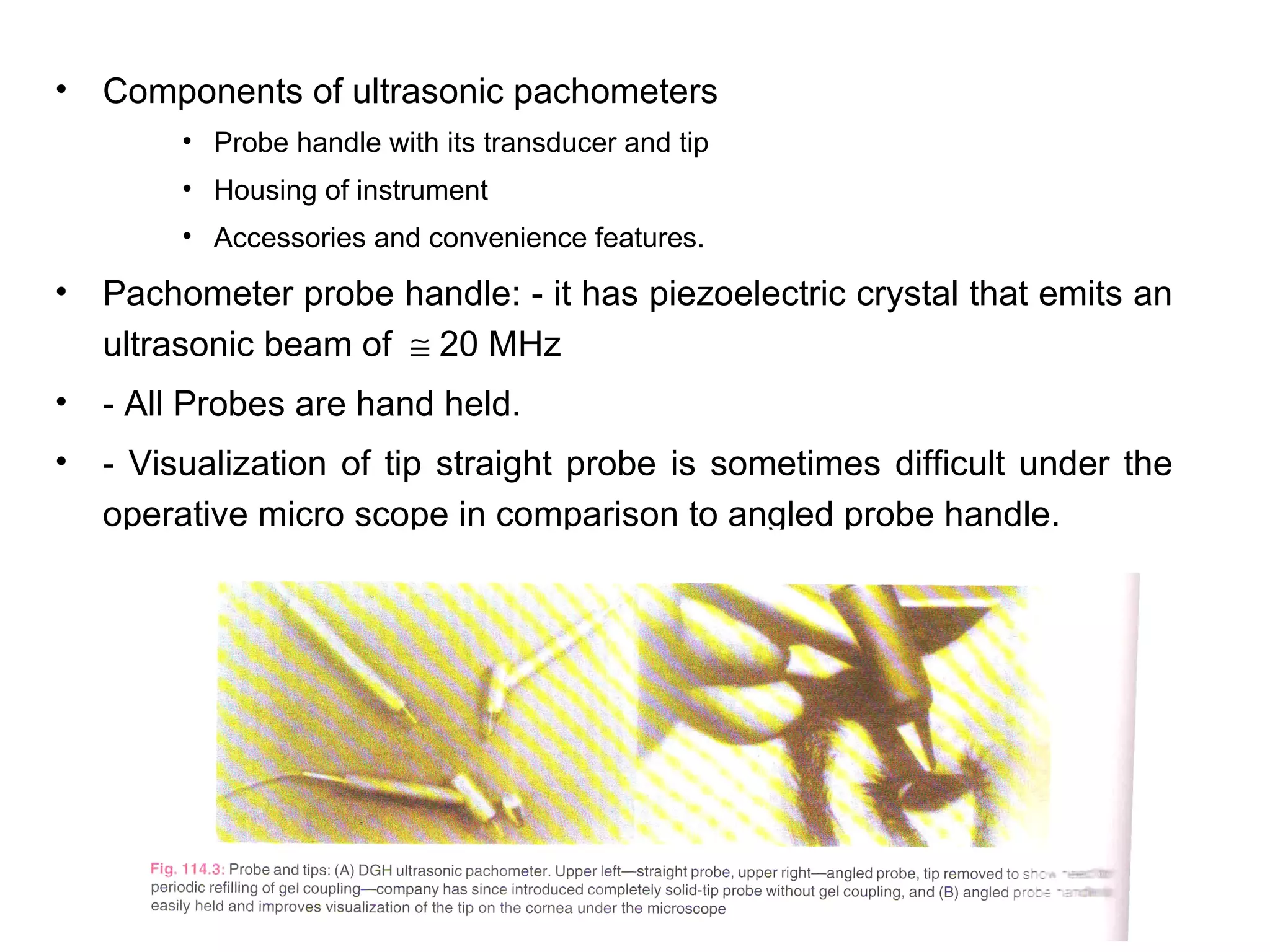
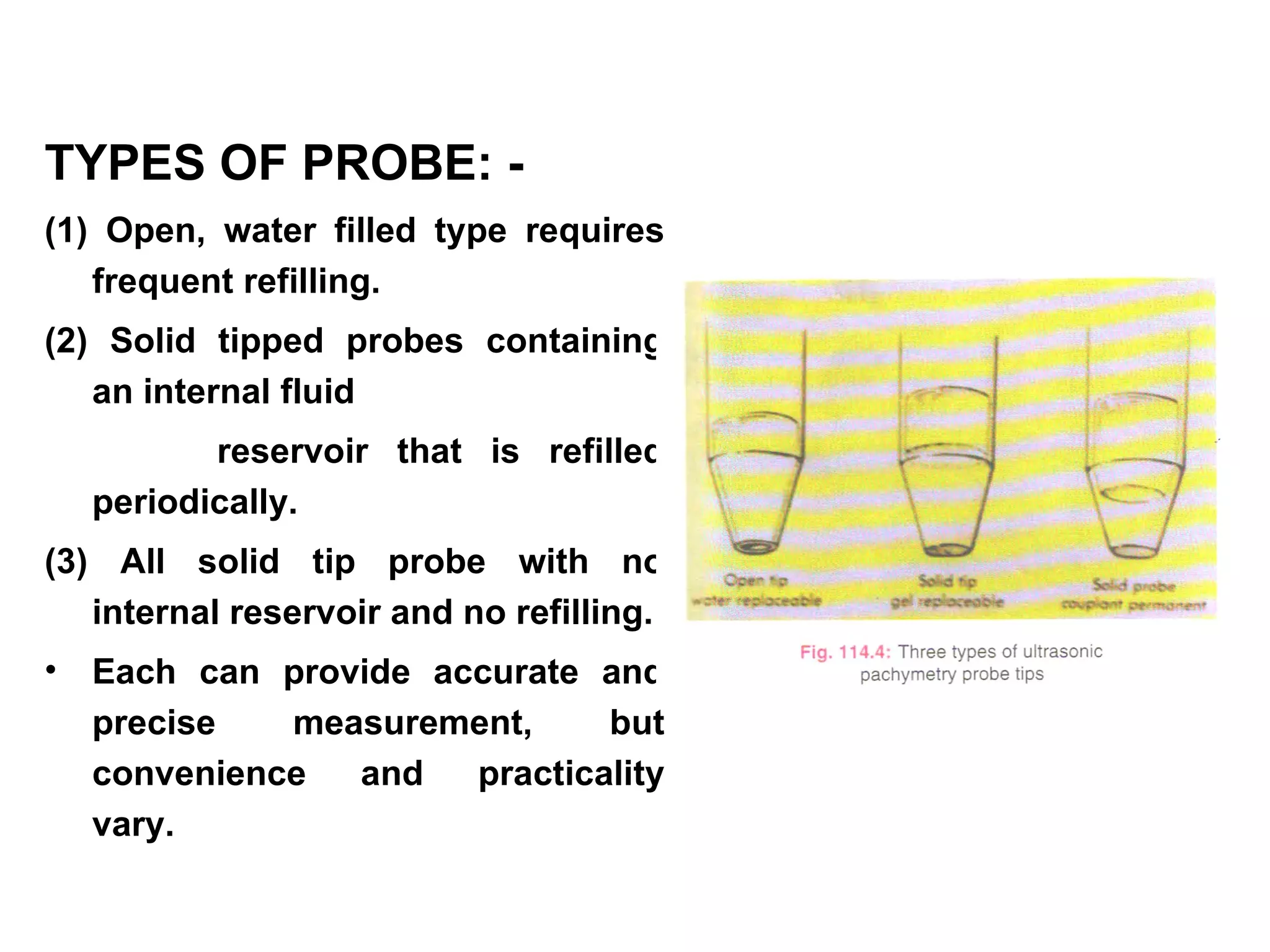
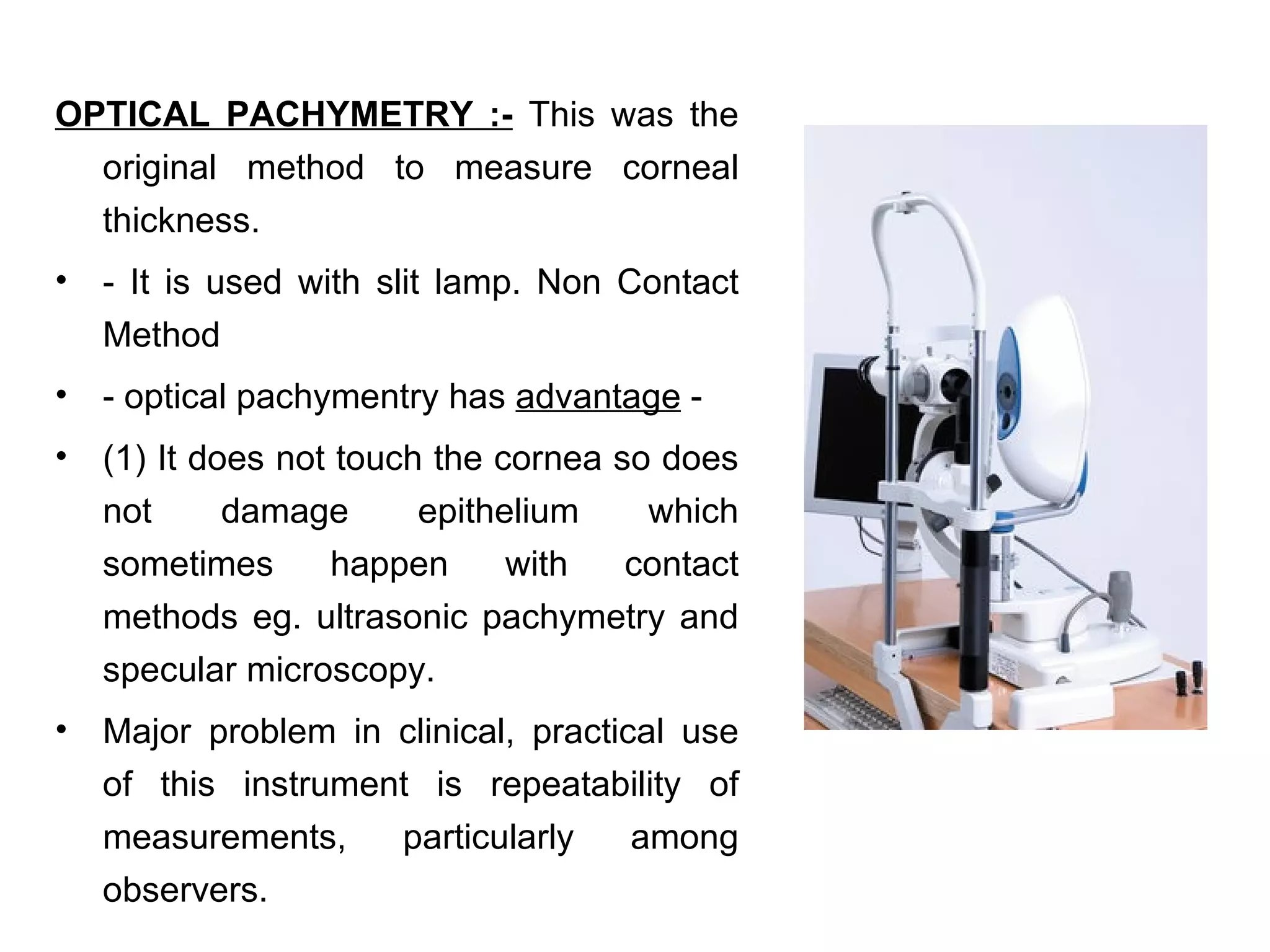
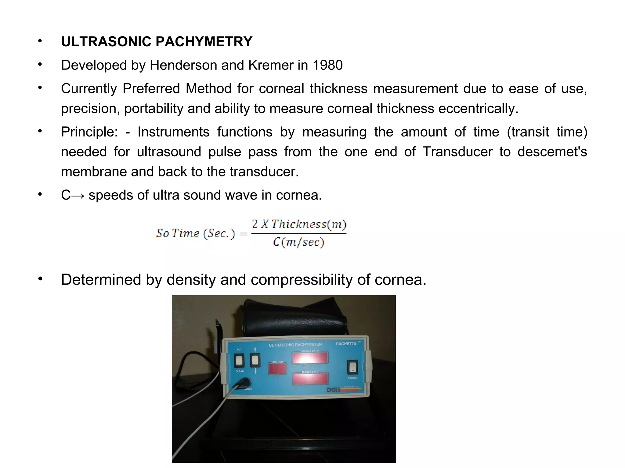
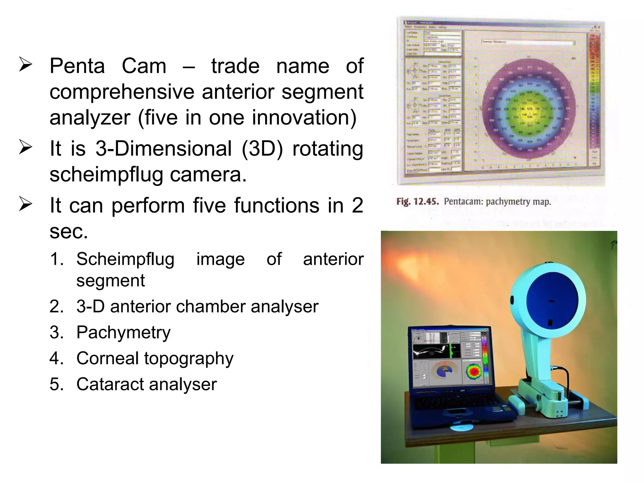
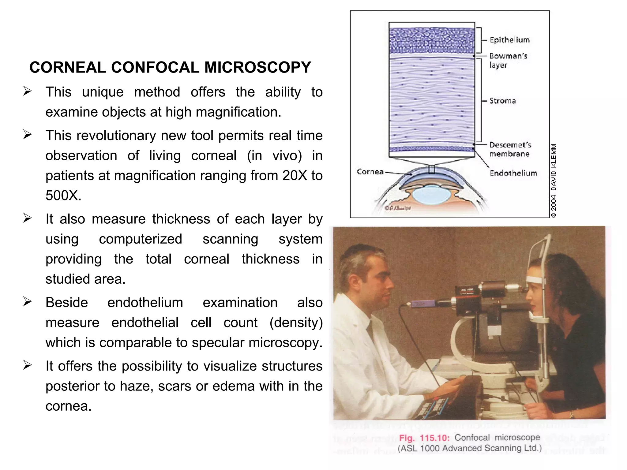
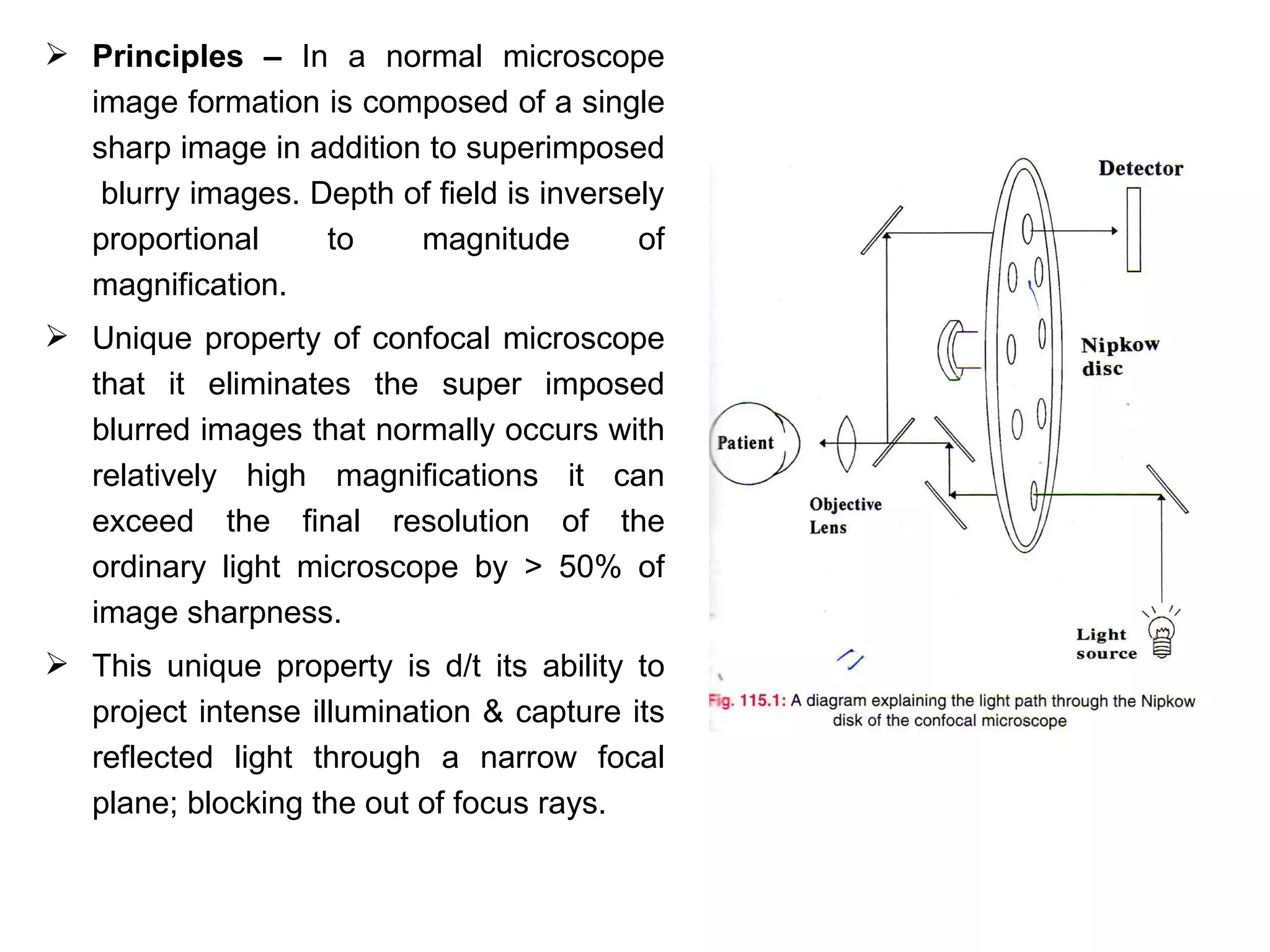
This document discusses techniques for measuring corneal thickness and examining the cornea using confocal microscopy. It describes pachymetry methods including optical, ultrasonic, and specular microscopy pachymetry. Ultrasonic pachymetry is now the preferred method due to its ease of use, precision and ability to measure thickness eccentrically. Confocal microscopy allows high magnification examination of the living cornea and measurement of thickness for each layer.
Accommodation/ Accommodation of Eye, Measurement of Accommodation of Eye (hea...Bikash Sapkota
CLICK HERE TO DOWNLOAD FULL PPT ❤❤ https://healthkura.com/measurement-of-accommodation-of-eye/ ❤❤
Dear viewers Check Out my other piece of works at ❤❤❤ https://healthkura.com ❤❤❤
Measurement of Accommodation of eye:
Amplitude, Facility,
Relative Accommodation, Fatigue, Lag,
Dynamic Retinoscopy
Presentation Layout:
-Introduction to accommodation of eye
-Mechanism
-Components
-Measurement of accommodation of eye
- Amplitude
- Facility
- Relative accommodation
- Lag
-Dynamic Retinoscopy
Accommodation
-dioptric adjustment of the crystalline lens of the eye
- to obtain clear vision for a given target of regard
-process by which the refractive power of eye is altered
- to ensure a clear retinal image
For further reading
-Clinical Procedures in Optometry by J.D. Bartlett, J.B. Eskridge, J.F. Amos
-Primary Care Optometry by Theodere Grosvenor
-Borish’s Clinical Refraction by W.J. Benjamin
-Clinical Procedures for Ocular examination by Carlson et al
-American Academy of Ophthalmology
-Optometric Clinical Practice Guideline by American Optometric Association
-Internet
Follow me to get in touch with optometric and ophthalmic updates
Ophthalmic Prisms: Prismatic Effects and DecentrationRabindraAdhikary
Ophthalmic Prisms: Prismatic Effects and Decentration
here we discuss about the ophthalmic prisms, the prismatic effects as caused by the decentration( moving the optical center away from the visual axis)
DIRECT DOWNLOAD LINK ❤❤https://healthkura.com/retinoscopy/❤❤
Dear viewers Check Out my other piece of works at ❤❤❤ https://healthkura.com ❤❤❤
Retinoscopy and Objective Refraction and Subjective Refraction in spherical ametropia and astigmatism
Retinoscopy (Principle & Techniques of Retinoscopy) and objective refraction, Subjective Refracition
Best presentation about retinoscopy and objective refraction techniques, and basis of subjective refraction. If you want to master the technique of retinoscopy, this presentation can be your guidance and partner in your journey to retinoscopy, objective refraction and subjective refraction.
Presentation Layout:
Retinoscope, types of retinoscope and uses of retinoscope
-Introduction to retinoscopy and objective refraction
-Retinoscopy
- In spherical ametropia
- In astigmatism
- Others: strabismus, amblyopia, pediatric pt.,
cycloplegic refraction
-Static and Dynamic Retinoscopy
-Problems seeing reflex during retinoscopy
-Errors in retinoscopy
Objective of retinoscopy and objective refraction
-To locate the far point of the eye conjugate to the retina
- Myopia or hyperopia
-Bring far point to the infinity by using appropriate lenses
- Determines amount of ametropia by retinoscopy and objective refraction
References:
-Clinical Procedures in Optometry by Eskridge, Amos and Bartlett ,
-Primary Care Optometry by Grosvenor T.,
-Borish’s Clinical Refraction by Benjamin W. J.,
-Theory And Practice Of Optics And Refraction by AK Khurana
-Retinoscopy-Student Manual by ICEE Refractive Error Training Package (2009)
-Clinical Optics and Refraction By Andrew Keirl, Caroline Christie
-Clinical Refraction Guide - A Kumar Bhootra
-Clinical Procedures in Primary Eye Care by David B. Elliott
-Internet
Follow me to get in touch with optometric and ophthalmic updates.
Review of the imaging modalities in Glaucoma. Structural loss precedes functional loss. Presentation includes a review of OCT, HRT and GDxVcc for posterior segment as well as AS-OCT and UBM for anterior segment.
This presentation is mainly focused on the clinical diagnosis and interpretation of oct macula.This is presented on 4th year optometry as topic presentation.
These simplified slides by Dr. Sidra Arshad present an overview of the non-respiratory functions of the respiratory tract.
Learning objectives:
1. Enlist the non-respiratory functions of the respiratory tract
2. Briefly explain how these functions are carried out
3. Discuss the significance of dead space
4. Differentiate between minute ventilation and alveolar ventilation
5. Describe the cough and sneeze reflexes
Study Resources:
1. Chapter 39, Guyton and Hall Textbook of Medical Physiology, 14th edition
2. Chapter 34, Ganong’s Review of Medical Physiology, 26th edition
3. Chapter 17, Human Physiology by Lauralee Sherwood, 9th edition
4. Non-respiratory functions of the lungs https://academic.oup.com/bjaed/article/13/3/98/278874
Tom Selleck Health: A Comprehensive Look at the Iconic Actor’s Wellness Journeygreendigital
Tom Selleck, an enduring figure in Hollywood. has captivated audiences for decades with his rugged charm, iconic moustache. and memorable roles in television and film. From his breakout role as Thomas Magnum in Magnum P.I. to his current portrayal of Frank Reagan in Blue Bloods. Selleck's career has spanned over 50 years. But beyond his professional achievements. fans have often been curious about Tom Selleck Health. especially as he has aged in the public eye.
Follow us on: Pinterest
Introduction
Many have been interested in Tom Selleck health. not only because of his enduring presence on screen but also because of the challenges. and lifestyle choices he has faced and made over the years. This article delves into the various aspects of Tom Selleck health. exploring his fitness regimen, diet, mental health. and the challenges he has encountered as he ages. We'll look at how he maintains his well-being. the health issues he has faced, and his approach to ageing .
Early Life and Career
Childhood and Athletic Beginnings
Tom Selleck was born on January 29, 1945, in Detroit, Michigan, and grew up in Sherman Oaks, California. From an early age, he was involved in sports, particularly basketball. which played a significant role in his physical development. His athletic pursuits continued into college. where he attended the University of Southern California (USC) on a basketball scholarship. This early involvement in sports laid a strong foundation for his physical health and disciplined lifestyle.
Transition to Acting
Selleck's transition from an athlete to an actor came with its physical demands. His first significant role in "Magnum P.I." required him to perform various stunts and maintain a fit appearance. This role, which he played from 1980 to 1988. necessitated a rigorous fitness routine to meet the show's demands. setting the stage for his long-term commitment to health and wellness.
Fitness Regimen
Workout Routine
Tom Selleck health and fitness regimen has evolved. adapting to his changing roles and age. During his "Magnum, P.I." days. Selleck's workouts were intense and focused on building and maintaining muscle mass. His routine included weightlifting, cardiovascular exercises. and specific training for the stunts he performed on the show.
Selleck adjusted his fitness routine as he aged to suit his body's needs. Today, his workouts focus on maintaining flexibility, strength, and cardiovascular health. He incorporates low-impact exercises such as swimming, walking, and light weightlifting. This balanced approach helps him stay fit without putting undue strain on his joints and muscles.
Importance of Flexibility and Mobility
In recent years, Selleck has emphasized the importance of flexibility and mobility in his fitness regimen. Understanding the natural decline in muscle mass and joint flexibility with age. he includes stretching and yoga in his routine. These practices help prevent injuries, improve posture, and maintain mobilit
Recomendações da OMS sobre cuidados maternos e neonatais para uma experiência pós-natal positiva.
Em consonância com os ODS – Objetivos do Desenvolvimento Sustentável e a Estratégia Global para a Saúde das Mulheres, Crianças e Adolescentes, e aplicando uma abordagem baseada nos direitos humanos, os esforços de cuidados pós-natais devem expandir-se para além da cobertura e da simples sobrevivência, de modo a incluir cuidados de qualidade.
Estas diretrizes visam melhorar a qualidade dos cuidados pós-natais essenciais e de rotina prestados às mulheres e aos recém-nascidos, com o objetivo final de melhorar a saúde e o bem-estar materno e neonatal.
Uma “experiência pós-natal positiva” é um resultado importante para todas as mulheres que dão à luz e para os seus recém-nascidos, estabelecendo as bases para a melhoria da saúde e do bem-estar a curto e longo prazo. Uma experiência pós-natal positiva é definida como aquela em que as mulheres, pessoas que gestam, os recém-nascidos, os casais, os pais, os cuidadores e as famílias recebem informação consistente, garantia e apoio de profissionais de saúde motivados; e onde um sistema de saúde flexível e com recursos reconheça as necessidades das mulheres e dos bebês e respeite o seu contexto cultural.
Estas diretrizes consolidadas apresentam algumas recomendações novas e já bem fundamentadas sobre cuidados pós-natais de rotina para mulheres e neonatos que recebem cuidados no pós-parto em unidades de saúde ou na comunidade, independentemente dos recursos disponíveis.
É fornecido um conjunto abrangente de recomendações para cuidados durante o período puerperal, com ênfase nos cuidados essenciais que todas as mulheres e recém-nascidos devem receber, e com a devida atenção à qualidade dos cuidados; isto é, a entrega e a experiência do cuidado recebido. Estas diretrizes atualizam e ampliam as recomendações da OMS de 2014 sobre cuidados pós-natais da mãe e do recém-nascido e complementam as atuais diretrizes da OMS sobre a gestão de complicações pós-natais.
O estabelecimento da amamentação e o manejo das principais intercorrências é contemplada.
Recomendamos muito.
Vamos discutir essas recomendações no nosso curso de pós-graduação em Aleitamento no Instituto Ciclos.
Esta publicação só está disponível em inglês até o momento.
Prof. Marcus Renato de Carvalho
www.agostodourado.com
New Drug Discovery and Development .....NEHA GUPTA
The "New Drug Discovery and Development" process involves the identification, design, testing, and manufacturing of novel pharmaceutical compounds with the aim of introducing new and improved treatments for various medical conditions. This comprehensive endeavor encompasses various stages, including target identification, preclinical studies, clinical trials, regulatory approval, and post-market surveillance. It involves multidisciplinary collaboration among scientists, researchers, clinicians, regulatory experts, and pharmaceutical companies to bring innovative therapies to market and address unmet medical needs.
Flu Vaccine Alert in Bangalore Karnatakaaddon Scans
As flu season approaches, health officials in Bangalore, Karnataka, are urging residents to get their flu vaccinations. The seasonal flu, while common, can lead to severe health complications, particularly for vulnerable populations such as young children, the elderly, and those with underlying health conditions.
Dr. Vidisha Kumari, a leading epidemiologist in Bangalore, emphasizes the importance of getting vaccinated. "The flu vaccine is our best defense against the influenza virus. It not only protects individuals but also helps prevent the spread of the virus in our communities," he says.
This year, the flu season is expected to coincide with a potential increase in other respiratory illnesses. The Karnataka Health Department has launched an awareness campaign highlighting the significance of flu vaccinations. They have set up multiple vaccination centers across Bangalore, making it convenient for residents to receive their shots.
To encourage widespread vaccination, the government is also collaborating with local schools, workplaces, and community centers to facilitate vaccination drives. Special attention is being given to ensuring that the vaccine is accessible to all, including marginalized communities who may have limited access to healthcare.
Residents are reminded that the flu vaccine is safe and effective. Common side effects are mild and may include soreness at the injection site, mild fever, or muscle aches. These side effects are generally short-lived and far less severe than the flu itself.
Healthcare providers are also stressing the importance of continuing COVID-19 precautions. Wearing masks, practicing good hand hygiene, and maintaining social distancing are still crucial, especially in crowded places.
Protect yourself and your loved ones by getting vaccinated. Together, we can help keep Bangalore healthy and safe this flu season. For more information on vaccination centers and schedules, residents can visit the Karnataka Health Department’s official website or follow their social media pages.
Stay informed, stay safe, and get your flu shot today!
Knee anatomy and clinical tests 2024.pdfvimalpl1234
This includes all relevant anatomy and clinical tests compiled from standard textbooks, Campbell,netter etc..It is comprehensive and best suited for orthopaedicians and orthopaedic residents.
Title: Sense of Smell
Presenter: Dr. Faiza, Assistant Professor of Physiology
Qualifications:
MBBS (Best Graduate, AIMC Lahore)
FCPS Physiology
ICMT, CHPE, DHPE (STMU)
MPH (GC University, Faisalabad)
MBA (Virtual University of Pakistan)
Learning Objectives:
Describe the primary categories of smells and the concept of odor blindness.
Explain the structure and location of the olfactory membrane and mucosa, including the types and roles of cells involved in olfaction.
Describe the pathway and mechanisms of olfactory signal transmission from the olfactory receptors to the brain.
Illustrate the biochemical cascade triggered by odorant binding to olfactory receptors, including the role of G-proteins and second messengers in generating an action potential.
Identify different types of olfactory disorders such as anosmia, hyposmia, hyperosmia, and dysosmia, including their potential causes.
Key Topics:
Olfactory Genes:
3% of the human genome accounts for olfactory genes.
400 genes for odorant receptors.
Olfactory Membrane:
Located in the superior part of the nasal cavity.
Medially: Folds downward along the superior septum.
Laterally: Folds over the superior turbinate and upper surface of the middle turbinate.
Total surface area: 5-10 square centimeters.
Olfactory Mucosa:
Olfactory Cells: Bipolar nerve cells derived from the CNS (100 million), with 4-25 olfactory cilia per cell.
Sustentacular Cells: Produce mucus and maintain ionic and molecular environment.
Basal Cells: Replace worn-out olfactory cells with an average lifespan of 1-2 months.
Bowman’s Gland: Secretes mucus.
Stimulation of Olfactory Cells:
Odorant dissolves in mucus and attaches to receptors on olfactory cilia.
Involves a cascade effect through G-proteins and second messengers, leading to depolarization and action potential generation in the olfactory nerve.
Quality of a Good Odorant:
Small (3-20 Carbon atoms), volatile, water-soluble, and lipid-soluble.
Facilitated by odorant-binding proteins in mucus.
Membrane Potential and Action Potential:
Resting membrane potential: -55mV.
Action potential frequency in the olfactory nerve increases with odorant strength.
Adaptation Towards the Sense of Smell:
Rapid adaptation within the first second, with further slow adaptation.
Psychological adaptation greater than receptor adaptation, involving feedback inhibition from the central nervous system.
Primary Sensations of Smell:
Camphoraceous, Musky, Floral, Pepperminty, Ethereal, Pungent, Putrid.
Odor Detection Threshold:
Examples: Hydrogen sulfide (0.0005 ppm), Methyl-mercaptan (0.002 ppm).
Some toxic substances are odorless at lethal concentrations.
Characteristics of Smell:
Odor blindness for single substances due to lack of appropriate receptor protein.
Behavioral and emotional influences of smell.
Transmission of Olfactory Signals:
From olfactory cells to glomeruli in the olfactory bulb, involving lateral inhibition.
Primitive, less old, and new olfactory systems with different path
Ozempic: Preoperative Management of Patients on GLP-1 Receptor Agonists Saeid Safari
Preoperative Management of Patients on GLP-1 Receptor Agonists like Ozempic and Semiglutide
ASA GUIDELINE
NYSORA Guideline
2 Case Reports of Gastric Ultrasound
TEST BANK for Operations Management, 14th Edition by William J. Stevenson, Ve...kevinkariuki227
TEST BANK for Operations Management, 14th Edition by William J. Stevenson, Verified Chapters 1 - 19, Complete Newest Version.pdf
ARTIFICIAL INTELLIGENCE IN HEALTHCARE.pdfAnujkumaranit
Artificial intelligence (AI) refers to the simulation of human intelligence processes by machines, especially computer systems. It encompasses tasks such as learning, reasoning, problem-solving, perception, and language understanding. AI technologies are revolutionizing various fields, from healthcare to finance, by enabling machines to perform tasks that typically require human intelligence.
MANAGEMENT OF ATRIOVENTRICULAR CONDUCTION BLOCK.pdfJim Jacob Roy
Cardiac conduction defects can occur due to various causes.
Atrioventricular conduction blocks ( AV blocks ) are classified into 3 types.
This document describes the acute management of AV block.
NVBDCP.pptx Nation vector borne disease control programSapna Thakur
NVBDCP was launched in 2003-2004 . Vector-Borne Disease: Disease that results from an infection transmitted to humans and other animals by blood-feeding arthropods, such as mosquitoes, ticks, and fleas. Examples of vector-borne diseases include Dengue fever, West Nile Virus, Lyme disease, and malaria.
1. Corneal Pachymetry &
confocal microscopy
Dr. Ram Singh
(Department of Ophthalmology)
S.P. Medical College, Bikaner
2. Pachymetry: - Literally meaning of this word– Thickness
• Measurement of corneal thickness is important factor in
deciding different kind of refractive surgeries.
• It is can be performed with optical pachometer or an ultra
sonic pachometer.
• Certain specular's microscopes are calibrated in such a
way so that they can measure corneal thickness while
focusing corneal endothelium.
3. OPTICAL PACHYMETRY :- This was the
original method to measure corneal
thickness.
• - It is used with slit lamp. Non Contact
Method
• - optical pachymentry has advantage -
• (1) It does not touch the cornea so does
not damage epithelium which
sometimes happen with contact
methods eg. ultrasonic pachymetry and
specular microscopy.
• Major problem in clinical, practical use
of this instrument is repeatability of
measurements, particularly among
observers.
4. Major sources for these problems.
(1) Lack of small fixation target for the pt, which is located in a fixed
position to the instrument.
(2) Lack of known alignment of the pachymeter with the cornea in a
reproducible position so that the slit beam intersects the cornea of
the same angle for consistent thickness reading.
• Location on the cornea that is being measured can be identified
visually.
• It is not easy to return to that specific point without auxiliary fixation
devices.
• Errors in accuracy and precision inherent in this method are
minimized when the instrument is used by single observer & errors
are <10µm, an acceptable error for practical a clinical refractive
keratotomy.
• Errors can increases up to 20µm or more when a multiple users and
this is unacceptable.
5. There are two criteria for measurement of
corneal
thickness.
(1) "Just touch" criterion: - Alignment is
made in such a way that an imaginary line
extends from the endothelial border of upper
image to the epithelial border of lower
image.
(2) Overlap – Method: - imaginary extension of
the bright portion of endothelial image is
over lapped with bright portion of epithelial
image. Because bright portion is actually
produced by the finite width of the slit lamp
as it passes through each surface of cornea.
6. JUST TOUCH METHOD IS EASIER, POPULAR A PRACTICAL
AMONG MOST OBSERVERS.
• Five different studies using five different optical pachometers all
showed mean central corneal thickness values 0.51 to 0.52 mm
(S.D. 0.02 to 0.04mm)
• Kremer – Ultrasonic pachometer – (sound speed in cornea 1640m/
sec.) – central average corneal thickness – 0.512+ 0.035mm in 175
eyes.
• Novak et al. compared optical (Haag streit 900 Ĉ mishima Hedbys
attachments).
• Specular microscope (Pro-koster) and
• Ultrasonic (Accutome, 1630 ± 10m/sec.) Pachymetry in 93 pts in
study – using mean value of 3 method corneal thickness
measurements reading for each instrument on each eye.
• Optical – 0.554 ± 0.028mm
• Specular microscopy 0.551 ± 0.37mm
• Ultrasonic 0.542 ± 0.035mm.
7. • ULTRASONIC PACHYMETRY
• Developed by Henderson and Kremer in 1980
• Currently Preferred Method for corneal thickness measurement due to ease of use,
precision, portability and ability to measure corneal thickness eccentrically.
• Principle: - Instruments functions by measuring the amount of time (transit time)
needed for ultrasound pulse pass from the one end of Transducer to descemet's
membrane and back to the transducer.
• C→ speeds of ultra sound wave in cornea.
• Determined by density and compressibility of cornea.
8. • Cornea is made of 78% mater.
• Propagation Velocity → Water 1524m/sec.
→Steel – 6000m/sec.
• Thus it is imp. that propagation velocity of cornea be known
accurately because this variable can be set on many ultrasonic
pachometer and different setting will change than thickness of
cornea.
• = Speed of sound in cornea: - Current standard is 1640 m/sec.
• Kremer selected 1640 m/sec., because that measurement gave
corneal thickness of 0.512 ± 0.035 in 175 Eyes.
9. • Components of ultrasonic pachometers
• Probe handle with its transducer and tip
• Housing of instrument
• Accessories and convenience features.
• Pachometer probe handle: - it has piezoelectric crystal that emits an
ultrasonic beam of ≅ 20 MHz
• - All Probes are hand held.
• - Visualization of tip straight probe is sometimes difficult under the
operative micro scope in comparison to angled probe handle.
10. • Transducers: - Transducer sends the beam of
ultrasound wave through the probe tips into the cornea
and receives them on return.
• Width of transducer beam is related to the size of the
emitting crystal and of the width and configuration of the
probe through which it passes.
# #:- A wide probe tip and wide transducer beam reduces the
accuracy of the corneal thickness reading at a single point.
• Transducer has limited lifetime approximately 150-200
cases. It loses its accuracy and precision.
• The reading becomes increasingly variable on the
calibration block. The probe should be changed.
11. • Probe tip: - it is interface between the cornea and
transducer.
• Material in the probe should not attenuate the ultrasound
beam and the geometric design of the probe tip should
facilitate its optimal transmission.
• Diameter of probe should be <2mm. to diminish the area
over which the ultrasound beam is spread (to allow)
observer to see exactly where the tip is placed on the
cornea.
• Surface of tip should be smooth to avoid any injury to
corneal epithelium.
12. TYPES OF PROBE: -
(1) Open, water filled type requires
frequent refilling.
(2) Solid tipped probes containing
an internal fluid
reservoir that is refilled
periodically.
(3) All solid tip probe with no
internal reservoir and no refilling.
• Each can provide accurate and
precise measurement, but
convenience and practicality
vary.
13. (1) Open water filled tips: - Used Earlier
Inconvenient to use:-
• As fluid pulled out of the tip by surface tension & capillary action, air
bubble enter and give erroneons readings.
(2) The solid tipped probe – have replaceable couplet (glue or oil)
• and are more convenient.
• - Frequency of refilling varies from once a week to once a year,
depending on design.
• Tekner optha sonic pachometer has oil interface and has to
changes once a year.
(3) All Solid tipped – No replaceable couplants
• Tips are made of polystyrene.
• More convenient to handle a requires less maintenance eg.
accutome corneometer, pach – pen, sonogaga and Humphreys
Pachometers
14. - All pachomaters average a series of thickness measurements
to give the single, final read out display of corneal thickness.
- Instruments take 30-500 reading in a fraction of second.
There are two methods by which pachometer create an average
reading.
1. Pulse locked method: - Unit will record all readings that are
within 5 to 10 0 of perpendicularity or within 5 to 10µ of each
other, rejecting those outside the range.
2. Fixed no. of consecutive reading must be within 5 to 100 of
perpendicularity on 5 to 10µ of each other, before there are
averaged. If the probe is not perpendicular or the readings are
too disparate, the series is rejected and must be began again.
15. Resolution of instrument is smallest unit
measurable by machine - 1µ
Clinical accuracy of most instrument ± 5 to 10µ
Ultrasonic corneal pachometer can measure thickness
range 200 to 2000 µ
Most pachometers have a selected speed of 1640m/sec.
Some units allow adjustments of sound speed, so
operators can select faster or slower speed.
** Selection of faster speed will produced a thicker
corneal reading
16. Other methods for corneal thickness measurement.
High frequency ultrasound corneal pachymetry
70MHz is used frequency
This technique produces B-scan images in real time, by on the fly
analog processing, involving rectifying averaging reflected
ultrasound waves.
Pachymetry using the ORBSCAN topography system
Orbscan technique results in pictorial representation of corneal
topography in true as opposed to derivative terms.
The creation of a surface of orbascan topographic measurement
provides the basis for the derivation of pachymetric & radius of
curvature maps.
Pachymetry by laser Doppler interferometry (LDI)
17. Penta Cam – trade name of
comprehensive anterior segment
analyzer (five in one innovation)
It is 3-Dimensional (3D) rotating
scheimpflug camera.
It can perform five functions in 2
sec.
1. Scheimpflug image of anterior
segment
2. 3-D anterior chamber analyser
3. Pachymetry
4. Corneal topography
5. Cataract analyser
18. Pachymetry by pentacam is displayed as a color image
over its entire area from limbus to limbus.
Actual thickness can be measured individually by a
mouse click at any locations.
Thickness in the pupil centre
Thickness in the apex
Thinnest location
Corneal volume
Applications –
1. Preoperative planning for corneal refractive surgery
2. Glaucoma screening
3. IOP modification with regard to corneal thickness
4. Keratoconus detection & quantification.
19. CORNEAL CONFOCAL MICROSCOPY
This unique method offers the ability to
examine objects at high magnification.
This revolutionary new tool permits real time
observation of living corneal (in vivo) in
patients at magnification ranging from 20X to
500X.
It also measure thickness of each layer by
using computerized scanning system
providing the total corneal thickness in
studied area.
Beside endothelium examination also
measure endothelial cell count (density)
which is comparable to specular microscopy.
It offers the possibility to visualize structures
posterior to haze, scars or edema with in the
cornea.
20. Principles – In a normal microscope
image formation is composed of a single
sharp image in addition to superimposed
blurry images. Depth of field is inversely
proportional to magnitude of
magnification.
Unique property of confocal microscope
that it eliminates the super imposed
blurred images that normally occurs with
relatively high magnifications it can
exceed the final resolution of the
ordinary light microscope by > 50% of
image sharpness.
This unique property is d/t its ability to
project intense illumination & capture its
reflected light through a narrow focal
plane; blocking the out of focus rays.
21. first study with clinical approach using this
instrument was done by Ichijima in 1992 to
document the changes in superficial oepithelial
cells after extended wear rigid contact lenses.
Later Cavanagh used a tendon scanning
confocal microscope & examined various
corneal diseases.
Confocal microscope uses white light or a
focused laser beam but clinical white light is safe
becuase laser having risk for damaging living
tissue.
22. Procedure – After topical anaesthesia patient is
guided to the chin rest.
A clear visoelastic solution is applied to the cover tip
of the microscope to avoid corneal abrasion.
Machine is approximated until a bright image is
seen & the then the corneal scanning is done.
Once the epithelium is well focused, the zooming
examination of all corneal layers can be fulfilled in
only 30 sec. examination is stored in computer
software.
23. NORMAL CORNEA -
Epithelium – Superficial
layers – large surface cells
arranged in irregular
polygonal mosaic. 1. At the superficial epithelium, poorly demarcated roundish cells
demonstrate hyperreflective nuclei (arrows) on confocal
These cells demonstrate microscopy (original magnification 210).
hyper reflective nuclei.
Basal epithelial cells –
Immature cells appear without
nuclei reflectivity or faint.
2. Bowman's Layer – Acellular
Bowman’s layer is an acellular hyperreflective structure, where subepithelial nerve
hyper reflective structure plexus (arrows) may be identified easily (original magnification 210).
subepithelial nerve plexus
may be seen.
In normal cornea, vessels are
not present in epithelium &
Bowman's layer.
Basal epithelial cells appear hyporeflective and have hyperreflective borders
(original magnification 250).
24. 3. Stroma – Hyper reflective keratocyte nuclei are scattered against a dark
background.
Ketatocyte density is maximum (800 cells/mm2) immediately under
Bowman's membrane and decreased (65 cells/mm2)sharply towards
posterior cornea.
Nerves – Which may present branching images, are found in the stroma
and are thicker than at the subepithelial level.
In normal eyes vessles are not found in stroma.
4. Descemet's Membrane – Acellular layer of moderate reflectivity : however
nerve plexus is absent.
This layer is rather difficult to see under normal circumstances.
In the stroma, hyperreflective keratocyte nuclei (arrows) are scattered against a dark
background (original magnification 250).
25. 5. Endothelium – Regular, hexaogonal, hyperreflective
shape surrounded by hyporeflective borders and the
absence of any nuclei reflection.
Endothelial count with confocal & specular microscopy
are comparable.
Negative correlation b/w age & endothelial count.
No vessels or nerves are present in this layer.
The physiologic responses of the corneal to different
stimuli may by analyzed by confocal microscopy.
Activated keratocytes presenting as cells with increased
reflectivity in the stroma, are seen when cellular
metabolic activity is increased.
Scar tissue, infection, inflammation – Hyperreflective
images.
Vessels – Lumen – Hyporeflective
Wall – Hyperreflective
26. Stroma – Hyper reflective keratocyte nuclei are scattered
against a dark background.
Ketatocyte density is maximum (800 cells/mm2)
immediately under Bowman's membrane and decreased
(65 cells/mm2)sharply towards posterior cornea.
Nerves – Which may present branching images, are
found in the stroma and are thicker than at the
subepithelial level.
In normal eyes vessles are not found in stroma.
28. Vessel lumen appears hyporeflective on confocal microscopy,
whereas vessel wall demonstrates well-delineated
hyperreflectivity (arrows) on each side of the lumen (original
magnification 210).
29. Cotton candy-like hyperreflective material may be found at the subepithelial level in
amyloidosis with corneal deposits (original magnification 210).
30. Cystic epithelial lesions are demonstrated in a patient with Fuchs’ dystrophy.
Horizontal field width 5 610 mm. (Reprinted from Ophthalmology
Hernandez- Quintela et al82, Copyright 1998, with permission of American
Academy of Ophthalmology.)
31. Hyperreflective deposits (arrows) are found in area devoid of epithelium in an
eye treated with topical ciprofloxacin (original magnification 240).
(Reprinted from Essepian et al60 with permission of Cornea.)
32. Confocal microscopy in a case of corneal lattice dystrophy disclosed hyperreflective, linear, and
branching images (black arrows) in the stroma. The white arrows indicate some hyperreflective
keratocytes (original magnification 210). (Reprinted from Graefes Arch Clin Exp Ophthalmol. Chiou
AG, Beuerman RW, Kaufman SC, Kaufman HE: Confocal microscopy in lattice corneal dystrophy.
237:697--701, 1999, with kind permission of Springer Science and Business Media.)
33. Highly reflective and irregular material (*) is found at the level of Bowman’s
layer region and anterior stroma in Reis-Bu¨ckler dystrophy. Bar 5 50 mm.
(Reprinted from Ophthalmology Werner et al,222 Copyright 1999 with
permission of American Academy of Ophthalmology.)
34. In granular dystrophy, reflective diffuse deposits (arrows) may be found
in the stroma. Bar 5 50 mm. (Reprinted from Ophthalmology Werner
et al222 Copyright 1999, with permission of American Academy of
Ophthalmology.)
35. Stromal intracellular hyperreflective material is the hallmark of fleck
dystrophy. In the mid-stroma a cluster of hyperreflective dots is enclosed in
a cyst-like structure. Calibration bar 5 50 mm. (Reprinted from Frueh and
Bo¨hnke62 with permission of Cornea.)
36. Stromal crystalline accumulation is associated with Schnyder’s dystrophy
and is readily revealed by confocal microscopy (image is 250 170 mm).
(Reprinted from Ophthalmology Vesaluoma et al215 Copyright 1999, with
permission of American Academy of Ophthalmology.)
37. Subbasal nerve plexus presenting beads in a case of cornea plana (image is
390 290 mm). (Reprinted from Vesaluoma et al217 with permission of
Investigative Ophthalmology and Visual Science.)
38. Confocal microscopy (original magnification 210). Areas of highly abnormal cells characterized by marked
epithelial-like appearance and loss of regularity in size and shape were found. Hyperreflective structures
were found within and adjacent to these abnormal areas. Relatively normal appearing endothelial cells
were also detected (upper right corner of the photograph). (Reprinted from Chiou et al33 with permission of
British Journal of Ophthalmology.)
39. Cornea guttae (arrows) appear as roundish hyporeflective images with an
occasional central highlight at the level of the endothelium. Cell
pleomorphism shown is this picture is also a common feature (original
magnification 210).
40. Epithelial cells at the level of the corneal endothelium is pathognomonic of
epithelial downgrowth (original magnification 230). (Reprinted from Journal
of Cataract and Refractive Surgery Chiou et al36 Copyright 1999, with
permission of ASCRS & ESCRS.)
41. Confocal microscopy in a case of fibrous retrocorneal membrane after
penetrating keratoplasty (original magnification 210). At the endothelial cell
layer, a hyperreflective fibrous-appearing layer was demonstrated at the
periphery of the graft. (Reprinted from Chiou et al30 with permission of
Cornea.)
42. Subepithelial extracellular deposits (D) may be found 3 months after
photorefractive keratectomy at the epithelial-stromal interface (image is 382
382 mm). N 5 stromal nerve. (Reprinted from Ophthalmology, Corbett et al40
Copyright 1996 with permission of American Academy of Ophthalmology.)
43. Linear structures may be detected in the stroma years after photorefractive
keratectomy. Bar indicates 50 mm. (Reprinted from Archives of
Ophthalmology, Frueh et al63, Copyright 1998 with permission of
American Medical Association.)
44. Confocal microscopy of the cornea performed 10 days after the surgery.
Hyperreflective interface debris could be detected (arrows). A superficial
stromal nerve was also visualized (arrow heads) (original magnification
210).
45. Acanthamoeba cysts (black arrows) and trophozoite (white arrows) may be
visualized by confocal microscopy (marker 5 100 mm). (Reprinted from
Pfister et al177 with permission of Cornea.)
46. At a depth of 115 mm from the anterior corneal surface, linear and
branching structures are detected in a case of aspergillus keratitis
(original magnification 135).
47. Confocal microscopy can also resolve the double-walled structure of the
acanthamoeba ectocyst surrounding theendocyst (white arrow). Several
pear-shaped cysts are shown by the black arrows (marker 5 100 mm).
(Reprinted from Pfister et al177 with permission of American Journal of
Ophthalmology.)
48. Acanthamoeba radial keratoneuritis presents as swollen nerve (arrowheads). A bright
irregular body on the nerve (black arrow) is consistent with an acanthamoeba
trophozoite. A stromal keratocytes is shown by the white arrow. Inset: normal human
stromal nerve (arrowheads) and stromal keratocyte (white arrow). Arrows 5
keratocytes (marker 5 100 mm). (Reprinted from Pfister DR et al177 with permission of
American Journal of Ophthalmology.)
49. Refractive objects showing eight consecutive spots arranged in a straight
row in a case of Borrelia keratitis (horizontal field width 5 275 mm).
(Reprinted from Linna et al125 with permission of Cornea.)
50. Microsporidial keratitis has been reported to demonstrate images of epithelial cells of the
corneal surface containing intracellular spores upon confocal microscopic examination. An
enlargement of the two cells outlined shows numerous small, discrete, high-contrast,
intracellular microsporidial spores (white arrows), and an aggregate of tightly packed
microsporidial spores (gray arrows) (marker 5 100 mm). (Reprinted from Shah et al194 with
permission of American Journal of Ophthalmology.)
51. Degenerated fine stromal hyperreflective dots may be detected in long-term
contact lens wearers. Bar550 mm. (Reprinted from Ophthalmology, Bohnke
and Masters,11 Copyright 1997 with permission of American Academy of
Ophthalmology.)