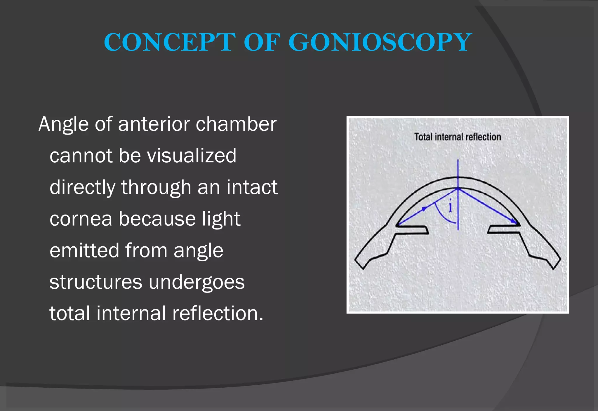
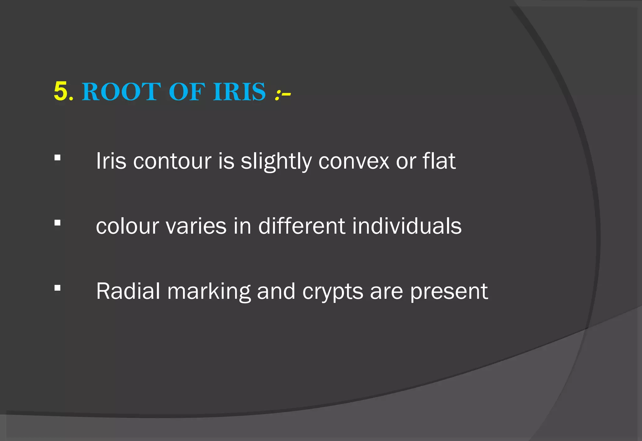
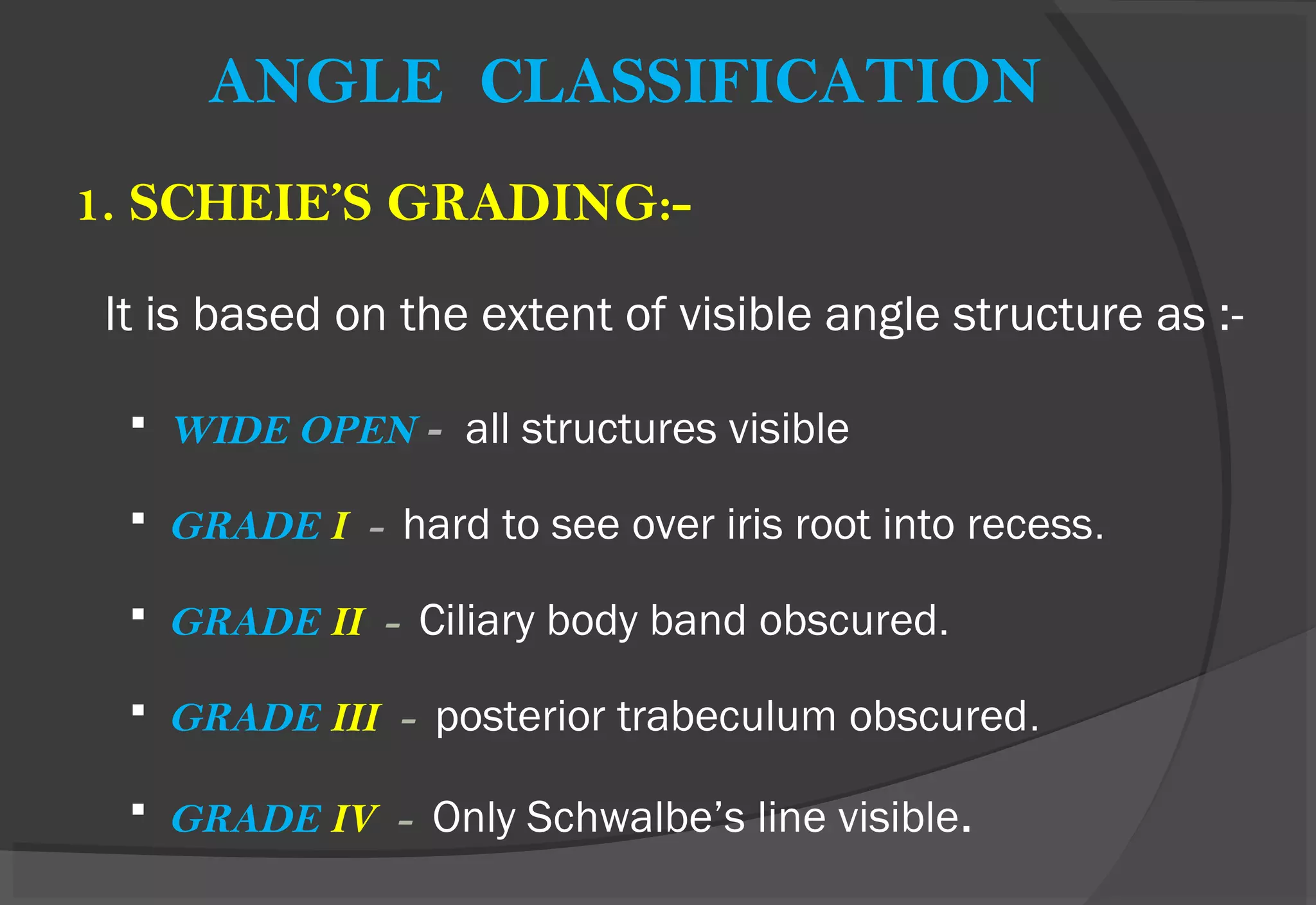
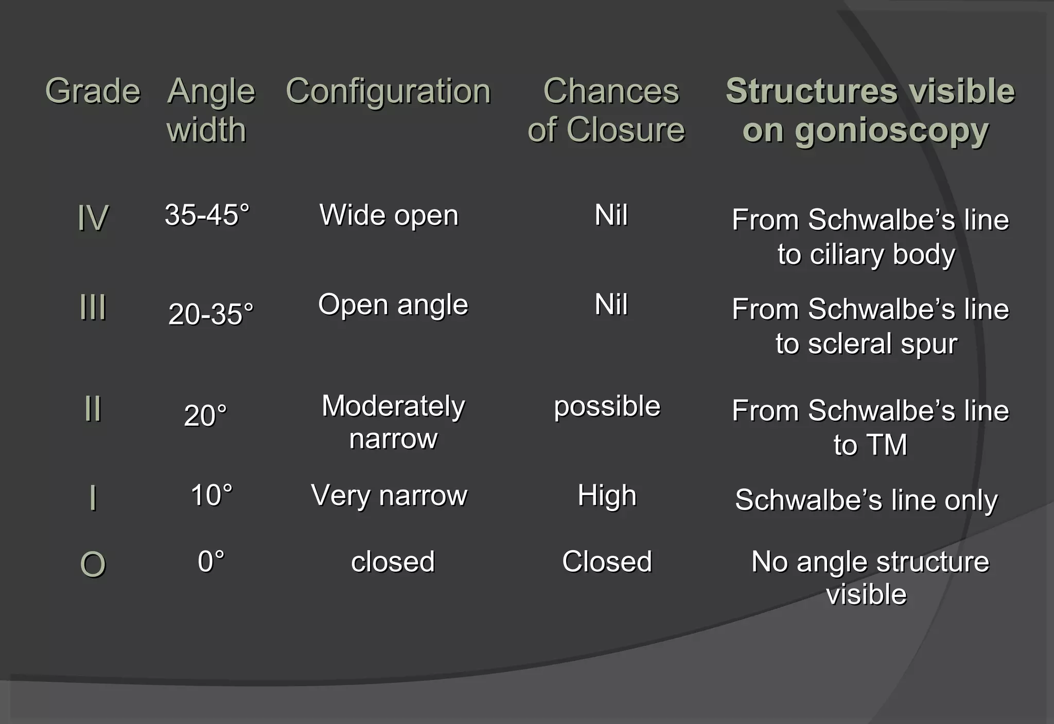
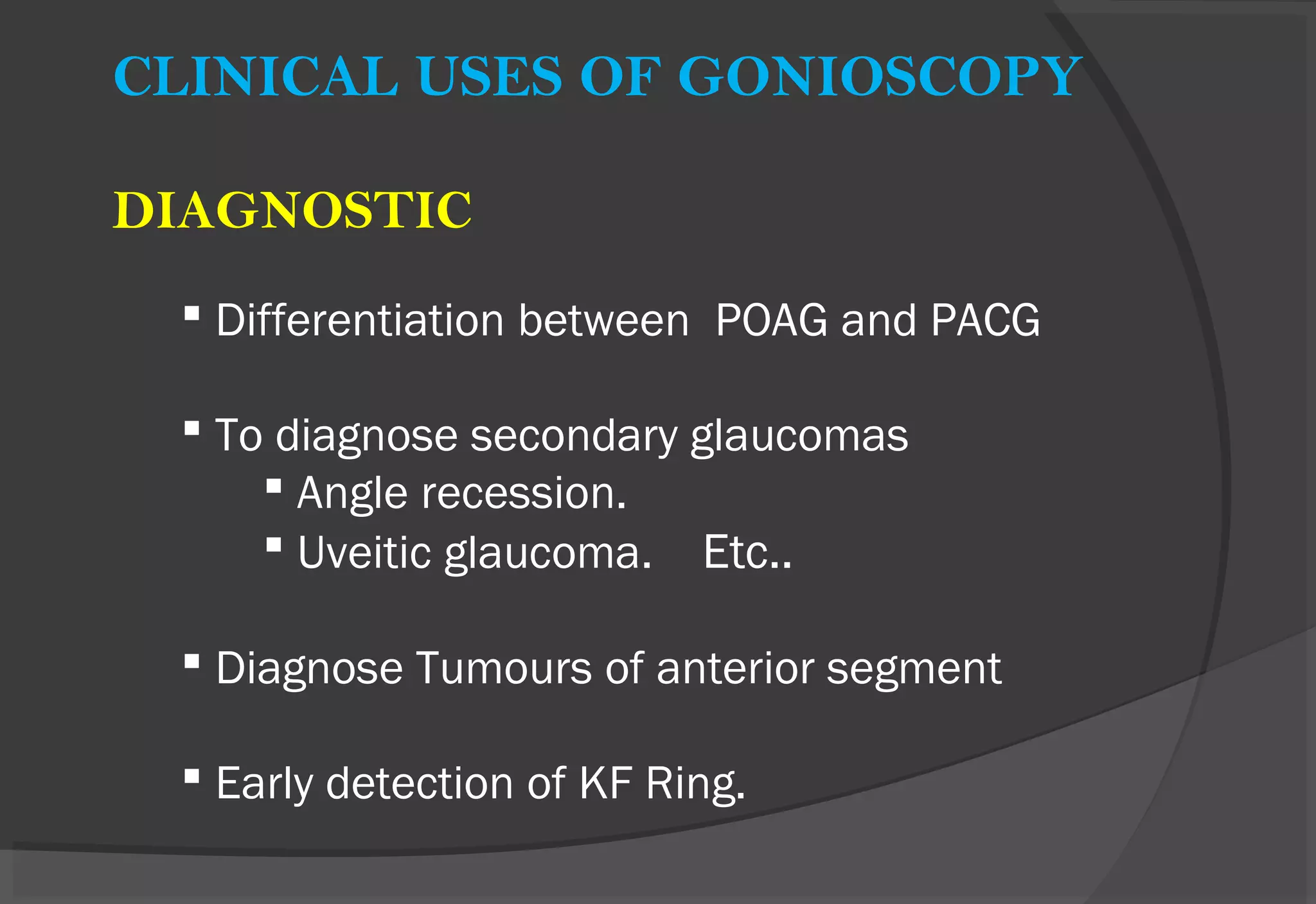
GONIOSCOPY
BY Dr. ASHISH GUPTA
MODERATOR - Dr. ANAMIKA TIWARI
HISTORY
TRANTAS (1907) :-
First to examine angle in the
keratoglobus eye and introduced the
term GONIOSCOPY.
SALSMANN (1914) :-
Introduced the goniolense to observe
the angle.
 GOLDMANN (1938) :- Introduced the
gonioprism.
 BARKAN :- Established the use of Gonioscopy
in management of Glaucoma.
CONCEPT OF GONIOSCOPY
Angle of anterior chamber
cannot be visualized
directly through an intact
cornea because light
emitted from angle
structures undergoes
total internal reflection.
Goniolenses eliminates
the total internal reflection by
replacing the cornea- air
interface by a new interface,
which has a refractive index
greater than that of cornea and
tears.
METHODS OF GONIOSCOPY
 1. DIRECT GONIOSCOPY
 2. INDIRECT GONIOSCOPY
- WITHOUT INDENTATION
- WITH INDENTATION
DIRECT GONIOSCOPY
 ANTERIOR curve of goniolense is such that the critical
angle is not reached and the light rays are
REFRACTED at contact lens-air surface.
 Provide a direct view of the angle. They do not require a
slit lamp & are used with in supine position.
DIRECT GONIOLENSES:
1. Koeppe goniolense
2. Richardson-shaffer
3. Huskins Barkan
4. Sieback
5. Swan jacob
6. Layden
KOEPPE GONIOLENS :-
 It is most commonly used for diagnostic direct
gonioscopy. It is easy to use & provides a
panoramic view of the angle.
 Richardson-Shaffer’s goniolens - It is basically a small
Koeppe lens used for
infants.
 Worth goniolens - It anchors to cornea by partial
vacuum.
 Sieback goniolens - It is a tiny goniolens which floats on
cornea.
 Huskins Barkans lens - It is the prototype surgical
goniolens used for goniotomy.
TECHNIQUE OF DIRECT GONIOSCOPY
 Patient lies supine with examiner sitting on the side of
the eye to be examined.
 After topical anesthesia, goniolens is positioned on
the cornea, using balanced salt solution or methyl
cellulose.
 Examiner holds the goniolense in one hand and the
light source in other and scans the anterior chamber
angle by shifting his or her position until all 360
degrees have been studied.
TECHNIQUE OF DIRECT GONIOSCOPY
ADVANTAGES-
 Greater flexibility as position of observer can be
changed.
 Panoramic view is obtained so one part of the
angle can be compared with other.
 Angle deep in supine position so it is easy to see
the angle.
 Can be used in sedated patient and also in infants.
 Provides a straight view rather than inverted view.
DISADVANTAGES:-
 Annoying light reflexes from cornea.
 Time consuming.
 Benefits of slit-lamp like variable
light & better clarity are not available.
INDIRECT GONIOSCOPY
 The light rays are REFLECTED by mirror in the contact
lense (gonioprism).
 Provide a mirror image of the opposite angle & used
in conjunction with a slit lamp.
INDIRECT GONIOSCOPY
Two types :-
1. Non indentation
2. Indentation
1. NON INDENTATION GONIOSCOPY .
TECHNIQUE
 After anaesthetising the cornea, the coupling gel
is inserted into the cup of the goniolens.
 goniolense is placed and tipped onto the
cornea.
 Slit-lamp beam is focused on the mirror which
shows the inverted angle image.
GONIOPRISMS (NON INDENTATION) -
Surface is slightly larger than cornea & require a
coupling gel.
1.Goldmann single mirror
2.Goldmann-3 mirror
3.Allen-Thorpe
GOLDMANN MIRRORS
GOLDMANN SINGLE MIRROR GOLDMANN 3 MIRROR
2. INDENTATION GONIOSCOPY
 Varying amount of pressure applied to the cornea with a tear
coupled indirect contact lens.
 Examiner observes the effect on angle width.
 Increased pressure indents the central cornea & displaces
fluid into the angle, opening it wider.
GONIOPRISMS (INDENTATION) -
1. Zeiss four mirror
2. Sussmman four mirror
3. Tokel single mirror
4. Posner four mirror
1. ZEISS -4 MIRROR GONIOPRISM -
-It has four identical mirrors angled at 64° which allow
examination of the four quadrants without rotation of
the lens.
-The contact surface of lens has a 9mm diameter.
2. SUSSMMAN LENS :- It is also similar to Zeiss
lense except it has no handle.
3.TOKEL GONIOLENS :- It is a single mirror goniolens
& has got a wider field of view.
4.POSNER GONIOLENS:- It is similar to Zeiss-goniolens
but it is made of plastic & also
has a fixed handle.
ADVANTAGE OF INDIRECT GONIOSCOPY:-
 Easy to perform when mastered
 All four quadrants can be seen when four mirror
lense used.
 With slit lamp, variable magnification and
illumination can be achieved.
 Indentation Gonioscopy can be performed, which
allows differentiation of appositional and synachial
angle closure.
 Coupling agent is not used, so visualization of
fundus & photography is possible.
 DISADVANTAGE-
 Difficult to master.
 Mirror image can be confusing.
 May open the angle artefactually if pressure applied.
GONIOSCOPIC VIEW OF ANGLE STRUCTURES
1. SCHWALBE’S LINE:-
 Most anterior structure
 Irregular elevation of 50 to 100 µm width.
 Peripheral termination of cornea where the Descemet’s
membrane ends.
 Marked only by a slight change in colour from
trabecular meshwork or by a faint white line.
 Best identified by locating the “Corneal Wedge”.
2. TRABECULAR MESHWORK :-
- It lies between the Schwalbe’s line anteriorly & scleral
spur posteriorly and has an average width of 600 Âľm.
- Smooth in infants but becomes coarse and pigmented
with age.
 The posterior functional pigmented part has a greyish-
blue translucent appearance.
 This posterior portion overlies the canal of Schlemm,
from where aqueous humour escapes from the anterior
chamber.
 Anterior part is non-functional and non-pigmented.
3. SCLERAL SPUR:-
- Anterior most projection of the sclera
- Situated just posterior to the TM and appears as a narrow,
dense, often shiny, whitish, band.
-May be obscured by iris process, and pigments.
4. CILIARY BODY BAND:-
-Lies just behind the scleral spur as a pink to dull brown or
slate grey band.
-Portion of ciliary body which is visible in the angle as a
result of insertion of the iris into the ciliary body.
5. ROOT OF IRIS :-
 Iris contour is slightly convex or flat
 colour varies in different individuals
 Radial marking and crypts are present
OTHER STRUCTURES
IRIS PROCESSES:-
 Normal variants
 Appear as fine strands extending from iris to the scleral
spur.
 These do not inhibit the
movement of the iris during
indentation Gonioscopy.
ANGLE CLASSIFICATION
1. SCHEIE’S GRADING:-
It is based on the extent of visible angle structure as :-
 WIDE OPEN - all structures visible
 GRADE I - hard to see over iris root into recess.
 GRADE II - Ciliary body band obscured.
 GRADE III - posterior trabeculum obscured.
 GRADE IV - Only Schwalbe’s line visible.
2. SHAFFER’S GRADING:-
 Estimation of the angle width is achieved by observing
the angle between two imaginary lines, constructed
tangential to inner surface of trabeculum and the
anterior iris surface.
GradeGrade AngleAngle
widthwidth
ConfigurationConfiguration ChancesChances
of Closureof Closure
Structures visibleStructures visible
on gonioscopyon gonioscopy
IVIV 35-45°35-45° Wide openWide open NilNil From SchwalbeFrom Schwalbe’’s lines line
to ciliary bodyto ciliary body
IIIIII 20-35°20-35° Open angleOpen angle NilNil From SchwalbeFrom Schwalbe’’s lines line
to scleral spurto scleral spur
IIII 20°20° ModeratelyModerately
narrownarrow
possiblepossible From SchwalbeFrom Schwalbe’’s lines line
to TMto TM
II 10°10° Very narrowVery narrow HighHigh SchwalbeSchwalbe’’s line onlys line only
OO 0°0° closedclosed ClosedClosed No angle structureNo angle structure
visiblevisible
3.SPAETH CLASSIFICATION:-
This classification elaborated a complex grading system
that captures a detailed three dimensional information
in coded form.

A- Anterior to trabecular meshwork.
(Schwalbe’s line)
B- Behind Schwalbe’s lines (at the
level of Trabecular meshwork).
C- Centered at the level of scleral
spur.
D- Deep to scleral spur (ant. Ciliary
body)
E- Extremely deep in the ciliary body
CLINICAL USES OF GONIOSCOPY
DIAGNOSTIC
 Differentiation between POAG and PACG
 To diagnose secondary glaucomas
 Angle recession.
 Uveitic glaucoma. Etc..
 Diagnose Tumours of anterior segment
 Early detection of KF Ring.
THERAPEUTIC -
 To perform argon laser trabeculoplasty
 Laser iridoplasty
 Laser sclerostomy
 Goniotomy & trabeculotomy
LIMITATIONS OF GONIOSCOPY
 Can not be performed in painful inflamed eyes.
 Can not be performed under mydriasis.
 Hyphaema
 Compromised cornea
 Lacerated or perforated globe
 COMPLICATION- Corneal abrasion.
GONIOSCOPY: PATHOLOGICAL CONDITIONS
ANGLE CLOSURE GLAUCOMA
 The angle is first occluded appositionally, followed
by a sticky adhesion & later on by peripheral ant.
synechiae over wide area.
.
PERIPHERAL ANTERIOR SYNECHIAE
 Not pathognomonic of any single disease entity
 Tend to form first
 Superiorly - Angle closure glaucoma
 Inferiorly – Uveitis
 Anywhere –post traumatic cases
PLATEAU IRIS CONFIGURATION
 Iris appears to be in plane to intersect with Schwalbes
line, a steep drop off occurs immediately before TM
 Potential for causing ACG
 Anteriorly positioned ciliary process
 On Indentation Gonioscopy -TM is visible & peripheral
hump can be seen in iris which is called
sine wave appearance.
PIGMENTARY GLAUCOMA
- Heavy pigment dispersion in Trabecular meshwork.
- Typical patients are young myopic males.
NEOVASCULAR GLAUCOMA
 New vessel formatiom occurs on iris known as RUBIOSIS
IRIDIS, which is frequently associated with severe form of
glaucoma, known as NEOVESCULAR GLAUCOMA.
PSEUDOEXFOLIATION GLAUCOMA
PXS Associated with Excessive pigment dispersion
which leads to increased pigmentation of trabecular
meshwork and schwalbe’s line (Sampaolesi’s line).
POSTERIOR EMBRYOTOXONE AXENFELE ANOMALY
AXENFELD ANOMALY
ANGLE RECESSION
Irregular widening of ciliary body band occurs sometimes
following trauma to the eye. It occurs due to tear between
longitudenal and circular muscles of cilliary body.
MISCELLENIOUS GONIOSCOPIC FINDINGS
IRIS MELANOMA LENSE HEPTIC

NEWER WAYs TO EVALUATE ANTERIOR
CHAMBER ANGLE
REFERENCE
S Textbook of glaucoma – M.Bruce Shields 5th
edition .
 Becker-Shaffer's, Diagnosis and therapy of Glaucoma 7th
edition.
 Jack J. Kanski, Clinical Ophthalmology 7th
edition.
 A.K. Khurana, Comprehensive ophthalmology.
 www.gonioscopy.org a website dedicated to gonioscopy -video atlas
GONIOSCOPY by

GONIOSCOPY by

  • 1.
    GONIOSCOPY BY Dr. ASHISHGUPTA MODERATOR - Dr. ANAMIKA TIWARI
  • 2.
    HISTORY TRANTAS (1907) :- Firstto examine angle in the keratoglobus eye and introduced the term GONIOSCOPY. SALSMANN (1914) :- Introduced the goniolense to observe the angle.
  • 3.
     GOLDMANN (1938):- Introduced the gonioprism.  BARKAN :- Established the use of Gonioscopy in management of Glaucoma.
  • 4.
    CONCEPT OF GONIOSCOPY Angleof anterior chamber cannot be visualized directly through an intact cornea because light emitted from angle structures undergoes total internal reflection.
  • 5.
    Goniolenses eliminates the totalinternal reflection by replacing the cornea- air interface by a new interface, which has a refractive index greater than that of cornea and tears.
  • 6.
    METHODS OF GONIOSCOPY 1. DIRECT GONIOSCOPY  2. INDIRECT GONIOSCOPY - WITHOUT INDENTATION - WITH INDENTATION
  • 7.
    DIRECT GONIOSCOPY  ANTERIORcurve of goniolense is such that the critical angle is not reached and the light rays are REFRACTED at contact lens-air surface.  Provide a direct view of the angle. They do not require a slit lamp & are used with in supine position.
  • 8.
    DIRECT GONIOLENSES: 1. Koeppegoniolense 2. Richardson-shaffer 3. Huskins Barkan 4. Sieback 5. Swan jacob 6. Layden
  • 9.
    KOEPPE GONIOLENS :- It is most commonly used for diagnostic direct gonioscopy. It is easy to use & provides a panoramic view of the angle.
  • 10.
     Richardson-Shaffer’s goniolens- It is basically a small Koeppe lens used for infants.  Worth goniolens - It anchors to cornea by partial vacuum.  Sieback goniolens - It is a tiny goniolens which floats on cornea.  Huskins Barkans lens - It is the prototype surgical goniolens used for goniotomy.
  • 11.
    TECHNIQUE OF DIRECTGONIOSCOPY  Patient lies supine with examiner sitting on the side of the eye to be examined.  After topical anesthesia, goniolens is positioned on the cornea, using balanced salt solution or methyl cellulose.  Examiner holds the goniolense in one hand and the light source in other and scans the anterior chamber angle by shifting his or her position until all 360 degrees have been studied.
  • 12.
  • 13.
    ADVANTAGES-  Greater flexibilityas position of observer can be changed.  Panoramic view is obtained so one part of the angle can be compared with other.  Angle deep in supine position so it is easy to see the angle.  Can be used in sedated patient and also in infants.  Provides a straight view rather than inverted view.
  • 14.
    DISADVANTAGES:-  Annoying lightreflexes from cornea.  Time consuming.  Benefits of slit-lamp like variable light & better clarity are not available.
  • 15.
    INDIRECT GONIOSCOPY  Thelight rays are REFLECTED by mirror in the contact lense (gonioprism).  Provide a mirror image of the opposite angle & used in conjunction with a slit lamp.
  • 16.
    INDIRECT GONIOSCOPY Two types:- 1. Non indentation 2. Indentation
  • 17.
    1. NON INDENTATIONGONIOSCOPY . TECHNIQUE  After anaesthetising the cornea, the coupling gel is inserted into the cup of the goniolens.  goniolense is placed and tipped onto the cornea.  Slit-lamp beam is focused on the mirror which shows the inverted angle image.
  • 19.
    GONIOPRISMS (NON INDENTATION)- Surface is slightly larger than cornea & require a coupling gel. 1.Goldmann single mirror 2.Goldmann-3 mirror 3.Allen-Thorpe
  • 20.
    GOLDMANN MIRRORS GOLDMANN SINGLEMIRROR GOLDMANN 3 MIRROR
  • 21.
    2. INDENTATION GONIOSCOPY Varying amount of pressure applied to the cornea with a tear coupled indirect contact lens.  Examiner observes the effect on angle width.  Increased pressure indents the central cornea & displaces fluid into the angle, opening it wider.
  • 22.
    GONIOPRISMS (INDENTATION) - 1.Zeiss four mirror 2. Sussmman four mirror 3. Tokel single mirror 4. Posner four mirror
  • 23.
    1. ZEISS -4MIRROR GONIOPRISM - -It has four identical mirrors angled at 64° which allow examination of the four quadrants without rotation of the lens. -The contact surface of lens has a 9mm diameter.
  • 24.
    2. SUSSMMAN LENS:- It is also similar to Zeiss lense except it has no handle.
  • 25.
    3.TOKEL GONIOLENS :-It is a single mirror goniolens & has got a wider field of view. 4.POSNER GONIOLENS:- It is similar to Zeiss-goniolens but it is made of plastic & also has a fixed handle.
  • 26.
    ADVANTAGE OF INDIRECTGONIOSCOPY:-  Easy to perform when mastered  All four quadrants can be seen when four mirror lense used.  With slit lamp, variable magnification and illumination can be achieved.  Indentation Gonioscopy can be performed, which allows differentiation of appositional and synachial angle closure.  Coupling agent is not used, so visualization of fundus & photography is possible.
  • 27.
     DISADVANTAGE-  Difficultto master.  Mirror image can be confusing.  May open the angle artefactually if pressure applied.
  • 28.
    GONIOSCOPIC VIEW OFANGLE STRUCTURES
  • 29.
    1. SCHWALBE’S LINE:- Most anterior structure  Irregular elevation of 50 to 100 µm width.  Peripheral termination of cornea where the Descemet’s membrane ends.
  • 30.
     Marked onlyby a slight change in colour from trabecular meshwork or by a faint white line.  Best identified by locating the “Corneal Wedge”.
  • 31.
    2. TRABECULAR MESHWORK:- - It lies between the Schwalbe’s line anteriorly & scleral spur posteriorly and has an average width of 600 µm. - Smooth in infants but becomes coarse and pigmented with age.
  • 32.
     The posteriorfunctional pigmented part has a greyish- blue translucent appearance.  This posterior portion overlies the canal of Schlemm, from where aqueous humour escapes from the anterior chamber.  Anterior part is non-functional and non-pigmented.
  • 33.
    3. SCLERAL SPUR:- -Anterior most projection of the sclera - Situated just posterior to the TM and appears as a narrow, dense, often shiny, whitish, band. -May be obscured by iris process, and pigments.
  • 34.
    4. CILIARY BODYBAND:- -Lies just behind the scleral spur as a pink to dull brown or slate grey band. -Portion of ciliary body which is visible in the angle as a result of insertion of the iris into the ciliary body.
  • 35.
    5. ROOT OFIRIS :-  Iris contour is slightly convex or flat  colour varies in different individuals  Radial marking and crypts are present
  • 36.
    OTHER STRUCTURES IRIS PROCESSES:- Normal variants  Appear as fine strands extending from iris to the scleral spur.  These do not inhibit the movement of the iris during indentation Gonioscopy.
  • 37.
    ANGLE CLASSIFICATION 1. SCHEIE’SGRADING:- It is based on the extent of visible angle structure as :-  WIDE OPEN - all structures visible  GRADE I - hard to see over iris root into recess.  GRADE II - Ciliary body band obscured.  GRADE III - posterior trabeculum obscured.  GRADE IV - Only Schwalbe’s line visible.
  • 38.
    2. SHAFFER’S GRADING:- Estimation of the angle width is achieved by observing the angle between two imaginary lines, constructed tangential to inner surface of trabeculum and the anterior iris surface.
  • 39.
    GradeGrade AngleAngle widthwidth ConfigurationConfiguration ChancesChances ofClosureof Closure Structures visibleStructures visible on gonioscopyon gonioscopy IVIV 35-45°35-45° Wide openWide open NilNil From SchwalbeFrom Schwalbe’’s lines line to ciliary bodyto ciliary body IIIIII 20-35°20-35° Open angleOpen angle NilNil From SchwalbeFrom Schwalbe’’s lines line to scleral spurto scleral spur IIII 20°20° ModeratelyModerately narrownarrow possiblepossible From SchwalbeFrom Schwalbe’’s lines line to TMto TM II 10°10° Very narrowVery narrow HighHigh SchwalbeSchwalbe’’s line onlys line only OO 0°0° closedclosed ClosedClosed No angle structureNo angle structure visiblevisible
  • 40.
    3.SPAETH CLASSIFICATION:- This classificationelaborated a complex grading system that captures a detailed three dimensional information in coded form. 
  • 41.
    A- Anterior totrabecular meshwork. (Schwalbe’s line) B- Behind Schwalbe’s lines (at the level of Trabecular meshwork). C- Centered at the level of scleral spur. D- Deep to scleral spur (ant. Ciliary body) E- Extremely deep in the ciliary body
  • 42.
    CLINICAL USES OFGONIOSCOPY DIAGNOSTIC  Differentiation between POAG and PACG  To diagnose secondary glaucomas  Angle recession.  Uveitic glaucoma. Etc..  Diagnose Tumours of anterior segment  Early detection of KF Ring.
  • 43.
    THERAPEUTIC -  Toperform argon laser trabeculoplasty  Laser iridoplasty  Laser sclerostomy  Goniotomy & trabeculotomy
  • 44.
    LIMITATIONS OF GONIOSCOPY Can not be performed in painful inflamed eyes.  Can not be performed under mydriasis.  Hyphaema  Compromised cornea  Lacerated or perforated globe  COMPLICATION- Corneal abrasion.
  • 45.
    GONIOSCOPY: PATHOLOGICAL CONDITIONS ANGLECLOSURE GLAUCOMA  The angle is first occluded appositionally, followed by a sticky adhesion & later on by peripheral ant. synechiae over wide area. .
  • 46.
    PERIPHERAL ANTERIOR SYNECHIAE Not pathognomonic of any single disease entity  Tend to form first  Superiorly - Angle closure glaucoma  Inferiorly – Uveitis  Anywhere –post traumatic cases
  • 48.
    PLATEAU IRIS CONFIGURATION Iris appears to be in plane to intersect with Schwalbes line, a steep drop off occurs immediately before TM  Potential for causing ACG  Anteriorly positioned ciliary process
  • 49.
     On IndentationGonioscopy -TM is visible & peripheral hump can be seen in iris which is called sine wave appearance.
  • 50.
    PIGMENTARY GLAUCOMA - Heavypigment dispersion in Trabecular meshwork. - Typical patients are young myopic males.
  • 51.
    NEOVASCULAR GLAUCOMA  Newvessel formatiom occurs on iris known as RUBIOSIS IRIDIS, which is frequently associated with severe form of glaucoma, known as NEOVESCULAR GLAUCOMA.
  • 52.
    PSEUDOEXFOLIATION GLAUCOMA PXS Associatedwith Excessive pigment dispersion which leads to increased pigmentation of trabecular meshwork and schwalbe’s line (Sampaolesi’s line).
  • 53.
    POSTERIOR EMBRYOTOXONE AXENFELEANOMALY AXENFELD ANOMALY
  • 54.
    ANGLE RECESSION Irregular wideningof ciliary body band occurs sometimes following trauma to the eye. It occurs due to tear between longitudenal and circular muscles of cilliary body.
  • 55.
    MISCELLENIOUS GONIOSCOPIC FINDINGS IRISMELANOMA LENSE HEPTIC 
  • 56.
    NEWER WAYs TOEVALUATE ANTERIOR CHAMBER ANGLE
  • 57.
    REFERENCE S Textbook ofglaucoma – M.Bruce Shields 5th edition .  Becker-Shaffer's, Diagnosis and therapy of Glaucoma 7th edition.  Jack J. Kanski, Clinical Ophthalmology 7th edition.  A.K. Khurana, Comprehensive ophthalmology.  www.gonioscopy.org a website dedicated to gonioscopy -video atlas