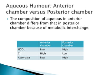
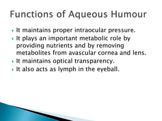
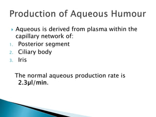
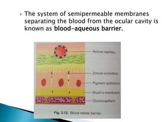
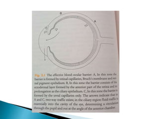
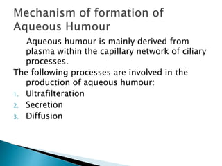
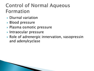
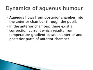
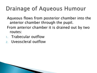
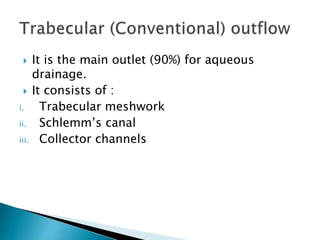
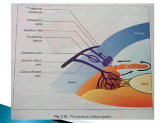
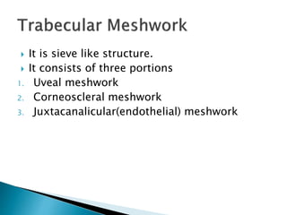
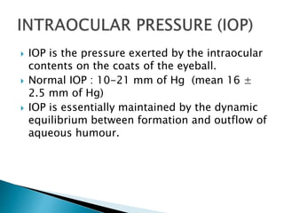
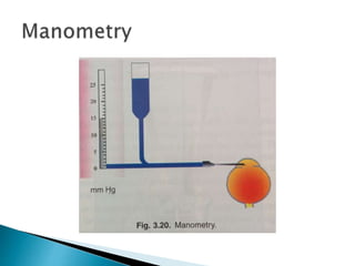
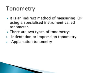
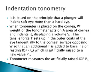
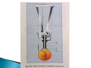
 It is a clear, colourless, watery fluid filling the
anterior chamber and posterior chamber of
the eyeball.
 Volume: 0.31ml
Anterior chamber- 0.25ml
Posterior chamber- 0.06ml
 Refractive Index: 1.336
 Density : 1.025-1.040 ( greater than water)
 pH : 7.2 (acidic)
 Rate of formation: 2.3µl/min
 Composition: constituents of normal aqueous humour are
 Water (99.9%)
 Solids (0.1%) which includes
1. Proteins (5-16mg%)
2. Amino acid (5mg/kg of water)
3. Non-colloid constituents –
a) Glucose (6.0 millimols/kg)
b) Urea (7 millimols/kg)
c) Ascorbate (0.9 millimols/kg)
d) Lactic acid (7.4 millimols/kg)
e) Inositol (0.1 millimols/kg)
f) Sodium (144 millimol/kg)
g) Potassium (4.5 millimols/kg)
h) Chloride (10 millimol/kg)
i) Carbonates (34 millimol/kg)
 Oxygen ( in dissolved state)
Composition of aqueous is similar to plasma
except:
 High concentration of : Ascorbate, pyruvate
and lactate.
 Low concentration of: Proteins, urea and
glucose.
 The composition of aqueous in anterior
chamber differs from that in posterior
chamber because of metabolic interchange:
Anterior
chamber
Posterior
chamber
HCO3
- Low High
Cl- High Low
Ascorbate Low High
 It maintains proper intraocular pressure.
 It plays an important metabolic role by
providing nutrients and by removing
metabolites from avascular cornea and lens.
 It maintains optical transparency.
 It also acts as lymph in the eyeball.
 Aqueous is derived from plasma within the
capillary network of:
1. Posterior segment
2. Ciliary body
3. Iris
The normal aqueous production rate is
2.3µl/min.
 The system of semipermeable membranes
separating the blood from the ocular cavity is
known as blood-aqueous barrier.
Aqueous humour is mainly derived from
plasma within the capillary network of ciliary
processes.
The following processes are involved in the
production of aqueous humour:
1. Ultrafilteration
2. Secretion
3. Diffusion
 Diurnal variation
 Blood pressure
 Plasma osmotic pressure
 Intraocular pressure
 Role of adrenergic innervation, vasopressin
and adenylcyclase
 Aqueous flows from posterior chamber into
the anterior chamber through the pupil.
 In the anterior chamber, there exist a
convection current which results from
temperature gradient between anterior and
posterior parts of anterior chamber.
Aqueous flows from posterior chamber into the
anterior chamber through the pupil.
From anterior chamber it is drained out by two
routes:
1. Trabecular outflow
2. Uveoscleral outflow
 It is the main outlet (90%) for aqueous
drainage.
 It consists of :
i. Trabecular meshwork
ii. Schlemm’s canal
iii. Collector channels
 It is sieve like structure.
 It consists of three portions
1. Uveal meshwork
2. Corneoscleral meshwork
3. Juxtacanalicular(endothelial) meshwork
 This an endothelial lined canal present
circumferentially in the sclearl sulcus.
 The endothelial cells present on its inner wall
are irregular, spindle shaped and contains
giant vacuoles.
 The outer wall contains smooth flat cells and
contains opening of collector channels.
 These are also called intra-scleral aqueous
vessels.
 They are about 25-35 in number.
 They leave the Schlemm’s cannal at oblique
angles to terminate in the episcleral veins.
 They do not have valves.
 They are divided into two systems:
1. Direct system
2. Indirect system
 It is responsible for 10% of aqueous drainage.
 Aqueous passes across the ciliary body into
the suprachoroidal space and is drained by
the venous circulation in the ciliary body,
choroid and sclera.
 Uveoscleral outflow is approximately around
0.3µl/min.
 Most of the aqueous drains into the episcleral
veins.
 These veins ultimately drain into the
cavernous sinus via the anterior ciliary and
superior ophthalmic veins.
 IOP is the pressure exerted by the intraocular
contents on the coats of the eyeball.
 Normal IOP : 10-21 mm of Hg (mean 16 ±
2.5 mm of Hg)
 IOP is essentially maintained by the dynamic
equilibrium between formation and outflow of
aqueous humour.
a) Local factors
b) General factors
1. Rate of aqueous formation
2. Resistance to aqueous outflow
3. Increased episcleral venous pressure
4. Dilation of pupil
1. Hereditary
2. Age
3. Sex
4. Diurnal variation
5. Postural variation
6. Seasonal variation
7. Blood pressure
8. Osmotic pressure of blood
9. Effects of Drugs
10. Effects of general anesthesia
11. Systemic hyperthermia
12. Refractive error
13. Mechanical pressure on globe
1. Manometry
2. Tonometry
 It is an indirect method of measuring IOP
using a specialised instrument called
tonometer.
 There are two types of tonometry:
1. Indentation or Impression tonometry
2. Applanation tonometry
 It is based on the principle that a plunger will
indent soft eye more than a hard eye.
 When tonometer is placed on the cornea, W
weight of the tonometer acts on A area of cornea
and indents it, displacing a volume Vc. The
tensile force T sets up in the outer coats of the
eye tangentially to the corneal surface opposing
W so that an additional T is added to baseline or
resting IOP (P0) which is artificially raised to a
new value P1.
 Tonometer measures the artificially raised IOP P1.
 Errors inherent in the instrument
 Errors due to contraction of extraocular
muscles
 Errors due to accomodation
 Errors due to ocular rigidity
 Errors due to variation in corneal curvature
 Errors in scale reading
 Blood volume alteration
 Moses effect
 It is based on Imbert-Fick law which states
that pressure inside a sphere P is equal to the
force W required to flatten its surface divided
by area of flattening A
P= W/A
 Two types:
1. Fixed Area (variable force) commenly used.
2. Fixed force (variable area).
 Other commenly used tonometers:
1. Perkin’s applanation tonometer
2. Pneumatic tonometer
3. Air-puff tonometer ( Non contact )
4. Pulse air tonometer
5. Tono pen
THANK YOU

Intraocular pressure and aqueous dynamics

  • 2.
     It isa clear, colourless, watery fluid filling the anterior chamber and posterior chamber of the eyeball.  Volume: 0.31ml Anterior chamber- 0.25ml Posterior chamber- 0.06ml  Refractive Index: 1.336  Density : 1.025-1.040 ( greater than water)  pH : 7.2 (acidic)  Rate of formation: 2.3µl/min
  • 3.
     Composition: constituentsof normal aqueous humour are  Water (99.9%)  Solids (0.1%) which includes 1. Proteins (5-16mg%) 2. Amino acid (5mg/kg of water) 3. Non-colloid constituents – a) Glucose (6.0 millimols/kg) b) Urea (7 millimols/kg) c) Ascorbate (0.9 millimols/kg) d) Lactic acid (7.4 millimols/kg) e) Inositol (0.1 millimols/kg) f) Sodium (144 millimol/kg) g) Potassium (4.5 millimols/kg) h) Chloride (10 millimol/kg) i) Carbonates (34 millimol/kg)  Oxygen ( in dissolved state)
  • 4.
    Composition of aqueousis similar to plasma except:  High concentration of : Ascorbate, pyruvate and lactate.  Low concentration of: Proteins, urea and glucose.
  • 5.
     The compositionof aqueous in anterior chamber differs from that in posterior chamber because of metabolic interchange: Anterior chamber Posterior chamber HCO3 - Low High Cl- High Low Ascorbate Low High
  • 6.
     It maintainsproper intraocular pressure.  It plays an important metabolic role by providing nutrients and by removing metabolites from avascular cornea and lens.  It maintains optical transparency.  It also acts as lymph in the eyeball.
  • 7.
     Aqueous isderived from plasma within the capillary network of: 1. Posterior segment 2. Ciliary body 3. Iris The normal aqueous production rate is 2.3µl/min.
  • 8.
     The systemof semipermeable membranes separating the blood from the ocular cavity is known as blood-aqueous barrier.
  • 10.
    Aqueous humour ismainly derived from plasma within the capillary network of ciliary processes. The following processes are involved in the production of aqueous humour: 1. Ultrafilteration 2. Secretion 3. Diffusion
  • 11.
     Diurnal variation Blood pressure  Plasma osmotic pressure  Intraocular pressure  Role of adrenergic innervation, vasopressin and adenylcyclase
  • 12.
     Aqueous flowsfrom posterior chamber into the anterior chamber through the pupil.  In the anterior chamber, there exist a convection current which results from temperature gradient between anterior and posterior parts of anterior chamber.
  • 13.
    Aqueous flows fromposterior chamber into the anterior chamber through the pupil. From anterior chamber it is drained out by two routes: 1. Trabecular outflow 2. Uveoscleral outflow
  • 14.
     It isthe main outlet (90%) for aqueous drainage.  It consists of : i. Trabecular meshwork ii. Schlemm’s canal iii. Collector channels
  • 16.
     It issieve like structure.  It consists of three portions 1. Uveal meshwork 2. Corneoscleral meshwork 3. Juxtacanalicular(endothelial) meshwork
  • 18.
     This anendothelial lined canal present circumferentially in the sclearl sulcus.  The endothelial cells present on its inner wall are irregular, spindle shaped and contains giant vacuoles.  The outer wall contains smooth flat cells and contains opening of collector channels.
  • 20.
     These arealso called intra-scleral aqueous vessels.  They are about 25-35 in number.  They leave the Schlemm’s cannal at oblique angles to terminate in the episcleral veins.  They do not have valves.  They are divided into two systems: 1. Direct system 2. Indirect system
  • 21.
     It isresponsible for 10% of aqueous drainage.  Aqueous passes across the ciliary body into the suprachoroidal space and is drained by the venous circulation in the ciliary body, choroid and sclera.  Uveoscleral outflow is approximately around 0.3µl/min.
  • 22.
     Most ofthe aqueous drains into the episcleral veins.  These veins ultimately drain into the cavernous sinus via the anterior ciliary and superior ophthalmic veins.
  • 23.
     IOP isthe pressure exerted by the intraocular contents on the coats of the eyeball.  Normal IOP : 10-21 mm of Hg (mean 16 ± 2.5 mm of Hg)  IOP is essentially maintained by the dynamic equilibrium between formation and outflow of aqueous humour.
  • 24.
    a) Local factors b)General factors
  • 25.
    1. Rate ofaqueous formation 2. Resistance to aqueous outflow 3. Increased episcleral venous pressure 4. Dilation of pupil
  • 26.
    1. Hereditary 2. Age 3.Sex 4. Diurnal variation 5. Postural variation 6. Seasonal variation 7. Blood pressure 8. Osmotic pressure of blood 9. Effects of Drugs 10. Effects of general anesthesia 11. Systemic hyperthermia 12. Refractive error 13. Mechanical pressure on globe
  • 27.
  • 29.
     It isan indirect method of measuring IOP using a specialised instrument called tonometer.  There are two types of tonometry: 1. Indentation or Impression tonometry 2. Applanation tonometry
  • 30.
     It isbased on the principle that a plunger will indent soft eye more than a hard eye.  When tonometer is placed on the cornea, W weight of the tonometer acts on A area of cornea and indents it, displacing a volume Vc. The tensile force T sets up in the outer coats of the eye tangentially to the corneal surface opposing W so that an additional T is added to baseline or resting IOP (P0) which is artificially raised to a new value P1.  Tonometer measures the artificially raised IOP P1.
  • 34.
     Errors inherentin the instrument  Errors due to contraction of extraocular muscles  Errors due to accomodation  Errors due to ocular rigidity  Errors due to variation in corneal curvature  Errors in scale reading  Blood volume alteration  Moses effect
  • 35.
     It isbased on Imbert-Fick law which states that pressure inside a sphere P is equal to the force W required to flatten its surface divided by area of flattening A P= W/A  Two types: 1. Fixed Area (variable force) commenly used. 2. Fixed force (variable area).
  • 38.
     Other commenlyused tonometers: 1. Perkin’s applanation tonometer 2. Pneumatic tonometer 3. Air-puff tonometer ( Non contact ) 4. Pulse air tonometer 5. Tono pen
  • 39.