Downloaded 35 times
![Ocular scans
• Probe positions
• Transverse : on sclera (@ TS)
• Longitudinal :at Limbus (@ LL)
• Axial : on cornea (@ AC)
• [NOTE: Markers aims nasally while screening superior &
Inferior and Superiorly for nasal & temporal]](https://image.slidesharecdn.com/ophthalmicultrasound1-200512132749/75/A-quick-guide-to-Ophthalmic-Ultrasound-B-Scan-interpretation-10-2048.jpg)


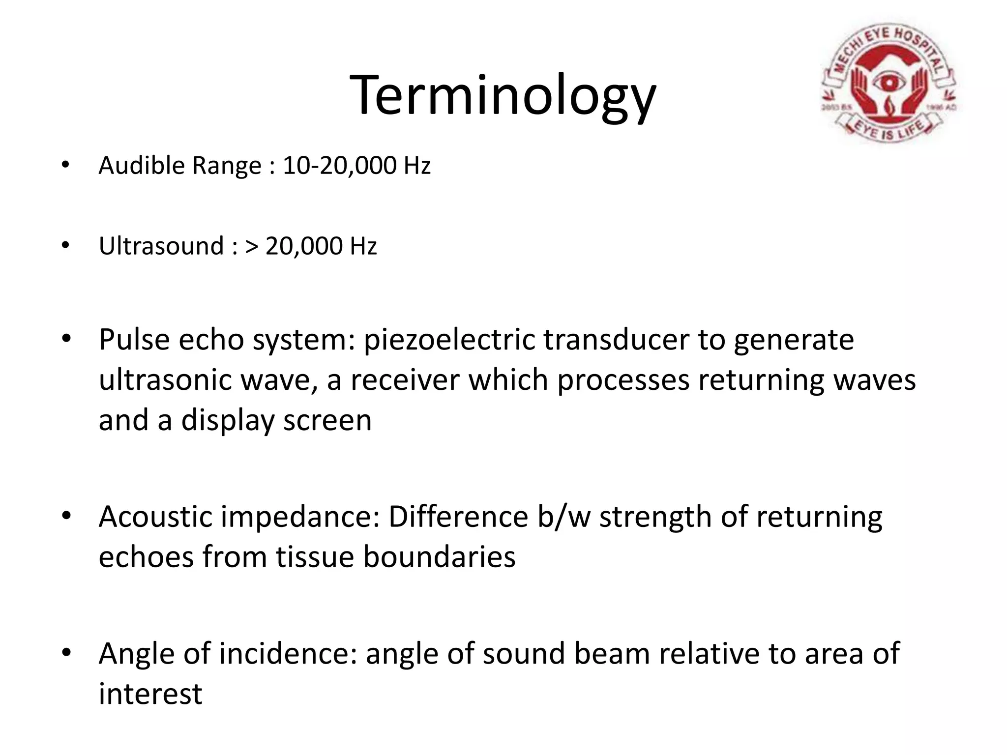
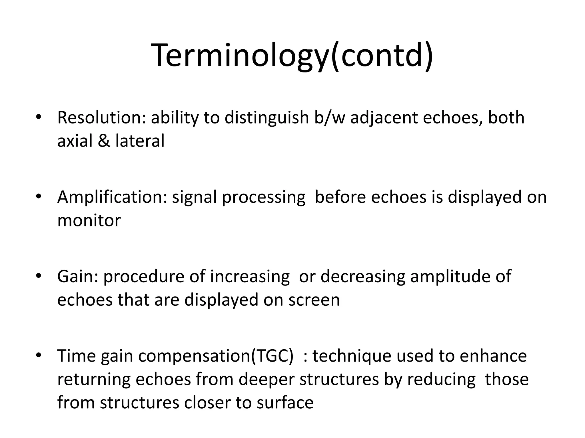
This document is a comprehensive guide to ophthalmic ultrasound and B-scan interpretation, providing detailed terminology, principles, and techniques for evaluating various ocular conditions. It covers ultrasound frequency ranges, indications for scans in both ocular and orbital assessments, normal appearances, and descriptions of lesions. The guide also includes information on different ocular pathologies, their ultrasound characteristics, and distinctions between similar conditions.


![Apporach to lung biopsy [Auto-saved].pptx latest](https://cdn.slidesharecdn.com/ss_thumbnails/apporachtolungbiopsyauto-saved-251211225655-93258539-thumbnail.jpg?width=640&height=640&fit=bounds)


