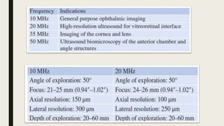
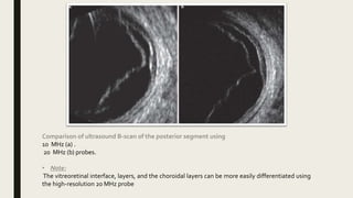
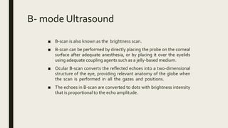
SlideShare a Scribd company logo
B- SCAN
By
Dr. Alshymaa Moustafa
Ohthalmology specialist
Principles of Ocular Ultrasonography
■ Ultrasonography utilizes sound waves to capture images.
■ Sound has a better penetration profile than light and can be used in patients with
media opacities.
■ generation of sound waves at a high frequency and back reflection to the
transducer from the tissue in its path.
■ When the sound waves return, the piezoelectric crystal in the transducer
vibrates, resulting in electrical impulses that are translated into an image or
other data.
■ Higher-frequency waves penetrate less into tissue but have better resolution.
In contrast, lower-frequency waves penetrate more deeply but have
suboptimal resolution.
Transducers and Probes
■ The basic element of an ultrasound probe, the piezoelectric crystal, usually consists of
lead zirconate titanate.
■ Recently, polymeric fluoride-based crystals have also been used, and lead-free and
fluoride-free transducers are being developed.
■ In the current practice, probes of various frequencies, including 10, 20, and 50 MHz,
are available for ocular ultrasonography.
Comparison of ultrasound B-scan of the posterior segment using
10 MHz (a) .
20 MHz (b) probes.
• Note:
The vitreoretinal interface, layers, and the choroidal layers can be more easily differentiated using
the high-resolution 20 MHz probe
B- mode Ultrasound
■ B-scan is also known as the brightness scan.
■ B-scan can be performed by directly placing the probe on the corneal
surface after adequate anesthesia, or by placing it over the eyelids
using adequate coupling agents such as a jelly-based medium.
■ Ocular B-scan converts the reflected echoes into a two-dimensional
structure of the eye, providing relevant anatomy of the globe when
the scan is performed in all the gazes and positions.
■ The echoes in B-scan are converted to dots with brightness intensity
that is proportional to the echo amplitude.
Gain
■ Gain is a frequently used index in ocular examination.
1. Higher gain increases the signal strength and thereby improves the sensitivity of the
examination.
2. Gain can be used to detect vitreous hemorrhages, syneresis, posterior hyaloid
interface, and inflammatory cells.
3. Low gain values improve the resolution in order to detect various layers such as
retina, choroid, and hyaloid; retinal breaks and tears; macular edema; or vascularity
of tumors.
Positions of B-scan
■ Axial scan (horizontal or vertical):
1. In this scan, the orientation of display is anterior-to-posterior with the patient
looking straight ahead.
2. The probe marker is nasal (horizontal, labeled HAX) or upwards (vertical, labeled
VAX).
■ Transverse scan:
1. In this scan, the scan plane traverses several clock hours.
2. This technique is used for scanning different quadrants (superior, nasal,
inferior, or temporal).
3. The probe marker is nasal (for S,I ) , or upwards (for N,T).
4. The scan sweeps the acoustic section posterior to anterior.
■ Longitudinal scan:
1. In this mode, each clock hour is scanned one by one.
2. The orientation of display is anterior to posterior.
3. The posterior part (ie, the optic nerve) is always at the bottom of the scan.
4. The probe is placed at the limbus and the marker is pointing towards the
clock hour.
5. This scan is used for localizing foreign bodies or retinal tears and for
documenting the macula.
Ultrasound B-scan in a case of intraocular foreign body shows a hyperechoic shadow in
the posterior vitreous cavity.
A-scan (superimposed on the B-scan) shows high-amplitude spikes suggestive of a metallic
foreign body.
This scan is a longitudinal scan at the 6 o’clock position
High-resolution ultrasound B-scan of the posterior segment shows macular
edema (yellow arrow) in a patient with diabetic maculopathy
Ocular UltrasoundTechnique
■ One can examine the entire globe in just five maneuvers, i.e. four dynamic quadrant
views and one more static slice through the macula and optic disc, also known as
longitudinal macula (LMAC).
■ The quadrants views are designatedT12,T3,T6, andT9.
■ These numbered quadrants correspond to a clock face superimposed on the eye.
■ For example, T12 is a view through the superior quadrant of the eye, T3 the nasal
quadrant of the right eye (temporal quadrant of the left eye), and so on.
■ Ultrasound images can be obtained through the patient's eyelids or with the
probe directly on the surface of the eye with appropriate topical anesthesia.
■ Begin with the gain on high. The patient should look in the direction of the
quadrant to be evaluated.
■ The marker on the probe is always oriented superiorly or nasally by convention.
■ Use a limbus-to-fornix rocking, rotational motion so that the tip of the probe
moves a small distance, while the base of the probe moves a larger distance.
■ The probe rotates around the globe so that the sound waves always pass through
the center of the eye.
■ This rotational motion will maximize the amount of retina visualized during the
scan.
Schematic of ultrasound quadrants
Limbus-to-fornix rotational motion
TransverseView 1:T12
(quadrant centered at 12 o'clock)
■ Ask the patient to look up.
■ Place your probe on the inferior aspect of the globe with the marker
oriented nasally.
■ Begin at the limbus (L) and locate the optic nerve shadow, both to
orient yourself and assure you are imaging the posterior segment.
■ Slowly sweep your probe toward the inferior fornix (F) until
visualization of theT12 quadrant is complete.
■ Repeat if necessary.
■ Remember to center any pathology along the equatorial plane of the
image for the best resolution.
TransverseView 2:T6
(quadrant centered at 6 o'clock)
■ Ask the patient to look down.
■ Place your probe on the superior aspect of the globe with the marker aimed nasally.
Again, begin at the limbus (L)).
■ Ensure you have an image of the retina and optic nerve before sweeping the probe
toward the superior fornix (F).
■ Repeat if necessary.
■ centering any pathology.
TransverseView 3:T3
(quadrant centered at 3 o'clock)
■ Remember, to scan the medial and lateral quadrants of the eye, the probe marker
should point superiorly.
■ For theT3 quadrant of the patient's right eye, instruct the patient to look left.
■ Place the probe on the temporal limbus (L).
■ After obtaining an image of the retina and optic nerve, gently sweep the probe to the
fornix (F) to complete evaluation of this quadrant.
■ To view theT3 quadrant of the left eye, the patient should still gaze to the left, but the
probe will be placed at the medial limbus, with the marker oriented superiorly.
TransverseView 4:T9View
(quadrant centered at 9 o'clock)
■ Scanning the T9 quadrant of the right eye is simply the reverse scan of the T3
quadrant.
■ With the probe marker oriented superiorly, instruct the patient to direct their gaze to
the right.
■ Place the probe on the globe at the nasal limbus (L).
■ For the T9 quadrant of the left eye, place the probe at the temporal limbus. Proceed,
again with a limbus-to-fornix (F) rotational sweeping movement.
Longitudinal Macula (LMAC)View
■ The LMAC view allows for proper visualization of the macula and optic nerve.
■ Gently place the probe on the nasal aspect of the eye with the patient's gaze directed
temporally.
■ Note: For this position, the marker of the probe should be directed toward the pupil,
instead of superiorly.
■ A longitudinal scan is the only scan where this occurs! In this view, the optic nerve will
be below the macula.
■ Maneuver the probe to bring the macula into the center of the image to obtain the
best resolution.
Thanks

More Related Content

What's hot

AC/A
AC/AAC/A
AC/A
zarin45
 
Specular microscopy
Specular microscopySpecular microscopy
Specular microscopy
Ruchi sood
 
Hess screen test mhr
Hess screen test mhrHess screen test mhr
Hess screen test mhr
Mehedi Hasan
 
Tangent screen
Tangent screenTangent screen
Tangent screen
sana butt
 
Stereopsis
Stereopsis  Stereopsis
Duochrome.pptx
Duochrome.pptxDuochrome.pptx
Duochrome.pptx
Vishnu645963
 
Fundus fluorescein angiography and B-scan by vijay
Fundus  fluorescein angiography  and B-scan by vijayFundus  fluorescein angiography  and B-scan by vijay
Fundus fluorescein angiography and B-scan by vijay
Dr. vijay pratap
 
IMAGING TECHNIQUES IN GLAUCOMA
IMAGING TECHNIQUES IN GLAUCOMAIMAGING TECHNIQUES IN GLAUCOMA
IMAGING TECHNIQUES IN GLAUCOMA
Laxmi Eye Institute
 
Corneal topography
Corneal topographyCorneal topography
Corneal topography
Dr Saurabh Kushwaha
 
OPTICS OF CONTACT LENSES
OPTICS OF CONTACT LENSESOPTICS OF CONTACT LENSES
OPTICS OF CONTACT LENSES
GREESHMA G
 
Recumbent prisms and fresnel prisms
Recumbent prisms and fresnel prismsRecumbent prisms and fresnel prisms
Recumbent prisms and fresnel prisms
Loknath Goswami
 
Lensometry.
Lensometry.Lensometry.
Lensometry.
ANUJA DHAKAL
 
Anterior segment OCT & UBM
Anterior segment OCT & UBMAnterior segment OCT & UBM
Anterior segment OCT & UBM
Dinesh Madduri
 
Tinted Contact lenses.pptx
Tinted Contact lenses.pptxTinted Contact lenses.pptx
Tinted Contact lenses.pptx
Ananya Chowdhury
 
HRT and GDx VCC
HRT and GDx VCCHRT and GDx VCC
HRT and GDx VCC
SSSIHMS-PG
 
Decentration and prismatic effect in lens (1)
Decentration and prismatic effect in lens (1)Decentration and prismatic effect in lens (1)
Decentration and prismatic effect in lens (1)
Sachitanand Singh
 
Accommodation
AccommodationAccommodation
Accommodation
Laxmi Eye Institute
 
Role of oct in glaucoma
Role of oct in glaucomaRole of oct in glaucoma
Role of oct in glaucoma
Dr Samarth Mishra
 
Gonioscopy
GonioscopyGonioscopy
Gonioscopy
Rashmi Ranjan
 
AS-OCT
AS-OCTAS-OCT

What's hot (20)

AC/A
AC/AAC/A
AC/A
 
Specular microscopy
Specular microscopySpecular microscopy
Specular microscopy
 
Hess screen test mhr
Hess screen test mhrHess screen test mhr
Hess screen test mhr
 
Tangent screen
Tangent screenTangent screen
Tangent screen
 
Stereopsis
Stereopsis  Stereopsis
Stereopsis
 
Duochrome.pptx
Duochrome.pptxDuochrome.pptx
Duochrome.pptx
 
Fundus fluorescein angiography and B-scan by vijay
Fundus  fluorescein angiography  and B-scan by vijayFundus  fluorescein angiography  and B-scan by vijay
Fundus fluorescein angiography and B-scan by vijay
 
IMAGING TECHNIQUES IN GLAUCOMA
IMAGING TECHNIQUES IN GLAUCOMAIMAGING TECHNIQUES IN GLAUCOMA
IMAGING TECHNIQUES IN GLAUCOMA
 
Corneal topography
Corneal topographyCorneal topography
Corneal topography
 
OPTICS OF CONTACT LENSES
OPTICS OF CONTACT LENSESOPTICS OF CONTACT LENSES
OPTICS OF CONTACT LENSES
 
Recumbent prisms and fresnel prisms
Recumbent prisms and fresnel prismsRecumbent prisms and fresnel prisms
Recumbent prisms and fresnel prisms
 
Lensometry.
Lensometry.Lensometry.
Lensometry.
 
Anterior segment OCT & UBM
Anterior segment OCT & UBMAnterior segment OCT & UBM
Anterior segment OCT & UBM
 
Tinted Contact lenses.pptx
Tinted Contact lenses.pptxTinted Contact lenses.pptx
Tinted Contact lenses.pptx
 
HRT and GDx VCC
HRT and GDx VCCHRT and GDx VCC
HRT and GDx VCC
 
Decentration and prismatic effect in lens (1)
Decentration and prismatic effect in lens (1)Decentration and prismatic effect in lens (1)
Decentration and prismatic effect in lens (1)
 
Accommodation
AccommodationAccommodation
Accommodation
 
Role of oct in glaucoma
Role of oct in glaucomaRole of oct in glaucoma
Role of oct in glaucoma
 
Gonioscopy
GonioscopyGonioscopy
Gonioscopy
 
AS-OCT
AS-OCTAS-OCT
AS-OCT
 

Similar to B scan

BScan and Ascan in ophthalmology and eye field
BScan and Ascan  in ophthalmology and eye fieldBScan and Ascan  in ophthalmology and eye field
BScan and Ascan in ophthalmology and eye field
Asif469093
 
Bscanmataryia22
Bscanmataryia22Bscanmataryia22
Bscanmataryia22
Mohamed Abdel-Aziz
 
B-Scan.pptx
B-Scan.pptxB-Scan.pptx
B-Scan.pptx
Asif469093
 
Extra-oral Radiographic Techniques
Extra-oral Radiographic TechniquesExtra-oral Radiographic Techniques
Extra-oral Radiographic Techniques
Arun Panwar
 
Ophthalmic ultrasonography
Ophthalmic ultrasonographyOphthalmic ultrasonography
Ophthalmic ultrasonography
kajal bhagat
 
Pa ceph analysis
Pa ceph analysisPa ceph analysis
Pa ceph analysis
Kumar Adarsh
 
B scan
B scanB scan
OPTHALMOSCOPY.pdf
OPTHALMOSCOPY.pdfOPTHALMOSCOPY.pdf
OPTHALMOSCOPY.pdf
shamsudheenpp1
 
Basic principles of ocular ultrasonography
Basic principles of ocular ultrasonographyBasic principles of ocular ultrasonography
Basic principles of ocular ultrasonography
Rohit Rao
 
OPG and Extraoral radiography
OPG and Extraoral radiographyOPG and Extraoral radiography
OPG and Extraoral radiography
Neha Sharma
 
Optic Nerve Sheath Diameter (ONSD) Measurement for Intracranial Pressure Moni...
Optic Nerve Sheath Diameter (ONSD) Measurement for Intracranial Pressure Moni...Optic Nerve Sheath Diameter (ONSD) Measurement for Intracranial Pressure Moni...
Optic Nerve Sheath Diameter (ONSD) Measurement for Intracranial Pressure Moni...
Ade Wijaya
 
Ophthalmoscopy
OphthalmoscopyOphthalmoscopy
Ophthalmoscopy
AIMS Education
 
Ophthalmoscopy
OphthalmoscopyOphthalmoscopy
Ophthalmoscopy
AIMS Education
 
L2 Gynaecological usg (TAUS part 2).pptx
L2 Gynaecological usg (TAUS part 2).pptxL2 Gynaecological usg (TAUS part 2).pptx
L2 Gynaecological usg (TAUS part 2).pptx
iqra saeed
 
Neuroimaging in Psychiatry
Neuroimaging in PsychiatryNeuroimaging in Psychiatry
Neuroimaging in Psychiatry
Enoch R G
 
Introduction to echocardiography
Introduction to echocardiography Introduction to echocardiography
Introduction to echocardiography
Bibini Bab
 
ULTRASONOGRAPHY (USG) AND ULTRASOUND BIOMICROSCOPY(UBM)
ULTRASONOGRAPHY (USG)  AND ULTRASOUND BIOMICROSCOPY(UBM)ULTRASONOGRAPHY (USG)  AND ULTRASOUND BIOMICROSCOPY(UBM)
ULTRASONOGRAPHY (USG) AND ULTRASOUND BIOMICROSCOPY(UBM)
Dr. Gaurav Shukla
 
E fast scan for surgeons- skill lab procedure- osce - copy
E fast scan for surgeons- skill lab procedure- osce - copyE fast scan for surgeons- skill lab procedure- osce - copy
E fast scan for surgeons- skill lab procedure- osce - copy
Selvaraj Balasubramani
 
B scan
B scanB scan
DOC-20230615-WA0003..pptx
DOC-20230615-WA0003..pptxDOC-20230615-WA0003..pptx
DOC-20230615-WA0003..pptx
Divya785180
 

Similar to B scan (20)

BScan and Ascan in ophthalmology and eye field
BScan and Ascan  in ophthalmology and eye fieldBScan and Ascan  in ophthalmology and eye field
BScan and Ascan in ophthalmology and eye field
 
Bscanmataryia22
Bscanmataryia22Bscanmataryia22
Bscanmataryia22
 
B-Scan.pptx
B-Scan.pptxB-Scan.pptx
B-Scan.pptx
 
Extra-oral Radiographic Techniques
Extra-oral Radiographic TechniquesExtra-oral Radiographic Techniques
Extra-oral Radiographic Techniques
 
Ophthalmic ultrasonography
Ophthalmic ultrasonographyOphthalmic ultrasonography
Ophthalmic ultrasonography
 
Pa ceph analysis
Pa ceph analysisPa ceph analysis
Pa ceph analysis
 
B scan
B scanB scan
B scan
 
OPTHALMOSCOPY.pdf
OPTHALMOSCOPY.pdfOPTHALMOSCOPY.pdf
OPTHALMOSCOPY.pdf
 
Basic principles of ocular ultrasonography
Basic principles of ocular ultrasonographyBasic principles of ocular ultrasonography
Basic principles of ocular ultrasonography
 
OPG and Extraoral radiography
OPG and Extraoral radiographyOPG and Extraoral radiography
OPG and Extraoral radiography
 
Optic Nerve Sheath Diameter (ONSD) Measurement for Intracranial Pressure Moni...
Optic Nerve Sheath Diameter (ONSD) Measurement for Intracranial Pressure Moni...Optic Nerve Sheath Diameter (ONSD) Measurement for Intracranial Pressure Moni...
Optic Nerve Sheath Diameter (ONSD) Measurement for Intracranial Pressure Moni...
 
Ophthalmoscopy
OphthalmoscopyOphthalmoscopy
Ophthalmoscopy
 
Ophthalmoscopy
OphthalmoscopyOphthalmoscopy
Ophthalmoscopy
 
L2 Gynaecological usg (TAUS part 2).pptx
L2 Gynaecological usg (TAUS part 2).pptxL2 Gynaecological usg (TAUS part 2).pptx
L2 Gynaecological usg (TAUS part 2).pptx
 
Neuroimaging in Psychiatry
Neuroimaging in PsychiatryNeuroimaging in Psychiatry
Neuroimaging in Psychiatry
 
Introduction to echocardiography
Introduction to echocardiography Introduction to echocardiography
Introduction to echocardiography
 
ULTRASONOGRAPHY (USG) AND ULTRASOUND BIOMICROSCOPY(UBM)
ULTRASONOGRAPHY (USG)  AND ULTRASOUND BIOMICROSCOPY(UBM)ULTRASONOGRAPHY (USG)  AND ULTRASOUND BIOMICROSCOPY(UBM)
ULTRASONOGRAPHY (USG) AND ULTRASOUND BIOMICROSCOPY(UBM)
 
E fast scan for surgeons- skill lab procedure- osce - copy
E fast scan for surgeons- skill lab procedure- osce - copyE fast scan for surgeons- skill lab procedure- osce - copy
E fast scan for surgeons- skill lab procedure- osce - copy
 
B scan
B scanB scan
B scan
 
DOC-20230615-WA0003..pptx
DOC-20230615-WA0003..pptxDOC-20230615-WA0003..pptx
DOC-20230615-WA0003..pptx
 

More from Sheim Elteb

CASE REVIEW.pptx
CASE REVIEW.pptxCASE REVIEW.pptx
CASE REVIEW.pptx
Sheim Elteb
 
VF in glaucoma.pptx
VF in glaucoma.pptxVF in glaucoma.pptx
VF in glaucoma.pptx
Sheim Elteb
 
EUA-WPS Office.pptx
EUA-WPS Office.pptxEUA-WPS Office.pptx
EUA-WPS Office.pptx
Sheim Elteb
 
GLAUCOMA MEDICATION.pptx
GLAUCOMA MEDICATION.pptxGLAUCOMA MEDICATION.pptx
GLAUCOMA MEDICATION.pptx
Sheim Elteb
 
POAG.pptx
POAG.pptxPOAG.pptx
POAG.pptx
Sheim Elteb
 
Exotropia cases
Exotropia casesExotropia cases
Exotropia cases
Sheim Elteb
 
Exotropia
ExotropiaExotropia
Exotropia
Sheim Elteb
 
Incomitant strabismus
Incomitant strabismusIncomitant strabismus
Incomitant strabismus
Sheim Elteb
 
INDIRECT OPHTHALOMOSCOPE
INDIRECT OPHTHALOMOSCOPEINDIRECT OPHTHALOMOSCOPE
INDIRECT OPHTHALOMOSCOPE
Sheim Elteb
 
APHACIC IOL
APHACIC IOLAPHACIC IOL
APHACIC IOL
Sheim Elteb
 
Keratometer
KeratometerKeratometer
Keratometer
Sheim Elteb
 
Non accommodative et
Non  accommodative etNon  accommodative et
Non accommodative et
Sheim Elteb
 
PHACIC IOL
PHACIC IOLPHACIC IOL
PHACIC IOL
Sheim Elteb
 
Nystagmus
NystagmusNystagmus
Nystagmus
Sheim Elteb
 
Pupil examination
Pupil examinationPupil examination
Pupil examination
Sheim Elteb
 
Retinoscopy
RetinoscopyRetinoscopy
Retinoscopy
Sheim Elteb
 
UWS
UWSUWS
Yag post capsulotomy
Yag post capsulotomyYag post capsulotomy
Yag post capsulotomy
Sheim Elteb
 
Et cases
Et casesEt cases
Et cases
Sheim Elteb
 
EOM test
EOM testEOM test
EOM test
Sheim Elteb
 

More from Sheim Elteb (20)

CASE REVIEW.pptx
CASE REVIEW.pptxCASE REVIEW.pptx
CASE REVIEW.pptx
 
VF in glaucoma.pptx
VF in glaucoma.pptxVF in glaucoma.pptx
VF in glaucoma.pptx
 
EUA-WPS Office.pptx
EUA-WPS Office.pptxEUA-WPS Office.pptx
EUA-WPS Office.pptx
 
GLAUCOMA MEDICATION.pptx
GLAUCOMA MEDICATION.pptxGLAUCOMA MEDICATION.pptx
GLAUCOMA MEDICATION.pptx
 
POAG.pptx
POAG.pptxPOAG.pptx
POAG.pptx
 
Exotropia cases
Exotropia casesExotropia cases
Exotropia cases
 
Exotropia
ExotropiaExotropia
Exotropia
 
Incomitant strabismus
Incomitant strabismusIncomitant strabismus
Incomitant strabismus
 
INDIRECT OPHTHALOMOSCOPE
INDIRECT OPHTHALOMOSCOPEINDIRECT OPHTHALOMOSCOPE
INDIRECT OPHTHALOMOSCOPE
 
APHACIC IOL
APHACIC IOLAPHACIC IOL
APHACIC IOL
 
Keratometer
KeratometerKeratometer
Keratometer
 
Non accommodative et
Non  accommodative etNon  accommodative et
Non accommodative et
 
PHACIC IOL
PHACIC IOLPHACIC IOL
PHACIC IOL
 
Nystagmus
NystagmusNystagmus
Nystagmus
 
Pupil examination
Pupil examinationPupil examination
Pupil examination
 
Retinoscopy
RetinoscopyRetinoscopy
Retinoscopy
 
UWS
UWSUWS
UWS
 
Yag post capsulotomy
Yag post capsulotomyYag post capsulotomy
Yag post capsulotomy
 
Et cases
Et casesEt cases
Et cases
 
EOM test
EOM testEOM test
EOM test
 

Recently uploaded

Walmart Business+ and Spark Good for Nonprofits.pdf
Walmart Business+ and Spark Good for Nonprofits.pdfWalmart Business+ and Spark Good for Nonprofits.pdf
Walmart Business+ and Spark Good for Nonprofits.pdf
TechSoup
 
Film vocab for eal 3 students: Australia the movie
Film vocab for eal 3 students: Australia the movieFilm vocab for eal 3 students: Australia the movie
Film vocab for eal 3 students: Australia the movie
Nicholas Montgomery
 
ISO/IEC 27001, ISO/IEC 42001, and GDPR: Best Practices for Implementation and...
ISO/IEC 27001, ISO/IEC 42001, and GDPR: Best Practices for Implementation and...ISO/IEC 27001, ISO/IEC 42001, and GDPR: Best Practices for Implementation and...
ISO/IEC 27001, ISO/IEC 42001, and GDPR: Best Practices for Implementation and...
PECB
 
ANATOMY AND BIOMECHANICS OF HIP JOINT.pdf
ANATOMY AND BIOMECHANICS OF HIP JOINT.pdfANATOMY AND BIOMECHANICS OF HIP JOINT.pdf
ANATOMY AND BIOMECHANICS OF HIP JOINT.pdf
Priyankaranawat4
 
What is Digital Literacy? A guest blog from Andy McLaughlin, University of Ab...
What is Digital Literacy? A guest blog from Andy McLaughlin, University of Ab...What is Digital Literacy? A guest blog from Andy McLaughlin, University of Ab...
What is Digital Literacy? A guest blog from Andy McLaughlin, University of Ab...
GeorgeMilliken2
 
C1 Rubenstein AP HuG xxxxxxxxxxxxxx.pptx
C1 Rubenstein AP HuG xxxxxxxxxxxxxx.pptxC1 Rubenstein AP HuG xxxxxxxxxxxxxx.pptx
C1 Rubenstein AP HuG xxxxxxxxxxxxxx.pptx
mulvey2
 
Présentationvvvvvvvvvvvvvvvvvvvvvvvvvvvv2.pptx
Présentationvvvvvvvvvvvvvvvvvvvvvvvvvvvv2.pptxPrésentationvvvvvvvvvvvvvvvvvvvvvvvvvvvv2.pptx
Présentationvvvvvvvvvvvvvvvvvvvvvvvvvvvv2.pptx
siemaillard
 
LAND USE LAND COVER AND NDVI OF MIRZAPUR DISTRICT, UP
LAND USE LAND COVER AND NDVI OF MIRZAPUR DISTRICT, UPLAND USE LAND COVER AND NDVI OF MIRZAPUR DISTRICT, UP
LAND USE LAND COVER AND NDVI OF MIRZAPUR DISTRICT, UP
RAHUL
 
The History of Stoke Newington Street Names
The History of Stoke Newington Street NamesThe History of Stoke Newington Street Names
The History of Stoke Newington Street Names
History of Stoke Newington
 
Advanced Java[Extra Concepts, Not Difficult].docx
Advanced Java[Extra Concepts, Not Difficult].docxAdvanced Java[Extra Concepts, Not Difficult].docx
Advanced Java[Extra Concepts, Not Difficult].docx
adhitya5119
 
Chapter wise All Notes of First year Basic Civil Engineering.pptx
Chapter wise All Notes of First year Basic Civil Engineering.pptxChapter wise All Notes of First year Basic Civil Engineering.pptx
Chapter wise All Notes of First year Basic Civil Engineering.pptx
Denish Jangid
 
Hindi varnamala | hindi alphabet PPT.pdf
Hindi varnamala | hindi alphabet PPT.pdfHindi varnamala | hindi alphabet PPT.pdf
Hindi varnamala | hindi alphabet PPT.pdf
Dr. Mulla Adam Ali
 
spot a liar (Haiqa 146).pptx Technical writhing and presentation skills
spot a liar (Haiqa 146).pptx Technical writhing and presentation skillsspot a liar (Haiqa 146).pptx Technical writhing and presentation skills
spot a liar (Haiqa 146).pptx Technical writhing and presentation skills
haiqairshad
 
Liberal Approach to the Study of Indian Politics.pdf
Liberal Approach to the Study of Indian Politics.pdfLiberal Approach to the Study of Indian Politics.pdf
Liberal Approach to the Study of Indian Politics.pdf
WaniBasim
 
Reimagining Your Library Space: How to Increase the Vibes in Your Library No ...
Reimagining Your Library Space: How to Increase the Vibes in Your Library No ...Reimagining Your Library Space: How to Increase the Vibes in Your Library No ...
Reimagining Your Library Space: How to Increase the Vibes in Your Library No ...
Diana Rendina
 
NEWSPAPERS - QUESTION 1 - REVISION POWERPOINT.pptx
NEWSPAPERS - QUESTION 1 - REVISION POWERPOINT.pptxNEWSPAPERS - QUESTION 1 - REVISION POWERPOINT.pptx
NEWSPAPERS - QUESTION 1 - REVISION POWERPOINT.pptx
iammrhaywood
 
Wound healing PPT
Wound healing PPTWound healing PPT
Wound healing PPT
Jyoti Chand
 
বাংলাদেশ অর্থনৈতিক সমীক্ষা (Economic Review) ২০২৪ UJS App.pdf
বাংলাদেশ অর্থনৈতিক সমীক্ষা (Economic Review) ২০২৪ UJS App.pdfবাংলাদেশ অর্থনৈতিক সমীক্ষা (Economic Review) ২০২৪ UJS App.pdf
বাংলাদেশ অর্থনৈতিক সমীক্ষা (Economic Review) ২০২৪ UJS App.pdf
eBook.com.bd (প্রয়োজনীয় বাংলা বই)
 
Beyond Degrees - Empowering the Workforce in the Context of Skills-First.pptx
Beyond Degrees - Empowering the Workforce in the Context of Skills-First.pptxBeyond Degrees - Empowering the Workforce in the Context of Skills-First.pptx
Beyond Degrees - Empowering the Workforce in the Context of Skills-First.pptx
EduSkills OECD
 
BÀI TẬP DẠY THÊM TIẾNG ANH LỚP 7 CẢ NĂM FRIENDS PLUS SÁCH CHÂN TRỜI SÁNG TẠO ...
BÀI TẬP DẠY THÊM TIẾNG ANH LỚP 7 CẢ NĂM FRIENDS PLUS SÁCH CHÂN TRỜI SÁNG TẠO ...BÀI TẬP DẠY THÊM TIẾNG ANH LỚP 7 CẢ NĂM FRIENDS PLUS SÁCH CHÂN TRỜI SÁNG TẠO ...
BÀI TẬP DẠY THÊM TIẾNG ANH LỚP 7 CẢ NĂM FRIENDS PLUS SÁCH CHÂN TRỜI SÁNG TẠO ...
Nguyen Thanh Tu Collection
 

Recently uploaded (20)

Walmart Business+ and Spark Good for Nonprofits.pdf
Walmart Business+ and Spark Good for Nonprofits.pdfWalmart Business+ and Spark Good for Nonprofits.pdf
Walmart Business+ and Spark Good for Nonprofits.pdf
 
Film vocab for eal 3 students: Australia the movie
Film vocab for eal 3 students: Australia the movieFilm vocab for eal 3 students: Australia the movie
Film vocab for eal 3 students: Australia the movie
 
ISO/IEC 27001, ISO/IEC 42001, and GDPR: Best Practices for Implementation and...
ISO/IEC 27001, ISO/IEC 42001, and GDPR: Best Practices for Implementation and...ISO/IEC 27001, ISO/IEC 42001, and GDPR: Best Practices for Implementation and...
ISO/IEC 27001, ISO/IEC 42001, and GDPR: Best Practices for Implementation and...
 
ANATOMY AND BIOMECHANICS OF HIP JOINT.pdf
ANATOMY AND BIOMECHANICS OF HIP JOINT.pdfANATOMY AND BIOMECHANICS OF HIP JOINT.pdf
ANATOMY AND BIOMECHANICS OF HIP JOINT.pdf
 
What is Digital Literacy? A guest blog from Andy McLaughlin, University of Ab...
What is Digital Literacy? A guest blog from Andy McLaughlin, University of Ab...What is Digital Literacy? A guest blog from Andy McLaughlin, University of Ab...
What is Digital Literacy? A guest blog from Andy McLaughlin, University of Ab...
 
C1 Rubenstein AP HuG xxxxxxxxxxxxxx.pptx
C1 Rubenstein AP HuG xxxxxxxxxxxxxx.pptxC1 Rubenstein AP HuG xxxxxxxxxxxxxx.pptx
C1 Rubenstein AP HuG xxxxxxxxxxxxxx.pptx
 
Présentationvvvvvvvvvvvvvvvvvvvvvvvvvvvv2.pptx
Présentationvvvvvvvvvvvvvvvvvvvvvvvvvvvv2.pptxPrésentationvvvvvvvvvvvvvvvvvvvvvvvvvvvv2.pptx
Présentationvvvvvvvvvvvvvvvvvvvvvvvvvvvv2.pptx
 
LAND USE LAND COVER AND NDVI OF MIRZAPUR DISTRICT, UP
LAND USE LAND COVER AND NDVI OF MIRZAPUR DISTRICT, UPLAND USE LAND COVER AND NDVI OF MIRZAPUR DISTRICT, UP
LAND USE LAND COVER AND NDVI OF MIRZAPUR DISTRICT, UP
 
The History of Stoke Newington Street Names
The History of Stoke Newington Street NamesThe History of Stoke Newington Street Names
The History of Stoke Newington Street Names
 
Advanced Java[Extra Concepts, Not Difficult].docx
Advanced Java[Extra Concepts, Not Difficult].docxAdvanced Java[Extra Concepts, Not Difficult].docx
Advanced Java[Extra Concepts, Not Difficult].docx
 
Chapter wise All Notes of First year Basic Civil Engineering.pptx
Chapter wise All Notes of First year Basic Civil Engineering.pptxChapter wise All Notes of First year Basic Civil Engineering.pptx
Chapter wise All Notes of First year Basic Civil Engineering.pptx
 
Hindi varnamala | hindi alphabet PPT.pdf
Hindi varnamala | hindi alphabet PPT.pdfHindi varnamala | hindi alphabet PPT.pdf
Hindi varnamala | hindi alphabet PPT.pdf
 
spot a liar (Haiqa 146).pptx Technical writhing and presentation skills
spot a liar (Haiqa 146).pptx Technical writhing and presentation skillsspot a liar (Haiqa 146).pptx Technical writhing and presentation skills
spot a liar (Haiqa 146).pptx Technical writhing and presentation skills
 
Liberal Approach to the Study of Indian Politics.pdf
Liberal Approach to the Study of Indian Politics.pdfLiberal Approach to the Study of Indian Politics.pdf
Liberal Approach to the Study of Indian Politics.pdf
 
Reimagining Your Library Space: How to Increase the Vibes in Your Library No ...
Reimagining Your Library Space: How to Increase the Vibes in Your Library No ...Reimagining Your Library Space: How to Increase the Vibes in Your Library No ...
Reimagining Your Library Space: How to Increase the Vibes in Your Library No ...
 
NEWSPAPERS - QUESTION 1 - REVISION POWERPOINT.pptx
NEWSPAPERS - QUESTION 1 - REVISION POWERPOINT.pptxNEWSPAPERS - QUESTION 1 - REVISION POWERPOINT.pptx
NEWSPAPERS - QUESTION 1 - REVISION POWERPOINT.pptx
 
Wound healing PPT
Wound healing PPTWound healing PPT
Wound healing PPT
 
বাংলাদেশ অর্থনৈতিক সমীক্ষা (Economic Review) ২০২৪ UJS App.pdf
বাংলাদেশ অর্থনৈতিক সমীক্ষা (Economic Review) ২০২৪ UJS App.pdfবাংলাদেশ অর্থনৈতিক সমীক্ষা (Economic Review) ২০২৪ UJS App.pdf
বাংলাদেশ অর্থনৈতিক সমীক্ষা (Economic Review) ২০২৪ UJS App.pdf
 
Beyond Degrees - Empowering the Workforce in the Context of Skills-First.pptx
Beyond Degrees - Empowering the Workforce in the Context of Skills-First.pptxBeyond Degrees - Empowering the Workforce in the Context of Skills-First.pptx
Beyond Degrees - Empowering the Workforce in the Context of Skills-First.pptx
 
BÀI TẬP DẠY THÊM TIẾNG ANH LỚP 7 CẢ NĂM FRIENDS PLUS SÁCH CHÂN TRỜI SÁNG TẠO ...
BÀI TẬP DẠY THÊM TIẾNG ANH LỚP 7 CẢ NĂM FRIENDS PLUS SÁCH CHÂN TRỜI SÁNG TẠO ...BÀI TẬP DẠY THÊM TIẾNG ANH LỚP 7 CẢ NĂM FRIENDS PLUS SÁCH CHÂN TRỜI SÁNG TẠO ...
BÀI TẬP DẠY THÊM TIẾNG ANH LỚP 7 CẢ NĂM FRIENDS PLUS SÁCH CHÂN TRỜI SÁNG TẠO ...
 

B scan

  • 1. B- SCAN By Dr. Alshymaa Moustafa Ohthalmology specialist
  • 2. Principles of Ocular Ultrasonography ■ Ultrasonography utilizes sound waves to capture images. ■ Sound has a better penetration profile than light and can be used in patients with media opacities.
  • 3. ■ generation of sound waves at a high frequency and back reflection to the transducer from the tissue in its path. ■ When the sound waves return, the piezoelectric crystal in the transducer vibrates, resulting in electrical impulses that are translated into an image or other data. ■ Higher-frequency waves penetrate less into tissue but have better resolution. In contrast, lower-frequency waves penetrate more deeply but have suboptimal resolution.
  • 4. Transducers and Probes ■ The basic element of an ultrasound probe, the piezoelectric crystal, usually consists of lead zirconate titanate. ■ Recently, polymeric fluoride-based crystals have also been used, and lead-free and fluoride-free transducers are being developed. ■ In the current practice, probes of various frequencies, including 10, 20, and 50 MHz, are available for ocular ultrasonography.
  • 5.
  • 6.
  • 7. Comparison of ultrasound B-scan of the posterior segment using 10 MHz (a) . 20 MHz (b) probes. • Note: The vitreoretinal interface, layers, and the choroidal layers can be more easily differentiated using the high-resolution 20 MHz probe
  • 8. B- mode Ultrasound ■ B-scan is also known as the brightness scan. ■ B-scan can be performed by directly placing the probe on the corneal surface after adequate anesthesia, or by placing it over the eyelids using adequate coupling agents such as a jelly-based medium. ■ Ocular B-scan converts the reflected echoes into a two-dimensional structure of the eye, providing relevant anatomy of the globe when the scan is performed in all the gazes and positions. ■ The echoes in B-scan are converted to dots with brightness intensity that is proportional to the echo amplitude.
  • 9. Gain ■ Gain is a frequently used index in ocular examination. 1. Higher gain increases the signal strength and thereby improves the sensitivity of the examination. 2. Gain can be used to detect vitreous hemorrhages, syneresis, posterior hyaloid interface, and inflammatory cells. 3. Low gain values improve the resolution in order to detect various layers such as retina, choroid, and hyaloid; retinal breaks and tears; macular edema; or vascularity of tumors.
  • 10. Positions of B-scan ■ Axial scan (horizontal or vertical): 1. In this scan, the orientation of display is anterior-to-posterior with the patient looking straight ahead. 2. The probe marker is nasal (horizontal, labeled HAX) or upwards (vertical, labeled VAX).
  • 11. ■ Transverse scan: 1. In this scan, the scan plane traverses several clock hours. 2. This technique is used for scanning different quadrants (superior, nasal, inferior, or temporal). 3. The probe marker is nasal (for S,I ) , or upwards (for N,T). 4. The scan sweeps the acoustic section posterior to anterior.
  • 12. ■ Longitudinal scan: 1. In this mode, each clock hour is scanned one by one. 2. The orientation of display is anterior to posterior. 3. The posterior part (ie, the optic nerve) is always at the bottom of the scan. 4. The probe is placed at the limbus and the marker is pointing towards the clock hour. 5. This scan is used for localizing foreign bodies or retinal tears and for documenting the macula.
  • 13. Ultrasound B-scan in a case of intraocular foreign body shows a hyperechoic shadow in the posterior vitreous cavity. A-scan (superimposed on the B-scan) shows high-amplitude spikes suggestive of a metallic foreign body. This scan is a longitudinal scan at the 6 o’clock position
  • 14. High-resolution ultrasound B-scan of the posterior segment shows macular edema (yellow arrow) in a patient with diabetic maculopathy
  • 15. Ocular UltrasoundTechnique ■ One can examine the entire globe in just five maneuvers, i.e. four dynamic quadrant views and one more static slice through the macula and optic disc, also known as longitudinal macula (LMAC). ■ The quadrants views are designatedT12,T3,T6, andT9. ■ These numbered quadrants correspond to a clock face superimposed on the eye. ■ For example, T12 is a view through the superior quadrant of the eye, T3 the nasal quadrant of the right eye (temporal quadrant of the left eye), and so on.
  • 16. ■ Ultrasound images can be obtained through the patient's eyelids or with the probe directly on the surface of the eye with appropriate topical anesthesia. ■ Begin with the gain on high. The patient should look in the direction of the quadrant to be evaluated. ■ The marker on the probe is always oriented superiorly or nasally by convention. ■ Use a limbus-to-fornix rocking, rotational motion so that the tip of the probe moves a small distance, while the base of the probe moves a larger distance. ■ The probe rotates around the globe so that the sound waves always pass through the center of the eye. ■ This rotational motion will maximize the amount of retina visualized during the scan.
  • 17. Schematic of ultrasound quadrants Limbus-to-fornix rotational motion
  • 18. TransverseView 1:T12 (quadrant centered at 12 o'clock) ■ Ask the patient to look up. ■ Place your probe on the inferior aspect of the globe with the marker oriented nasally. ■ Begin at the limbus (L) and locate the optic nerve shadow, both to orient yourself and assure you are imaging the posterior segment. ■ Slowly sweep your probe toward the inferior fornix (F) until visualization of theT12 quadrant is complete. ■ Repeat if necessary. ■ Remember to center any pathology along the equatorial plane of the image for the best resolution.
  • 19.
  • 20.
  • 21. TransverseView 2:T6 (quadrant centered at 6 o'clock) ■ Ask the patient to look down. ■ Place your probe on the superior aspect of the globe with the marker aimed nasally. Again, begin at the limbus (L)). ■ Ensure you have an image of the retina and optic nerve before sweeping the probe toward the superior fornix (F). ■ Repeat if necessary. ■ centering any pathology.
  • 22.
  • 23.
  • 24. TransverseView 3:T3 (quadrant centered at 3 o'clock) ■ Remember, to scan the medial and lateral quadrants of the eye, the probe marker should point superiorly. ■ For theT3 quadrant of the patient's right eye, instruct the patient to look left. ■ Place the probe on the temporal limbus (L). ■ After obtaining an image of the retina and optic nerve, gently sweep the probe to the fornix (F) to complete evaluation of this quadrant. ■ To view theT3 quadrant of the left eye, the patient should still gaze to the left, but the probe will be placed at the medial limbus, with the marker oriented superiorly.
  • 25.
  • 26.
  • 27. TransverseView 4:T9View (quadrant centered at 9 o'clock) ■ Scanning the T9 quadrant of the right eye is simply the reverse scan of the T3 quadrant. ■ With the probe marker oriented superiorly, instruct the patient to direct their gaze to the right. ■ Place the probe on the globe at the nasal limbus (L). ■ For the T9 quadrant of the left eye, place the probe at the temporal limbus. Proceed, again with a limbus-to-fornix (F) rotational sweeping movement.
  • 28.
  • 29.
  • 30. Longitudinal Macula (LMAC)View ■ The LMAC view allows for proper visualization of the macula and optic nerve. ■ Gently place the probe on the nasal aspect of the eye with the patient's gaze directed temporally. ■ Note: For this position, the marker of the probe should be directed toward the pupil, instead of superiorly. ■ A longitudinal scan is the only scan where this occurs! In this view, the optic nerve will be below the macula. ■ Maneuver the probe to bring the macula into the center of the image to obtain the best resolution.
  • 31.
  • 32.