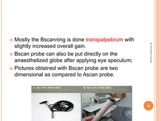
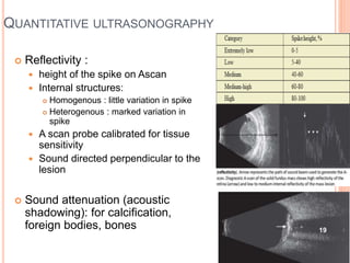
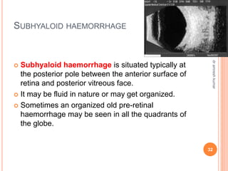
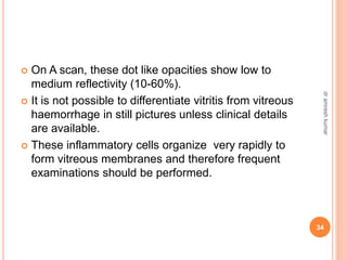
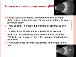
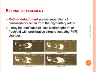
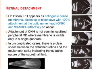
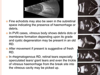
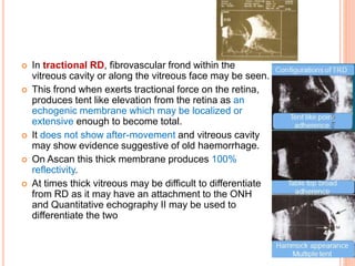
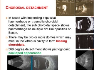
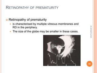
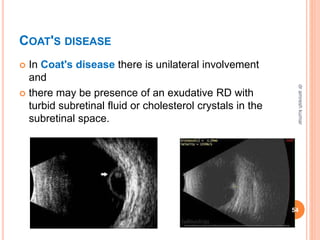
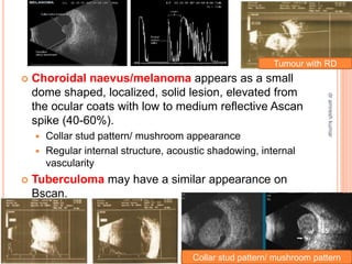
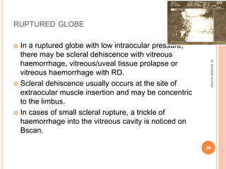
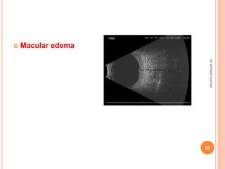
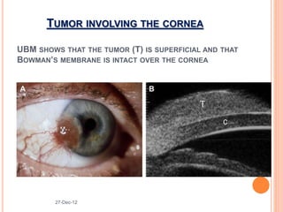
B-SCAN IN
OPHTHALMOLOGY
Dr. Amresh Kumar
Associate Professor
Department Of Ophthalmology
SBMCH, Hazaribag, Jharkhand
 B-scan or brightness modulation scan provides two
dimensional images of a series of dots and lines.
 B-scan provides the topographic information of
shape, location, extension , mobility, and gross
estimation of thickness of the tissue
2
dr
amresh
kumar
HISTORY
 It was first used in the field of ophthalmology by Mundt and
Hughes .
 Oksala et al reported the sound velocities in the various
components of the eye
 Baum and Greenwood came up with two dimensional,
immersion scan
 First commercially available B scan was developed by
Coleman et al in seventies
 The importance of the instrument and standardization of
technique was emphasised by Karl Ossoinig 3
dr
amresh
kumar
ULTRASONOGRAPHY IS USED FOR
 Biometry (Ascan)
 for axial length and corneal thickness measurement.
 Standardized Ascan (diagnostic)
 for the echostructure assessment.
 It is a part of the Bscan in most of the contemporary machines with
cross vector facility.
 Diagnostic Bscan (two dimensional)
 has to be coupled with the standardized Ascan to arrive at a correct
diagnosis.
 Doppler ultrasonography
 is especially important in vascular lesions with different blood flow
rates.
 Ultrasound biomicroscopy 4
dr
amresh
kumar
BACKGROUND
 1-2 MHz :abdominal
ultrasound
 8-10 MHz :ophthalmic
ultrasound
 (b scan)
 is best for posterior segment
 35-80 MHz: ultrasound
biomicroscopy
 best for cornea and anterior
segment 5
dr
amresh
kumar
B MODE(BRIGHTNESS) -PRINCIPLE
 2 D acoustic section
 An oscillating focussed sound
beam is emitted, passing through
the eye and imaging a slice of
tissue ,
 the echoes of which are
represented as multitude of dots
that together form an image on the
screen.
 Stronger the echo, brighter the dot.
6
dr
amresh
kumar
THE PROBE
 Bscan probes are thick, with a mark
 They emit focussed sound beam at a frequency of
10MHz.
 The mark on the probe indicates beam orientation
 Area towards which the mark is directed appears at the
top of the echogram on display screen.
 Probe tip: the white line on the far left side of display
 Echoes to right side of this line –ocular structures
opposite the tip
7
dr
amresh
kumar
 Mostly the Bscanning is done transpalpebrum with
slightly increased overall gain.
 Bscan probe can also be put directly on the
anaesthetized globe after applying eye speculum;
 Pictures obtained with Bscan probe are two
dimensional as compared to Ascan probe.
8
dr
amresh
kumar
To obtain high quality B scan pictures
 Ensure that Lesions are placed in the center of the
scanning beam
 The beam is directed perpendicular to the
interfaces at the area of interest
 Use lowest possible decibel gain consistent with the
maintenance of adequate intensity to optimize the
resolution of images.
9
dr
amresh
kumar
 B scan pictures can be obtained by
 axial,
 transverse and
 longitudinal sections.
axial
axial longitudinal
transverse
10
dr
amresh
kumar
transverse
AXIAL SECTION
 The patient fixates in the primary gaze
 Probe is placed on the globe and directed axially towards the posterior
pole.
 Depending on the clock hour location of the marker, axial-horizontal,
axial-vertical and axial oblique pictures are obtained.
 These sections demonstrate lesions at the posterior pole and the optic
nerve head.
 Marked attenuation of the sound beam by the crystalline lens
 not suitable for macular thickness measurement.
 The lens is avoided by placing the probe at the limbus.
11
dr
amresh
kumar
TRANSVERSE SECTION:
 Transverse section:
 The mark is kept parallel to the limbus and probe is
shifted from limbus to the fornix and also sideways.
 Produces a circumferential slice through several
meridians
 This scan gives the lateral extent of the lesion.
12
dr
amresh
kumar
LONGITUDINAL SECTION:
 Longitudinal section:
 The mark is kept at right angle to the limbus towards the
centre of cornea
 Helps in determining the antero-posterior limit of the
lesion.
 Best to determine attachment of membranes to optic
disc
13
dr
amresh
kumar
 During the procedure the probe is moved from
limbus to fornix in different clock hour meridians
and the picture seen is of diagonally opposite
meridian as follows:
14
dr
amresh
kumar
 Probe can be moved antero-posteriorly as well as
sideways.
 Patient is instructed to fix the gaze so that the
probe is perpendicular to the area being examined
15
dr
amresh
kumar
 For macular screening, the four basic Bscan
probe positions that allow perpendicular sound
beam exposure to the macula are
 horizontal axial,
 vertical transverse,
 longitudinal and
 vertical macula approaches.
16
dr
amresh
kumar
 With contact type of scanning there is a dead zone
of about 7.5mm adjacent to the probe, so that the
lesions in this region are missed.
 To visualize this area, one can keep the probe on
the opposite side at right angle or use immersion
scan technique.
dead
zone
17
dr
amresh
kumar
TOPOGRAPHIC ULTRASONOGRAPHY
 To determine the location, shape and extent of the
lesion
 A transverse scan to determine the maximal height
and lateral basal dimension of the lesion
 A longitudinal scan is done to determine the
anterior to posterior topographic feature of the
lesion
18
dr
amresh
kumar
QUANTITATIVE ULTRASONOGRAPHY
 Reflectivity :
 height of the spike on Ascan
 Internal structures:
 Homogenous : little variation in spike
 Heterogenous : marked variation in
spike
 A scan probe calibrated for tissue
sensitivity
 Sound directed perpendicular to the
lesion
 Sound attenuation (acoustic
shadowing): for calcification,
foreign bodies, bones 19
dr
amresh
kumar
KINETIC ULTRASONOGRAPHY
 Motion of a lesion
 Within a lesion
 Mobility ,
 vascularity and
 convection movement
 Mobility
 Change in gaze
 PVDs, RDs and choroidal detachments all exhibit their
own distinctive pattern of movement
 Used in conjunction with colour Doppler
instruments 20
dr
amresh
kumar
21
dr
amresh
kumar
USE OF BSCAN:
Some of the common eye conditions where
diagnostic ultrasonography is helpful are:
A. Dense cataract
B. vitreous haemorrhage,
C. leucokoria
D. vitritis/endophthalmitis
E. painful blind eye
F. before penetrating keratoplasty
G. intraocular tumors
H. oculo-orbital trauma
I. postoperative cases 22
dr
amresh
kumar
DENSE CATARACT
Dense
cataract
marked and
rapid decrease
in visual acuity,
afferent
pupillary
defect,
Rubeosis
iridis
myopia
uveitis
trauma
diabetes
mellitus
Do bscan and look for
1. retinal detachment(RD),
2. intraocular tumor with
calcification,
3. posterior staphyloma,
4. vitreous haemorrhage,
5. asteroid hyalosis,
6. optic nerve head cupping,
7. abnormal growth over optic
nerve head or
8. axial length disparity.
23
dr
amresh
kumar
USE OF BSCAN
 In vitreous haemorrhage,
 on echoevaluation one may pick up
 retinal tear with detachment,
 disciform degeneration,
 melanoma,
 fibrovascular fronds with tractional RD or
 subhyloid haemorrhage.
 The cause of leucokoria whether due to
 retinoblastoma,
 PHPV,
 Coat's disease,
 Retinopathy of prematurity or
 old haemorrhage can be deduced.
24
dr
amresh
kumar
USE OF BSCAN
 In vitritis/endophthalmitis,
 it helps in ruling out foreign body (FB) and rupture of
intraocular cyst and
 helps in assessing the response to treatment.
 In a painful blind eye,
 it is indicated to rule out
 uveal melanoma,
 old RD with chronic uveitis,
 intraocular/subretinal cyst,
 lens dislocation,
 failed RD surgery,
 inflamed phthisical eye, e.t.c.
25
dr
amresh
kumar
USE OF BSCAN
 Patients planned for penetrating keratoplasty with
opaque anterior segment.
 Patients with clear media
 where on indirect ophthalmoscopy suspicious lesions
suggestive of intraocular tumors like
 choroidal melanoma,
 haemangioma,
 metastatic carcinoma,
 osteoma, etc are seen.
 Orbital screening should be performed in patients with
abnormal choroidal folds and posterior scleritis.
26
dr
amresh
kumar
USE OF BSCAN
 In oculo-orbital trauma,
 it is imperative to look for
 sclerochoroidal rupture with RD,
 intraocular/orbital FB,
 lens displacement,
 optic nerve avulsion and
 orbital haemorrhage.
 In postoperative cases to assess
 endophthalmitis/toxic anterior segment syndrome,
 lens fragment/ intraocular lens(IOL) displacement into
the vitreous cavity,
 choroidal detachment/ haemorrhage,
 status of retina post RD surgery, etc. 27
dr
amresh
kumar
ECHODESCRIPTION
OF
COMMON INTRAOCULAR CONDITIONS:
28
dr
amresh
kumar
VITREOUS FLOATERS
 Appear as one or more echo dots of less brightness in
the mid /posterior vitreous cavity
 They show mobility with after movement display on
Bscan.
 On Ascan, these echodots have extremely low to low
reflectivity (2-20%) and to appreciate them better overall
gain may be increased by 56db.
29
dr
amresh
kumar
VITREOUS HAEMORRHAGE
 They appear as multiple fine echo opacities dusting
the vitreous body which do not extend beyond the
posterior vitreous border.
 They are usually attached to the retinal surface
 They may be fresh, resolving, organizing or
organized with membrane formation.
 To pick up fresh vitreous haemorrhage, the overall
gain can be increased by 10 db.
30
dr
amresh
kumar
 In older haemorrhage,
 echodots are denser
 show higher reflectivity (up to 60%) on Ascan.
 In resolving vitreous haemorrhage,
 echodots on Bscan show decrease in brightness
and numbers.
 Old organized vitreous haemorrhage
 can result in vitreous-membrane formation
showing echogenic lines on Bscan mimicking
RD
 The attachment of the echomembrane on/upto
the optic nerve head and Quantitative
echography II help to differentiate RD from
vitreous membranes 31
dr
amresh
kumar
SUBHYALOID HAEMORRHAGE
 Subhyaloid haemorrhage is situated typically at
the posterior pole between the anterior surface of
retina and posterior vitreous face.
 It may be fluid in nature or may get organized.
 Sometimes an organized old pre-retinal
haemorrhage may be seen in all the quadrants of
the globe.
32
dr
amresh
kumar
ENDOPHTHALMITIS
 The inflammatory cells are seen dotlike on Bscan,
 These are multiple, scattered diffusely or
 may be localised to the anterior, mid or the posterior
one third of the vitreous cavity depending on the
etiology.
33
dr
amresh
kumar
 On A scan, these dot like opacities show low to
medium reflectivity (10-60%).
 It is not possible to differentiate vitritis from vitreous
haemorrhage in still pictures unless clinical details
are available.
 These inflammatory cells organize very rapidly to
form vitreous membranes and therefore frequent
examinations should be performed.
34
dr
amresh
kumar
ASTEROID HYALOSIS
 It is characterized clinically by presence of calcium
crystals embedded in an amorphous matrix on Bscan
 It appears as multiple, densely packed, homogeneously
distributed echodense dots of medium to high reflectivity
(50-100%)
 These are usually localized to the core of vitreous body.
 One may find clear retrovitreal or pre-retinal space
35
dr
amresh
kumar
POSTERIOR VITREOUS DETACHMENT (PVD):
 PVD is seen as echogenic membrane concentric to the
globe, infront of the retinochoroidoscleral complex with clear
subvitreal space.
 It may be small, interrupted, peripheral or continuous and
total.
 If lined with red blood cells its echo density increases.
 On A scan, the reflectivity of this membrane is low if the
PVD is thin but it may be high if it is thick and lined with red
blood cells.
 PVD usually does not show attachment to the optic nerve
head.
36
dr
amresh
kumar
RETINAL DETACHMENT
 Retinal detachment means separation of
neurosensory retina from the pigmentary retina.
 It may be total/subtotal, localized/peripheral or
fresh/old with proliferative vitreoretinopathy(PVR)
changes.
37
dr
amresh
kumar
RETINAL DETACHMENT
 On Bscan, RD appears as echogenic dense
membrane, biconvex or biconcave with 100%
attachment at the optic nerve head (ONH)
and 90-100% reflectivity on Ascan.
 Attachment at ONH is not seen in localized,
peripheral RD where membrane is visible
only in a single quadrant.
 In uncomplicated cases, there is a clear
space between the detached retina and the
ocular coat spike indicating transudative
nature of the subretinal fluid.
38
dr
amresh
kumar
 Fine echodots may also be seen in the subretinal
space indicating the presence of haemorrhage or
debris.
 In PVR cases, vitreous body shows debris dots or
membrane formation depending upon its grade
and cystic degeneration may be present in an old
RD.
 After movement if present is suggestive of fresh
RD.
 In rhegmatogenous RD, retinal tears especially
operculated tears/ giant tears and even the trickle
of vitreous haemorrhage from the break site into
the vitreous cavity may be picked up.
39
dr
amresh
kumar
 In tractional RD, fibrovascular frond within the
vitreous cavity or along the vitreous face may be seen.
 This frond when exerts tractional force on the retina,
produces tent like elevation from the retina as an
echogenic membrane which may be localized or
extensive enough to become total.
 It does not show after-movement and vitreous cavity
may show evidence suggestive of old haemorrhage.
 On Ascan this thick membrane produces 100%
reflectivity.
 At times thick vitreous may be difficult to differentiate
from RD as it may have an attachment to the ONH
and Quantitative echography II may be used to
differentiate the two
40
dr
amresh
kumar
41
dr
amresh
kumar
SCLERAL EXPLANTS
 Scleral Explants are used in rhegmatogenous RD
surgeries where buckle or sponge is applied to
indent the globe.
 On Bscan they appear as echogenic spots with the
globe indentation towards the vitreous body and
echolucent spot (shadowing) behind the scleral
explant.
 The explant shows high reflectivity on Ascan.
Silicone buckle is less echodense in comparison to
the sponge.
Silicon
buckle
sponge
Scleral erosion
42
dr
amresh
kumar
VITREOUS EXPANDERS
 Vitreous expanders like silicone oil or perfluorocarbons
may be seen in operated RD cases.
 Emulsified silicone oil produces marked sound
attenuation hindering the visualization of posterior
segment.
 It also results in a larger vitreous cavity which is
relatively echofree.
 Perfluorocarbons on the other hand show multiple,
highly reflective liquid bubbles in the posterior vitreous
Emulsified silicone oil Perfluorocarbons
43
dr
amresh
kumar
CHOROIDAL DETACHMENT
 Choroidal detachment is usually in the
periphery and may be localized or total.
 It is seen as dome shaped elevation with clear
sub choroidal space on Bscan
 and 90-100% double peaked tall spike on
Ascan.
 There is none or very little after movement on
kinetic echography.
44
dr
amresh
kumar
CHOROIDAL DETACHMENT
 In cases with impending expulsive
haemorrhage or traumatic choroidal
detachment, the sub choroidal space shows
haemorrhage as multiple dot like opacities on
Bscan.
 There may be two or more domes which may
meet in the vitreous cavity to form kissing
choroidals.
 360 degree detachment shows pathognomic
scalloped appearance
45
dr
amresh
kumar
46
dr
amresh
kumar
 Retinal tear
 Retinoschisis:
 Moderately elevated thin membrane shaped echo
Retinal tear with free
superior end
Vitreous attached to the
tear
tear
47
dr
amresh
kumar
INTRAOCULAR TUMORS
 Intraocular tumors which commonly require Bscan
evaluation are
 retinoblastoma,
 choroidal melanoma,
 hemangioma,
 metastasis,
 diktyoma and
 Osteoma
 Bscan helps in measurement of tumour dimensions,
differentiation ,extrascleral extension, size , assessing
tumour growth or regression
 help in distinguishing solid from cystic lesions
48
dr
amresh
kumar
RETINOBLASTOMA
 Retinoblastoma is seen as a solid tumor arising
from the retinal layer obliterating the vitreous
cavity.
 Calcification within the tumor mass is typical of
retinoblastoma.
 There may be shadowing effect behind the lesion
in the orbital mass.
 Concomitant RD may be sometimes present.
49
dr
amresh
kumar
RETINOBLASTOMA
 On A scan, spikes with moderate internal
reflectivity may be seen
 but in presence of necrosis and calcification,
highly reflective, irregular spikes are observed.
 Sound attenuation is moderate to high.
 The globe is usually normal in size except in
glaucomatous stage when it becomes enlarged,
50
dr
amresh
kumar
OSTEOMA
51
dr
amresh
kumar
 In persistent hyperplasic primary vitreous,
 the globe size may be smaller and
 the vitreous cavity shows persistence of the primary
vascular system seen as echo membranous track from
optic nerve head to the back of the lens
52
dr
amresh
kumar
RETINOPATHY OF PREMATURITY
 Retinopathy of prematurity
 is characterised by multiple vitreous membranes and
RD in the periphery.
 The size of the globe may be smaller in these cases.
53
dr
amresh
kumar
COAT'S DISEASE
 In Coat's disease there is unilateral involvement
and
 there may be presence of an exudative RD with
turbid subretinal fluid or cholesterol crystals in the
subretinal space.
54
dr
amresh
kumar
 Choroidal naevus/melanoma appears as a small
dome shaped, localized, solid lesion, elevated from
the ocular coats with low to medium reflective Ascan
spike (40-60%).
 Collar stud pattern/ mushroom appearance
 Regular internal structure, acoustic shadowing, internal
vascularity
 Tuberculoma may have a similar appearance on
Bscan.
Collar stud pattern/ mushroom pattern
Tumour with RD
55
dr
amresh
kumar
RUPTURED GLOBE
 In a ruptured globe with low intraocular pressure,
there may be scleral dehiscence with vitreous
haemorrhage, vitreous/uveal tissue prolapse or
vitreous haemorrhage with RD.
 Scleral dehiscence usually occurs at the site of
extraocular muscle insertion and may be concentric
to the limbus.
 In cases of small scleral rupture, a trickle of
haemorrhage into the vitreous cavity is noticed on
Bscan.
56
dr
amresh
kumar
HAEMOPHTHALMOS
 Hyphaema, vitreous haemorrhage with choroidal
haemorrhage and scleral rupture with orbital
haemorrhage may be seen in combination and the
condition may appear as haemophthalmos.
 Black eye (lid haemorrhage) may coexist with it.
57
dr
amresh
kumar
 Posteriorly dislocated crystalline lens into the vitreous
cavity
 It is seen as a biconvex body which may be mobile or fixed.
 Lens fragment in vitreous usually produces vitritis.
 The intraocular lens in vitreous cavity appears like a FB and
shows high reflectivity and shadowing effect behind it.
intraocular lens
crystalline lens
Lens fragment
58
dr
amresh
kumar
PHTHISIS BULBI
 Phthisis bulbi
 The globe is small, soft, deshaped and
 There is thickened retinochoroidal complex.
 Intraocular calcification or bone formation may
occur in choroidal layer in long standing cases
which is better appreciated on decreasing the gain
by 15-20db.
 Retro globe shadowing may also be visible.
59
dr
amresh
kumar
IOFB
 IOFB are seen as echodense spots with a 100%
reflectivity on Ascan spike irrespective of the nature
of the FB and
 ultrasonography enables its exact sizing and
localization.
 Shadowing effect is usually seen.
 Decreasing the gain on the machine by 10db helps
in differentiating it from dense blood clot and lens
fragment.
60
dr
amresh
kumar
IOFB
 Spherical FB like gunshot pellets, have an
anterior and posterior surface and between them
there are multiple internal
reverberations/echoes.
 These echoes are seen as echogenic opacities
with a wedge shaped trail of spikes.
 The trail disappears on decreasing the overall
gain of machine but the initial echodense spot
remains as such
61
dr
amresh
kumar
OPTIC NERVE EVALUATION
 general topography ,relationship to structures ,
optic disc anomalies and alteration in contour of the
globe
 the subarachnoid space surrounding optic nerve
appears as echoluscent crecentric or circle around
the nerve called ‘ DOUGHNUT SIGN
62
dr
amresh
kumar
 Macular edema
63
dr
amresh
kumar
 Retinal coloboma
 is a congenital abnormality seen in the inferonasal
quadrant as defect in the retinochoroidal layer of the
globe on Bscan.
 Optic disc coloboma :
 If the coloboma involves the ONH region, there is
absence of ONH.
Optic disc coloboma
64
dr
amresh
kumar
POSTERIOR STAPHYLOMA
 It appears as a sudden bowing backward of the
globe with thinning of the retinochoroidal layer.
 It is usually seen at the posterior pole and the axial
length of the globe is increased, indicating axial
myopia.
 There may be presence of vitreous debri.
Posterior staphyloma
65
dr
amresh
kumar
POST OPERATIVE ENDOPHTHALMITIS
 In endophthalmitis there is severe vitritis and
exudation in the vitreous cavity.
 Bscan is useful in evaluating the response to
intravitreal injection in endophthalmitis
66
dr
amresh
kumar
Cysticercosis
 It is common in vitreous cavity, subretinal space
and sub conjunctival space
 Bscan reveals a well defined cystic lesion with
clear contents and a hyperechoic area
suggestive of scolex.
 Serial echography helps in follow up
scolex
Cystic
lesion
67
dr
amresh
kumar
 Posterior scleritis :
 T-sign collection of fluid in sub tenon space
T-Sign in posterior scleritis 68
dr
amresh
kumar
 Reverberation artefacts
 insufficient coupling fluid
 entrapment of air between the probe and eye
 display of bright echoes representing multiple
signals
 ANGLE OF INCIDENCE ARTEFACT
69
dr
amresh
kumar
Thank you
70
dr
amresh
kumar
UBM
27-Dec-12
CILIARY BODY TUMOR
27-Dec-12
IRIS TUMOR
27-Dec-12
TUMOR INVOLVING THE CORNEA
UBM SHOWS THAT THE TUMOR (T) IS SUPERFICIAL AND THAT
BOWMAN’S MEMBRANE IS INTACT OVER THE CORNEA
27-Dec-12
UBM SHOWS DEEP INVOLVEMENT OF ALL
CORNEAL LAYERS (T)
27-Dec-12
IRIDOCILIARY CYST
27-Dec-12
PETER’S ANOMALY
27-Dec-12
IRIDODIALYSIS
27-Dec-12
CYCLODIALYSIS
27-Dec-12

B-SCAN IN OPHTHALMOLOGY.pptx

  • 1.
    B-SCAN IN OPHTHALMOLOGY Dr. AmreshKumar Associate Professor Department Of Ophthalmology SBMCH, Hazaribag, Jharkhand
  • 2.
     B-scan orbrightness modulation scan provides two dimensional images of a series of dots and lines.  B-scan provides the topographic information of shape, location, extension , mobility, and gross estimation of thickness of the tissue 2 dr amresh kumar
  • 3.
    HISTORY  It wasfirst used in the field of ophthalmology by Mundt and Hughes .  Oksala et al reported the sound velocities in the various components of the eye  Baum and Greenwood came up with two dimensional, immersion scan  First commercially available B scan was developed by Coleman et al in seventies  The importance of the instrument and standardization of technique was emphasised by Karl Ossoinig 3 dr amresh kumar
  • 4.
    ULTRASONOGRAPHY IS USEDFOR  Biometry (Ascan)  for axial length and corneal thickness measurement.  Standardized Ascan (diagnostic)  for the echostructure assessment.  It is a part of the Bscan in most of the contemporary machines with cross vector facility.  Diagnostic Bscan (two dimensional)  has to be coupled with the standardized Ascan to arrive at a correct diagnosis.  Doppler ultrasonography  is especially important in vascular lesions with different blood flow rates.  Ultrasound biomicroscopy 4 dr amresh kumar
  • 5.
    BACKGROUND  1-2 MHz:abdominal ultrasound  8-10 MHz :ophthalmic ultrasound  (b scan)  is best for posterior segment  35-80 MHz: ultrasound biomicroscopy  best for cornea and anterior segment 5 dr amresh kumar
  • 6.
    B MODE(BRIGHTNESS) -PRINCIPLE 2 D acoustic section  An oscillating focussed sound beam is emitted, passing through the eye and imaging a slice of tissue ,  the echoes of which are represented as multitude of dots that together form an image on the screen.  Stronger the echo, brighter the dot. 6 dr amresh kumar
  • 7.
    THE PROBE  Bscanprobes are thick, with a mark  They emit focussed sound beam at a frequency of 10MHz.  The mark on the probe indicates beam orientation  Area towards which the mark is directed appears at the top of the echogram on display screen.  Probe tip: the white line on the far left side of display  Echoes to right side of this line –ocular structures opposite the tip 7 dr amresh kumar
  • 8.
     Mostly theBscanning is done transpalpebrum with slightly increased overall gain.  Bscan probe can also be put directly on the anaesthetized globe after applying eye speculum;  Pictures obtained with Bscan probe are two dimensional as compared to Ascan probe. 8 dr amresh kumar
  • 9.
    To obtain highquality B scan pictures  Ensure that Lesions are placed in the center of the scanning beam  The beam is directed perpendicular to the interfaces at the area of interest  Use lowest possible decibel gain consistent with the maintenance of adequate intensity to optimize the resolution of images. 9 dr amresh kumar
  • 10.
     B scanpictures can be obtained by  axial,  transverse and  longitudinal sections. axial axial longitudinal transverse 10 dr amresh kumar transverse
  • 11.
    AXIAL SECTION  Thepatient fixates in the primary gaze  Probe is placed on the globe and directed axially towards the posterior pole.  Depending on the clock hour location of the marker, axial-horizontal, axial-vertical and axial oblique pictures are obtained.  These sections demonstrate lesions at the posterior pole and the optic nerve head.  Marked attenuation of the sound beam by the crystalline lens  not suitable for macular thickness measurement.  The lens is avoided by placing the probe at the limbus. 11 dr amresh kumar
  • 12.
    TRANSVERSE SECTION:  Transversesection:  The mark is kept parallel to the limbus and probe is shifted from limbus to the fornix and also sideways.  Produces a circumferential slice through several meridians  This scan gives the lateral extent of the lesion. 12 dr amresh kumar
  • 13.
    LONGITUDINAL SECTION:  Longitudinalsection:  The mark is kept at right angle to the limbus towards the centre of cornea  Helps in determining the antero-posterior limit of the lesion.  Best to determine attachment of membranes to optic disc 13 dr amresh kumar
  • 14.
     During theprocedure the probe is moved from limbus to fornix in different clock hour meridians and the picture seen is of diagonally opposite meridian as follows: 14 dr amresh kumar
  • 15.
     Probe canbe moved antero-posteriorly as well as sideways.  Patient is instructed to fix the gaze so that the probe is perpendicular to the area being examined 15 dr amresh kumar
  • 16.
     For macularscreening, the four basic Bscan probe positions that allow perpendicular sound beam exposure to the macula are  horizontal axial,  vertical transverse,  longitudinal and  vertical macula approaches. 16 dr amresh kumar
  • 17.
     With contacttype of scanning there is a dead zone of about 7.5mm adjacent to the probe, so that the lesions in this region are missed.  To visualize this area, one can keep the probe on the opposite side at right angle or use immersion scan technique. dead zone 17 dr amresh kumar
  • 18.
    TOPOGRAPHIC ULTRASONOGRAPHY  Todetermine the location, shape and extent of the lesion  A transverse scan to determine the maximal height and lateral basal dimension of the lesion  A longitudinal scan is done to determine the anterior to posterior topographic feature of the lesion 18 dr amresh kumar
  • 19.
    QUANTITATIVE ULTRASONOGRAPHY  Reflectivity:  height of the spike on Ascan  Internal structures:  Homogenous : little variation in spike  Heterogenous : marked variation in spike  A scan probe calibrated for tissue sensitivity  Sound directed perpendicular to the lesion  Sound attenuation (acoustic shadowing): for calcification, foreign bodies, bones 19 dr amresh kumar
  • 20.
    KINETIC ULTRASONOGRAPHY  Motionof a lesion  Within a lesion  Mobility ,  vascularity and  convection movement  Mobility  Change in gaze  PVDs, RDs and choroidal detachments all exhibit their own distinctive pattern of movement  Used in conjunction with colour Doppler instruments 20 dr amresh kumar
  • 21.
  • 22.
    USE OF BSCAN: Someof the common eye conditions where diagnostic ultrasonography is helpful are: A. Dense cataract B. vitreous haemorrhage, C. leucokoria D. vitritis/endophthalmitis E. painful blind eye F. before penetrating keratoplasty G. intraocular tumors H. oculo-orbital trauma I. postoperative cases 22 dr amresh kumar
  • 23.
    DENSE CATARACT Dense cataract marked and rapiddecrease in visual acuity, afferent pupillary defect, Rubeosis iridis myopia uveitis trauma diabetes mellitus Do bscan and look for 1. retinal detachment(RD), 2. intraocular tumor with calcification, 3. posterior staphyloma, 4. vitreous haemorrhage, 5. asteroid hyalosis, 6. optic nerve head cupping, 7. abnormal growth over optic nerve head or 8. axial length disparity. 23 dr amresh kumar
  • 24.
    USE OF BSCAN In vitreous haemorrhage,  on echoevaluation one may pick up  retinal tear with detachment,  disciform degeneration,  melanoma,  fibrovascular fronds with tractional RD or  subhyloid haemorrhage.  The cause of leucokoria whether due to  retinoblastoma,  PHPV,  Coat's disease,  Retinopathy of prematurity or  old haemorrhage can be deduced. 24 dr amresh kumar
  • 25.
    USE OF BSCAN In vitritis/endophthalmitis,  it helps in ruling out foreign body (FB) and rupture of intraocular cyst and  helps in assessing the response to treatment.  In a painful blind eye,  it is indicated to rule out  uveal melanoma,  old RD with chronic uveitis,  intraocular/subretinal cyst,  lens dislocation,  failed RD surgery,  inflamed phthisical eye, e.t.c. 25 dr amresh kumar
  • 26.
    USE OF BSCAN Patients planned for penetrating keratoplasty with opaque anterior segment.  Patients with clear media  where on indirect ophthalmoscopy suspicious lesions suggestive of intraocular tumors like  choroidal melanoma,  haemangioma,  metastatic carcinoma,  osteoma, etc are seen.  Orbital screening should be performed in patients with abnormal choroidal folds and posterior scleritis. 26 dr amresh kumar
  • 27.
    USE OF BSCAN In oculo-orbital trauma,  it is imperative to look for  sclerochoroidal rupture with RD,  intraocular/orbital FB,  lens displacement,  optic nerve avulsion and  orbital haemorrhage.  In postoperative cases to assess  endophthalmitis/toxic anterior segment syndrome,  lens fragment/ intraocular lens(IOL) displacement into the vitreous cavity,  choroidal detachment/ haemorrhage,  status of retina post RD surgery, etc. 27 dr amresh kumar
  • 28.
  • 29.
    VITREOUS FLOATERS  Appearas one or more echo dots of less brightness in the mid /posterior vitreous cavity  They show mobility with after movement display on Bscan.  On Ascan, these echodots have extremely low to low reflectivity (2-20%) and to appreciate them better overall gain may be increased by 56db. 29 dr amresh kumar
  • 30.
    VITREOUS HAEMORRHAGE  Theyappear as multiple fine echo opacities dusting the vitreous body which do not extend beyond the posterior vitreous border.  They are usually attached to the retinal surface  They may be fresh, resolving, organizing or organized with membrane formation.  To pick up fresh vitreous haemorrhage, the overall gain can be increased by 10 db. 30 dr amresh kumar
  • 31.
     In olderhaemorrhage,  echodots are denser  show higher reflectivity (up to 60%) on Ascan.  In resolving vitreous haemorrhage,  echodots on Bscan show decrease in brightness and numbers.  Old organized vitreous haemorrhage  can result in vitreous-membrane formation showing echogenic lines on Bscan mimicking RD  The attachment of the echomembrane on/upto the optic nerve head and Quantitative echography II help to differentiate RD from vitreous membranes 31 dr amresh kumar
  • 32.
    SUBHYALOID HAEMORRHAGE  Subhyaloidhaemorrhage is situated typically at the posterior pole between the anterior surface of retina and posterior vitreous face.  It may be fluid in nature or may get organized.  Sometimes an organized old pre-retinal haemorrhage may be seen in all the quadrants of the globe. 32 dr amresh kumar
  • 33.
    ENDOPHTHALMITIS  The inflammatorycells are seen dotlike on Bscan,  These are multiple, scattered diffusely or  may be localised to the anterior, mid or the posterior one third of the vitreous cavity depending on the etiology. 33 dr amresh kumar
  • 34.
     On Ascan, these dot like opacities show low to medium reflectivity (10-60%).  It is not possible to differentiate vitritis from vitreous haemorrhage in still pictures unless clinical details are available.  These inflammatory cells organize very rapidly to form vitreous membranes and therefore frequent examinations should be performed. 34 dr amresh kumar
  • 35.
    ASTEROID HYALOSIS  Itis characterized clinically by presence of calcium crystals embedded in an amorphous matrix on Bscan  It appears as multiple, densely packed, homogeneously distributed echodense dots of medium to high reflectivity (50-100%)  These are usually localized to the core of vitreous body.  One may find clear retrovitreal or pre-retinal space 35 dr amresh kumar
  • 36.
    POSTERIOR VITREOUS DETACHMENT(PVD):  PVD is seen as echogenic membrane concentric to the globe, infront of the retinochoroidoscleral complex with clear subvitreal space.  It may be small, interrupted, peripheral or continuous and total.  If lined with red blood cells its echo density increases.  On A scan, the reflectivity of this membrane is low if the PVD is thin but it may be high if it is thick and lined with red blood cells.  PVD usually does not show attachment to the optic nerve head. 36 dr amresh kumar
  • 37.
    RETINAL DETACHMENT  Retinaldetachment means separation of neurosensory retina from the pigmentary retina.  It may be total/subtotal, localized/peripheral or fresh/old with proliferative vitreoretinopathy(PVR) changes. 37 dr amresh kumar
  • 38.
    RETINAL DETACHMENT  OnBscan, RD appears as echogenic dense membrane, biconvex or biconcave with 100% attachment at the optic nerve head (ONH) and 90-100% reflectivity on Ascan.  Attachment at ONH is not seen in localized, peripheral RD where membrane is visible only in a single quadrant.  In uncomplicated cases, there is a clear space between the detached retina and the ocular coat spike indicating transudative nature of the subretinal fluid. 38 dr amresh kumar
  • 39.
     Fine echodotsmay also be seen in the subretinal space indicating the presence of haemorrhage or debris.  In PVR cases, vitreous body shows debris dots or membrane formation depending upon its grade and cystic degeneration may be present in an old RD.  After movement if present is suggestive of fresh RD.  In rhegmatogenous RD, retinal tears especially operculated tears/ giant tears and even the trickle of vitreous haemorrhage from the break site into the vitreous cavity may be picked up. 39 dr amresh kumar
  • 40.
     In tractionalRD, fibrovascular frond within the vitreous cavity or along the vitreous face may be seen.  This frond when exerts tractional force on the retina, produces tent like elevation from the retina as an echogenic membrane which may be localized or extensive enough to become total.  It does not show after-movement and vitreous cavity may show evidence suggestive of old haemorrhage.  On Ascan this thick membrane produces 100% reflectivity.  At times thick vitreous may be difficult to differentiate from RD as it may have an attachment to the ONH and Quantitative echography II may be used to differentiate the two 40 dr amresh kumar
  • 41.
  • 42.
    SCLERAL EXPLANTS  ScleralExplants are used in rhegmatogenous RD surgeries where buckle or sponge is applied to indent the globe.  On Bscan they appear as echogenic spots with the globe indentation towards the vitreous body and echolucent spot (shadowing) behind the scleral explant.  The explant shows high reflectivity on Ascan. Silicone buckle is less echodense in comparison to the sponge. Silicon buckle sponge Scleral erosion 42 dr amresh kumar
  • 43.
    VITREOUS EXPANDERS  Vitreousexpanders like silicone oil or perfluorocarbons may be seen in operated RD cases.  Emulsified silicone oil produces marked sound attenuation hindering the visualization of posterior segment.  It also results in a larger vitreous cavity which is relatively echofree.  Perfluorocarbons on the other hand show multiple, highly reflective liquid bubbles in the posterior vitreous Emulsified silicone oil Perfluorocarbons 43 dr amresh kumar
  • 44.
    CHOROIDAL DETACHMENT  Choroidaldetachment is usually in the periphery and may be localized or total.  It is seen as dome shaped elevation with clear sub choroidal space on Bscan  and 90-100% double peaked tall spike on Ascan.  There is none or very little after movement on kinetic echography. 44 dr amresh kumar
  • 45.
    CHOROIDAL DETACHMENT  Incases with impending expulsive haemorrhage or traumatic choroidal detachment, the sub choroidal space shows haemorrhage as multiple dot like opacities on Bscan.  There may be two or more domes which may meet in the vitreous cavity to form kissing choroidals.  360 degree detachment shows pathognomic scalloped appearance 45 dr amresh kumar
  • 46.
  • 47.
     Retinal tear Retinoschisis:  Moderately elevated thin membrane shaped echo Retinal tear with free superior end Vitreous attached to the tear tear 47 dr amresh kumar
  • 48.
    INTRAOCULAR TUMORS  Intraoculartumors which commonly require Bscan evaluation are  retinoblastoma,  choroidal melanoma,  hemangioma,  metastasis,  diktyoma and  Osteoma  Bscan helps in measurement of tumour dimensions, differentiation ,extrascleral extension, size , assessing tumour growth or regression  help in distinguishing solid from cystic lesions 48 dr amresh kumar
  • 49.
    RETINOBLASTOMA  Retinoblastoma isseen as a solid tumor arising from the retinal layer obliterating the vitreous cavity.  Calcification within the tumor mass is typical of retinoblastoma.  There may be shadowing effect behind the lesion in the orbital mass.  Concomitant RD may be sometimes present. 49 dr amresh kumar
  • 50.
    RETINOBLASTOMA  On Ascan, spikes with moderate internal reflectivity may be seen  but in presence of necrosis and calcification, highly reflective, irregular spikes are observed.  Sound attenuation is moderate to high.  The globe is usually normal in size except in glaucomatous stage when it becomes enlarged, 50 dr amresh kumar
  • 51.
  • 52.
     In persistenthyperplasic primary vitreous,  the globe size may be smaller and  the vitreous cavity shows persistence of the primary vascular system seen as echo membranous track from optic nerve head to the back of the lens 52 dr amresh kumar
  • 53.
    RETINOPATHY OF PREMATURITY Retinopathy of prematurity  is characterised by multiple vitreous membranes and RD in the periphery.  The size of the globe may be smaller in these cases. 53 dr amresh kumar
  • 54.
    COAT'S DISEASE  InCoat's disease there is unilateral involvement and  there may be presence of an exudative RD with turbid subretinal fluid or cholesterol crystals in the subretinal space. 54 dr amresh kumar
  • 55.
     Choroidal naevus/melanomaappears as a small dome shaped, localized, solid lesion, elevated from the ocular coats with low to medium reflective Ascan spike (40-60%).  Collar stud pattern/ mushroom appearance  Regular internal structure, acoustic shadowing, internal vascularity  Tuberculoma may have a similar appearance on Bscan. Collar stud pattern/ mushroom pattern Tumour with RD 55 dr amresh kumar
  • 56.
    RUPTURED GLOBE  Ina ruptured globe with low intraocular pressure, there may be scleral dehiscence with vitreous haemorrhage, vitreous/uveal tissue prolapse or vitreous haemorrhage with RD.  Scleral dehiscence usually occurs at the site of extraocular muscle insertion and may be concentric to the limbus.  In cases of small scleral rupture, a trickle of haemorrhage into the vitreous cavity is noticed on Bscan. 56 dr amresh kumar
  • 57.
    HAEMOPHTHALMOS  Hyphaema, vitreoushaemorrhage with choroidal haemorrhage and scleral rupture with orbital haemorrhage may be seen in combination and the condition may appear as haemophthalmos.  Black eye (lid haemorrhage) may coexist with it. 57 dr amresh kumar
  • 58.
     Posteriorly dislocatedcrystalline lens into the vitreous cavity  It is seen as a biconvex body which may be mobile or fixed.  Lens fragment in vitreous usually produces vitritis.  The intraocular lens in vitreous cavity appears like a FB and shows high reflectivity and shadowing effect behind it. intraocular lens crystalline lens Lens fragment 58 dr amresh kumar
  • 59.
    PHTHISIS BULBI  Phthisisbulbi  The globe is small, soft, deshaped and  There is thickened retinochoroidal complex.  Intraocular calcification or bone formation may occur in choroidal layer in long standing cases which is better appreciated on decreasing the gain by 15-20db.  Retro globe shadowing may also be visible. 59 dr amresh kumar
  • 60.
    IOFB  IOFB areseen as echodense spots with a 100% reflectivity on Ascan spike irrespective of the nature of the FB and  ultrasonography enables its exact sizing and localization.  Shadowing effect is usually seen.  Decreasing the gain on the machine by 10db helps in differentiating it from dense blood clot and lens fragment. 60 dr amresh kumar
  • 61.
    IOFB  Spherical FBlike gunshot pellets, have an anterior and posterior surface and between them there are multiple internal reverberations/echoes.  These echoes are seen as echogenic opacities with a wedge shaped trail of spikes.  The trail disappears on decreasing the overall gain of machine but the initial echodense spot remains as such 61 dr amresh kumar
  • 62.
    OPTIC NERVE EVALUATION general topography ,relationship to structures , optic disc anomalies and alteration in contour of the globe  the subarachnoid space surrounding optic nerve appears as echoluscent crecentric or circle around the nerve called ‘ DOUGHNUT SIGN 62 dr amresh kumar
  • 63.
  • 64.
     Retinal coloboma is a congenital abnormality seen in the inferonasal quadrant as defect in the retinochoroidal layer of the globe on Bscan.  Optic disc coloboma :  If the coloboma involves the ONH region, there is absence of ONH. Optic disc coloboma 64 dr amresh kumar
  • 65.
    POSTERIOR STAPHYLOMA  Itappears as a sudden bowing backward of the globe with thinning of the retinochoroidal layer.  It is usually seen at the posterior pole and the axial length of the globe is increased, indicating axial myopia.  There may be presence of vitreous debri. Posterior staphyloma 65 dr amresh kumar
  • 66.
    POST OPERATIVE ENDOPHTHALMITIS In endophthalmitis there is severe vitritis and exudation in the vitreous cavity.  Bscan is useful in evaluating the response to intravitreal injection in endophthalmitis 66 dr amresh kumar
  • 67.
    Cysticercosis  It iscommon in vitreous cavity, subretinal space and sub conjunctival space  Bscan reveals a well defined cystic lesion with clear contents and a hyperechoic area suggestive of scolex.  Serial echography helps in follow up scolex Cystic lesion 67 dr amresh kumar
  • 68.
     Posterior scleritis:  T-sign collection of fluid in sub tenon space T-Sign in posterior scleritis 68 dr amresh kumar
  • 69.
     Reverberation artefacts insufficient coupling fluid  entrapment of air between the probe and eye  display of bright echoes representing multiple signals  ANGLE OF INCIDENCE ARTEFACT 69 dr amresh kumar
  • 70.
  • 71.
  • 72.
  • 73.
  • 74.
    TUMOR INVOLVING THECORNEA UBM SHOWS THAT THE TUMOR (T) IS SUPERFICIAL AND THAT BOWMAN’S MEMBRANE IS INTACT OVER THE CORNEA 27-Dec-12
  • 75.
    UBM SHOWS DEEPINVOLVEMENT OF ALL CORNEAL LAYERS (T) 27-Dec-12
  • 76.
  • 77.
  • 78.
  • 79.