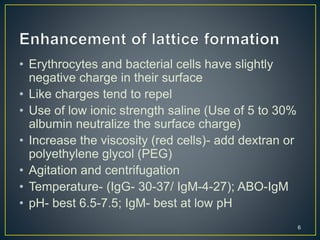
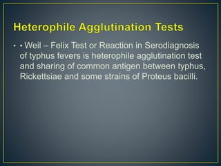
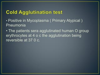
Downloaded 334 times


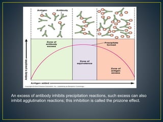
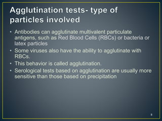
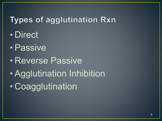
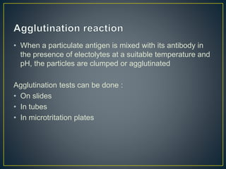

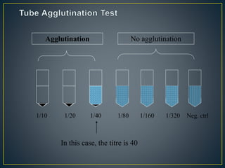
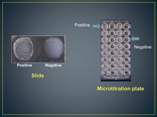
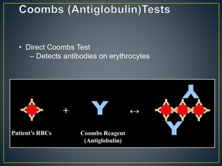
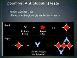
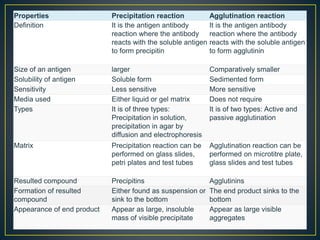


The document discusses the principles and methods of agglutination reactions, highlighting their relevance in serological testing for detecting antigens and antibodies. It explains various types of agglutination tests, including direct, passive, and reverse passive agglutination, with examples of their applications in diagnosing diseases. Additionally, it contrasts agglutination with precipitation reactions regarding their sensitivity and the nature of the formed complexes.


![PERI-PROSTHETIC FRACTURE NAIL-PLATE CONSTRUCT [NPC].pptx](https://cdn.slidesharecdn.com/ss_thumbnails/drarunkumardrmohamedashrafperiprostheticfrasturenail-plateconstructnpc-260209164459-7e9d15a1-thumbnail.jpg?width=640&height=640&fit=bounds)


