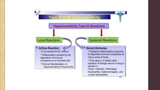
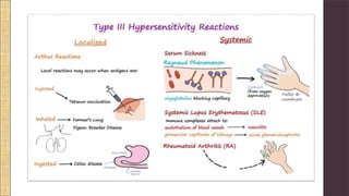
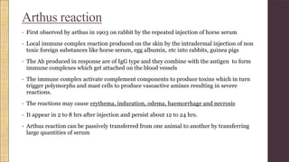
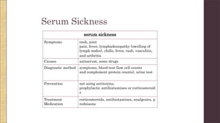
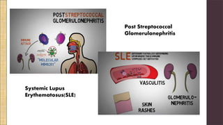
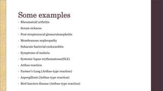
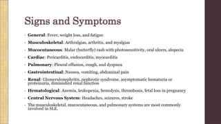
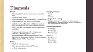
Hypersenstivity type3 is an Immune-complex mediated hypersensitivity.Hypersensitivity denotes a condition in which an exaggerated immune response of a host to non-harmful antigens that leads to destruction of host tissues.
3. Hypersensitivity
Disorder of our immune system
The term hypersensitivity denotes a condition in which an exaggerated immune response
of a host to non-harmful antigens that leads to destruction of host tissues.
Includes allergies and autoimmunity
Hypersensitivity reactions can be classified into four types.
• Type I - IgE mediated immediate reaction
• Type II- Antibody-mediated cytotoxic reaction (IgG or IgM
antibodies)
• Type III- Immune complex-mediated reaction
• Type IV- Cell-mediated, delayed hypersensitivity reaction
4. • Immediate- Hypersensitivity
reactions.
• theyoccurwithin24hours.
• Immunoglobulin-mediated
hypersensitivity (immediate)
reactions.
Type 1
Type 2
Type 3
Type 4
• Delayedhypersensitivityreactions.
• Itusuallyoccursmorethan12hoursafter
exposuretotheallergen,withamaximal
reactiontimebetween48and72hours.
• Lymphoidcell-mediated orsimplycell
mediatedhypersensitivity (delayed-type).
5.
6. • Immune-complex mediated hypersensitivity.
• In type III hypersensitivity reaction, an abnormal immune response is mediated by the
formation of antigen-antibody aggregates called "immune complexes."
• They can precipitate in various tissues such as skin, joints, vessels, or glomeruli, and
trigger the classical complement pathway.
• Complement activation leads to the recruitment of inflammatory cells (monocytes and
neutrophils) that release lysosomal enzymes and free radicals at the site of immune
complexes, causing tissue damage.
• “Antibody (IgG) binds to soluble antigen, forming a circulating immune complex. This is
often deposited in the vessel walls of the joints and kidney, initiating a local inflammatory
reaction”.
Hypersensitivity Type III
7. • The most common diseases involving a type III hypersensitivity reaction are
serum sickness, post-streptococcal glomerulonephritis, systemic lupus
erythematosus, farmers' lung (hypersensitivity pneumonitis), and
rheumatoid arthritis.
• The principle feature that separates type III reactions from other
hypersensitivity reactions is that- in type III reaction, the antigen-antibody
complexes are pre-formed in the circulation before their deposition in
tissues.
8. The process takes place in three steps:
PHASE 1 : Immune Complex Formation
PHASE 2 : Immune Complex Deposition
PHASE 3 : Inflammation and Tissue Injury
9. Immune complex formation:
• Endogenous or exogenous antigen exposure triggers an antibody formation.
• Exogenous antigens are foreign proteins such as an infectious microbe or a
pharmaceutical product.
• Endogenous antigens are self-antigens against which the autoantibodies are
generated (autoimmunity).
• In both cases, the antigens bind to antibodies, forming circulating immune
complexes, which can later migrate out of plasma and deposit in host tissues.
10. Immune complex
deposition:
• Excess of antigens cause formation of numerous
small sized aggregates of immune complexes.
• They are difficult to remove by phagocytosis and
get deposited in various tissue spaces and
activate our immune system by complement
system.
Inflammatory reaction:
• After the deposition of the immune complexes, the final step is the activation of the
classical pathway, leading to the release of C3a and C5a, which then recruit
macrophages and neutrophils, and causes inflammatory damage to tissues.
• Depending on the site, symptoms of vasculitis (blood vessels), arthritis (joints), or
glomerulonephritis (glomeruli) develop.
11.
12. Mechanism(summarized)
1. Antigen-antibody complexes are formed when antibodies bind to antigens.
2. Incase the complex is not cleared by normal process of phagocytosis, the immune complexes persist in
the circulation.
3. The immune complexes subsequently deposit in tissues.
4. The tissue deposited complexes activate the classical complement cascade.
5. The complement fragments (e.g. C3a and C5a) that form during complement activation activate a variety
of potent mediators of inflammation causing an influx of neutrophils and monocytes to the site of
deposition.
6. C3b as an opsonin attracts neutrophils, which then release lysosomal enzymes. C5a as a chemoattractant
brings in neutrophils.
7. The attracted neutrophils attempt to engulf the immune complexes. Since the complexes are deposited
over the tissues, the neutrophils do not succeed.
8. Consequently, the neutrophils release a number of substances like prostaglandins, lysosomal enzymes,
and free oxygen radicals over the complexes causing damage to the tissues at the site of immune
complex deposition.
9. Additionally, the binding of the Fc region of antibody in the immune complex may bind to the Fc receptor
on platelets causing aggregation, blood clots and blockage of blood vessels leading to hemorrhages at
the site.
16. Arthus reaction
• First observed by arthus in 1903 on rabbit by the repeated injection of horse serum
• Local immune complex reaction produced on the skin by the intradermal injection of non
toxic foreign substances like horse serum, egg albumin, etc into rabbits, guinea pigs
• The Ab produced in response are of IgG type and they combine with the antigen to form
immune complexes which get attached on the blood vessels
• The immune complex activate complement components to produce toxins which in turn
trigger polymorphs and mast cells to produce vasoactive amines resulting in severe
reactions.
• The reactions may cause erythema, induration, odema, haemorrhage and necrosis
• It appear in 2 to 8 hrs after injection and persist about 12 to 24 hrs.
• Arthus reaction can be passively transferred from one animal to another by transferring
large quantities of serum
17. Serum Sickness
serum sickness
Symptoms rash, joint
pain, fever, lymphadenopathy (swelling of
lymph nodes), chills, fever, rash, vasculitis,
and arthritis
Causes antiserum, some drugs
Diagnostic method symptoms, blood test (low cell counts
and complement protein counts), urine test
Prevention not using antitoxins,
prophylactic antihistamines or corticosteroid
s
Treatment
Medication
corticosteroids, antihistamines, analgesics, p
rednisone
20. • General: Fever, weight loss, and fatigue
• Musculoskeletal: Arthralgias, arthritis, and myalgias
• Mucocutaneous: Malar (butterfly) rash with photosensitivity, oral ulcers, alopecia
• Cardiac: Pericarditis, endocarditis, myocarditis
• Pulmonary: Pleural effusion, cough, and dyspnea
• Gastrointestinal: Nausea, vomiting, abdominal pain
• Renal: Glomerulonephritis, nephrotic syndrome, asymptomatic hematuria or
proteinuria, diminished renal function
• Hematological: Anemia, leukopenia, hemolysis, thrombosis, fetal loss in pregnancy
• Central Nervous System: Headaches, seizures, stroke
• The musculoskeletal, mucocutaneous, and pulmonary systems are most commonly
involved in SLE.
Signs and Symptoms
21. Blood
• CBC with a differential count, complete metabolic
panel
• Peripheral blood smear
• Quantitative serum Immunoglobulins - IgG and IgM
• Hepatitis serology and serum cryoglobulins
• Antibody testing -ANA, Anti-ds DNA, rheumatoid
factor, anti-histones, anti-Smith, anti-(SS-A), anti-
(SS-B), anti-RBC, antiplatelet, and anti-neutrophil
antibodies.
• Streptozyme test (measures five streptococcal
antibodies)- Antistreptolysin (ASO),
Antinicotinamide-adenine dinucleotidase (anti-
NAD), Antihyaluronidase (AHase), Antistreptokinase
(ASKase), Anti-DNAse B antibodies
Urine
• Urinalysis with microscopy
• Urine spot protein/creatinine (PC) ratio
• 24 -hour urine protein
Imaging Studies
• X-ray
• CT scan
Allergic Skin testing
• Skin prick tests are performed by using various
allergens from the animal, food, plants, pathogens,
and environmental pollutants.
Special Procedures
• Skin biopsy
• Renal biopsy
• Bronchoscopy
Cultures
• Blood cultures
• Skin culture
• Throat culture
Diagnosis
22. • Removal of the offending agent
• Antihistamines and nonsteroidal anti-inflammatory drugs
• Corticosteroids to suppress the inflammation.
• The avoidance of exposure to an allergen
• Corticosteroids are helpful in patients with inflammatory features.
• The patient must be hospitalized in cases of hemodynamic instability, life-threatening
symptoms, or unclear diagnosis.
• Treatment of autoimmune disorders (e.g., SLE) include one or a combination of
hydroxychloroquine, NSAIDs, azathioprine, cyclophosphamide, methotrexate, mycophenolate,
and tacrolimus.
Treatment