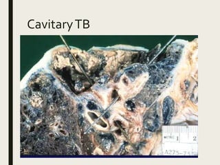
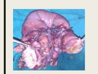
TUBERCULOSIS
Dr. Janani Mathialagan
Overview:
■ INTRODUCTION
■ TYPES
■ TRANSMISSION
■ PATHOGENESIS
■ GHON’S FOCUS
■ COMPLICATIONS
■ EXTRA- PULMONARY SITES
Tuberculosis
■ Chronic granulomatous inflammation
■ Predominant in developing countries
■ Predisposing factors:
 malnutrition,
 poverty,
 over-crowding,
 immunocompromised status
Causative organism
■ Mycobacterium tuberculosis (strict aerobe)
■ Tubercle bacillus
■ Also known as Koch’s bacillus
■ Mycobacterium tuberculosis complex-
Eg: M.tuberculosis hominis and M.bovis
Less common strains :
 M. africanum
 M. microti
 M. smegmatis
Atypical mycobacteria
■ Also know as non-tuberculous mycobacteria.
■ Or Environmental mycobacteria
1)Rapid growers (within 7 days)
M.abscessus,
M.fortitum,
M.chelonae.
2) Slow growers (2-3 weeks)
■ Photochromogens-yellow pigment produced in light
■ Scotochromogens-pigment produced in light or in dark
■ M.avium intracellulare,M.kansaii etc
■ No person to person contact
TYPES OFTB
■ PRIMARY
■ SECONDARY
SITES FOR PRIMARYTB:
■ LUNGAND HILAR LYMPHNODE
■ EXTRA PULMONARY
– CERVICAL LYMPH NODE (commonest)
– BONE
– TONSIL
– INTESTINE
– SKIN
Mode of transmission
 Inhalation
 Ingestion
 Inoculation into skin
 Transplacental route
Spread ofTB:
 local,
 lymphatic,
 hematogenous,
 natural passages.
Pathogenesis
■ Cell mediated immunity and
■ Delayed type of hypersensitivity
Bacterial wall contains the lipids:
A. Mycosides - cord factor of organism
B. Glycolipids - wax D
PATHOGENESIS:
Lymph node:
Fate of granuloma
1) Cold abscess
2) Sinus tract formation
3) Coalesce together enlarging the lesion
4) Dystrophic calcification
PrimaryTB
■ Ghon’s tuberculosis or childhood tuberculosis
■ Individuals who are not infected previously
■ Lungs and hilar lymph nodes
GHON’S FOCUS:
■ A primary lesion usually subpleural
■ Site : junction b/w the upper part of lower lobe and lower part
of upper lobe.
■ It is named for Anton Ghon (1866–1936), anAustrian
pathologist.
■ GHON’S COMPLEX:
 GHON’S FOCUS,
 HILAR LYMPHNODE INVOLVEMENT
 DRAINING LYMPHATICVESSEL INVOLVEMENT
Ghon’s focus
(sub pleural focus in the
upper part of the lower
lobe)
Microscopy- Ghon’s focus
Fate of primaryTB
■ Resolution and calcification
■ Progressive primary tuberculosis
■ Primary miliary tuberculosis
Secondary tuberculosis
■ Previously infected or sensitized individuals develop secondary
TB
■ Post-primary or re-infection or chronicTB
■ Endogenous source - dormant bacillus
■ Exogenous source - reinfection of bacillus
■ Commonly in lungs
Secondary pulmonaryTB
■ Commonest site - Apex of lung
■ Lympho-hematogenous spread
■ HIV patients are more prone
■ M.avium intracellulare occurs in the HIV patients too
Fate of secondary lungTB
■ Fibrosis and calcification
■ Tuberculous pneumonia
■ Miliary tuberculosis
■ Empyema
FibrocaseousTB
■ Fibrosed tubercle enter bronchus from a cavity -
cavitary or open fibrocaseousTB
■ Non cavitary lesion without entering bronchus -
chronic fibrocaseousTB
CavitaryTB
CavitaryTB
Cavitating granuloma
Complications of secondary
TB
■ Aneurysms of patent arteries
■ Bronchopleural fistula
■ Empyema
■ Adhesions on pleura
MiliaryTB
■ Spread to systemic organs
■ Via pulmonary vein or pulmonary artery
■ Millet sized lesions of 1 mm diameter
Gross-miliaryTB
Intestinal tuberculosis
■ Primary intestinalTB-
Ileocaecal region
Ingestion of M.bovis
Now a days, M.tuberculosis
Mesentric lymph nodes
■ Secondary intestinalTB-
Self swallowing of sputum in active disease
Terminal ileum
■ Hyperplastic ileocaecalTB
Secondary to pulmonaryTB
Common in India
Transverse ulcers in intestine:
Normal colonVSTB affected:
■ In the case of primaryTB, tabes mesentrica seen
■ Tabes mesentrica - enlarged mesentric lymph nodes
■ Mesentric lymphatic nodes rupture to causeTB
peritonitis
POTT’S SPINE:
Clinical features ofTB
■ Productive cough may be with hemoptysis
■ Dyspnoea, orthopnea
■ Fever
■ Night sweats
■ Fatigue
■ Loss of weight
■ Loss of appetite
Diagnosis
■ Clinical features are not confirmatory.
■ Zeil Neelson Stain
■ Adenosine deaminase test
■ Culture most sensitive and specific test.
– Conventional Lowenstein Jensen media 3-6 wks.
– Automated techniques within 9-16 days
■ PCR is available, but should only be
performed by experienced laboratories
■ Mantoux test
Demonstration of organism
■ Ziehl-neelsen staining or acid fast staining
■ Fluorescent dyes - auromine and rhodamine
■ LJ medium for sputum
■ Bactec culture, HPLC(high pressure liquid
chromatography)
■ Guinea pig inoculation
■ PCR
Ziehl-neelsen staining
immunisation
■ BCG - Bacillus Calmette Guerin
Mantoux test
■ Tuberculin skin test
■ 0.1 ml of tuberculoprotein,purified protein derivative
is administered intradermally
■ Induration more than 15 mm in 72 hours
■ DisseminatedTB-negative
■ False positive - atypical mycobacteria,
previous BCG vaccination
■ False negative - sarcoidosis,
recentTB,
hodgkins disease.
After 72 hrs…
MANAGEMENT:
■ ATT:

Tuberculosis