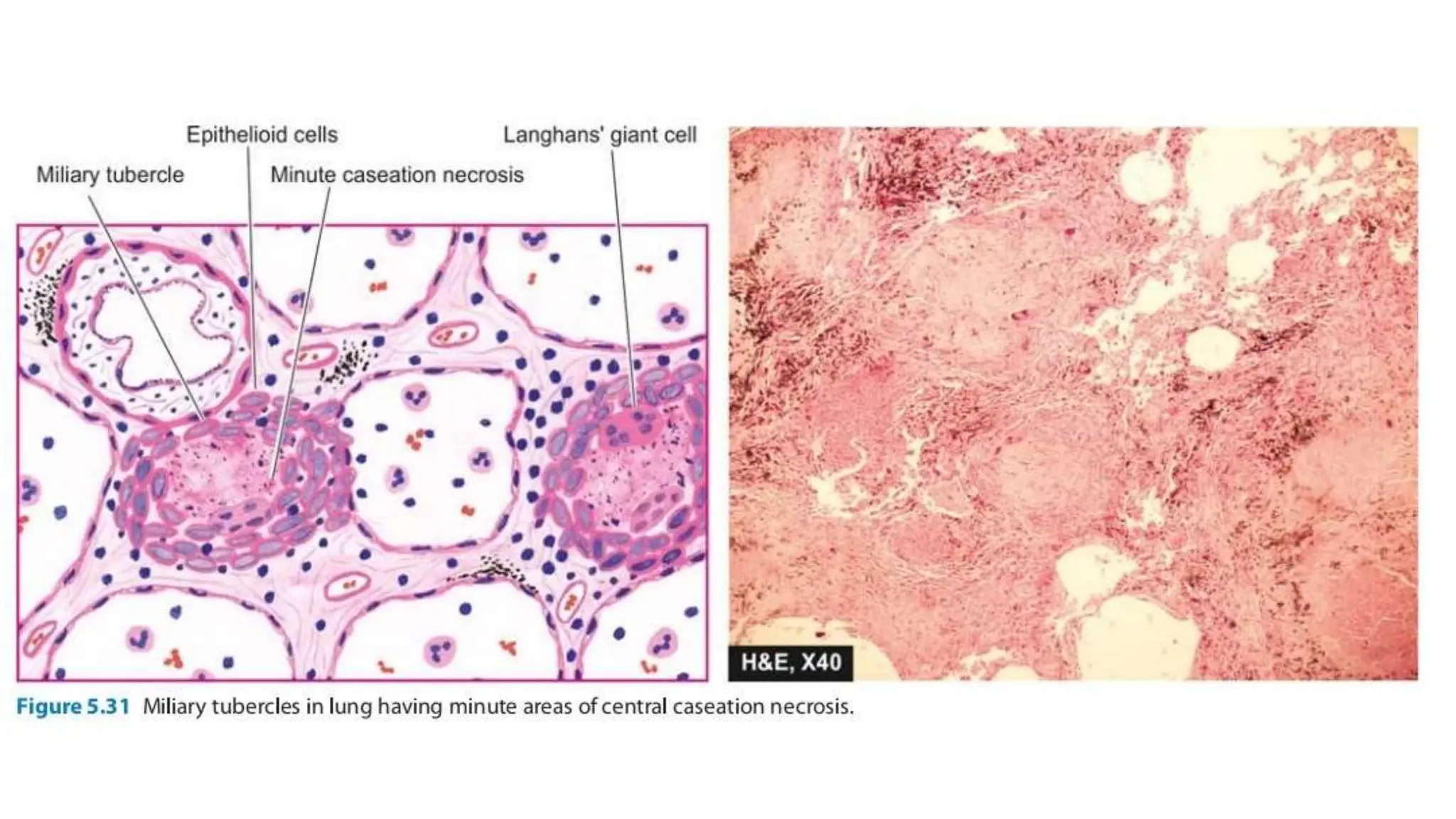
RESPIRATORY SYSTEM
Anatomy
• The normal adult right lung weighs 375 to 550 gm (average 450 gm) and is
divided by two fissures into three lobes - the upper, middle and lower lobes.
• The weight of the normal adult left lung is 325 to 450 gm (average 400 gm) and
has one fissure dividing it into two lobes - the upper and lower lobes, while the
middle lobe is represented by the lingula.
• The right main bronchus is more vertical so that aspirated foreign material tends to
pass down to the right lung rather than to the left.
TUBERCULOSIS
• Also known as – Koch’s disease.
• It is a chronic granulomatous disease caused by – mycobacterium
tuberculosis
ETIOLOGY
• Most common and important mycobacteria, causing human disease is
M. Tuberculosis.
• It is thin rod-shaped aerobic bacterium, which is not stained by
Gram’s staining.
• However, they are seen as pink bacteria, when stained by Ziehl-
Neelsen stain.
• When once stained, the bacilli cannot be decolorized by acid alcohol
and retain stains.
• Due to this characteristic,
• mycobacteria are classified as acid-fast (& alcohol fast) bacilli (AFB).
• Acid fastness is due to –
• The high content of mycolic acids, in the waxy (lipid) cell wall of the
mycobacteria.
PREDISPOSING FACTORS :
• Common in India.
• Incidence is high in poverty, overcrowding & chronic debilitating
illness.
• Some of the diseases are associated with increased risk –
• DM, malnutrition, immunosuppression, alcoholism, chronic lung
disease, chronic renal failure,
• HIV is the most important risk factor
Modes of transmission
1. Inhalation of organisms present in fresh cough droplets or in dried sputum from an open
case of pulmonary tuberculosis.
2. Ingestion of the organisms leads to development of tonsillar or intestinal tuberculosis.
This mode of infection of human tubercle bacilli is from self-swallowing of infected sputum
of an open case of pulmonary tuberculosis, or ingestion of bovine tubercle bacilli from milk
of diseased cows.
3. Inoculation of the organisms into the skin may rarely occur from infected postmortem
tissue.
4. Transplacental route results in development of congenital tuberculosis in foetus from
infected mother and is a rare mode of transmission.
PRIMARY TUBERCULOSIS
• Initial (first) infection with tubercle bacilli (mycobacterium
tuberculosis),
• In an unsensitized (previously unexposed) individual is known as –
primary tuberculosis.
• Source of organism is always exogenous.
MORPHOLOGY
SITES OF PRIMARY TB :
• Lung, intestine, tonsils, skin (very rare).
(1) LUNG :
• Commonest site of primary TB.
• Usually involved are – Lower part of upper lobe OR upper part of
lower lobe.
• This is because, most of the inspired air is distributed to the middle
and lower lung zones.
• The primary lesion is usually seen at the periphery of the lung, near
the pleural surface
About 2-4 weeks after the infection,
• A circumscribed gray-white area of about 1-1.5cm,
• Develops in the subpleural parenchymal region of the lung.
• This lung lesion is known as – Ghon’s focus.
• The centre of Ghon’s focus may undergo – caseous necrosis.
• Tubercle bacilli are carried along the lymphatics to the regional
draining nodes.
GHON COMPLEX :
• Combination of –
• Subpleural parenchymal primary lesion in the lung (Ghon focus),
• And regional lymph node involvement –
• Is known as – Ghon complex.
• In other organs, primary focus along with regional lymphadenitis is
called – primary complex.
FATE OF GHON COMPLEX :
In most (90%) individuals –
• Primary TB is healed by fibrosis and calcification.
In others (less than 10%) –
• It may develop into progressive primary TB OR
• May progress and spread by lymphatics and blood to other organs or
parts of the body.
OTHER SITES OF PRIMARY COMPLEX :
(2) Intestine :
• Primary focus in the small intestine (usually ileal region),
• Along with mesenteric lymphadenitis.
(3) Tonsils :
• Primary focus is in the pharynx and tonsils,
• With cervical lymph node enlargement.
(4) Skin :
• Primary focus in the skin along with regional lymph node involvement
MICROSCOPY :
• Granuloma in tuberculosis is called as – Tubercle.
• Granuloma with caseous necrosis is called – Soft tubercle.
• Granuloma without caseous necrosis is called – Hard tubercle.
Schematic evolution of tubercle. In fully formed
granuloma, the centre is composed of granular
caseation necrosis, surrounded by epithelioid
cells and Langhans’ giant cells and peripheral
rim of lymphocytes bounded by fibroblasts.
The typical caseating granuloma consists of –
Central area of caseous necrosis.
• The necrotic material resembles soft cheese, so known as caseous
necrosis.
It is surrounded by –
• Epithelioid cells (modified macrophages),
• Some of which may fuse to form, multinucleated giant cells.
The giant cells may be –
• Langhans type (nuclei arranged in horse-shoe pattern,
• OR foreign body type (nuclei in the center).
• The epithelioid cells are surrounded by – a rim of lymphocytes.
• These granulomas are usually enclosed by fibroblasts.
SECONDARY TUBERCULOSIS
• Also known as – Post-primary TB / Reactivation TB.
• Clinical illness, developing in a previously sensitized individual,
• Due to reactivation of initial infection OR Fresh infection, is called –
secondary (post primary) tuberculosis.
SOURCE OF INFECTION :
• Most common – reactivation of latent infection.
• Rarely – may be exogenous reinfection.
• Any organ may be involved in secondary tuberculosis,
• But the lungs are by far the most common site.
MORPHOLOGY
GROSSLY,
• In the lungs,
• Secondary TB usually involves the – Apex of the upper lobes of one or
both lungs.
• These areas have higher mean oxygen tension (compared to that of
lower zones),
• Which favours mycobacterial growth.
LESIONS are seen –
• Within 1-2cm of the apical pleura.
• The lesion may undergo – Cavitation.
• The erosion of cavities in to an airway is an important source of
infection,
• Because infected person expels bacilli, during coughing.
• The regional lymph nodes are less predominantly involved in
secondary tuberculosis, than primary TB.
MICROSCOPY,
• Active lesions show –
• Characteristic tubercles, with caseous necrosis, known as – Caseating
granulomas.
• Acid-fast stains may show tubercle bacilli.
FATE OF SECONDARY TUBERCLOSIS
Healing :
• With fibrosis and calcification.
• Rarely ossification.
• Progress :
• May progress along several different pathways.
SPREAD OF TB
• If the treatment is inadequate,
• Or if the host defense is impaired –
• The infection may spread via airways, lymphatics and blood vessels.
(1) PLEURAL SPREAD :
• It results in serous pleural effusion
• Or tuberculous empyema.
(2) SPREAD ALONG THE MUCOSAL LINING :
• Spread from expectorated infectious material may lead to –
• Endobronchial, endotracheal and laryngeal tuberculosis.
(3) MILIARY TUBERCULOSIS :
• It is the disseminated form of TB,
• And the lesions resemble millet seeds.
• So, named – “miliary TB”.
• It may occur when the organisms gain entry into systemic circulation.
• Most commonly involved organs are – liver, bone marrow, spleen,
adrenals, meninges, kidneys, fallopian tubes and epididymis.
(4) ISOLATED TB :
• Commonly involved organs are –
• Meninges (TB meningitis)
• Kidneys (Renal TB)
• Adrenals,
• Fallopian tubes (Salphingitis),
• Bones (osteomyelitis),
• Vertebrae (Pott’s disease)
(5) LYMPHADENITIS :
• Most frequent presentation of extrapulmonary TB.
• Usually occurs in the cervical region.
(6) INTESTINAL TUBERCULOSIS :
RESPIRATORY SYSTEM Tuberculosis pathology.pptx

RESPIRATORY SYSTEM Tuberculosis pathology.pptx

  • 1.
  • 2.
    Anatomy • The normaladult right lung weighs 375 to 550 gm (average 450 gm) and is divided by two fissures into three lobes - the upper, middle and lower lobes. • The weight of the normal adult left lung is 325 to 450 gm (average 400 gm) and has one fissure dividing it into two lobes - the upper and lower lobes, while the middle lobe is represented by the lingula. • The right main bronchus is more vertical so that aspirated foreign material tends to pass down to the right lung rather than to the left.
  • 4.
    TUBERCULOSIS • Also knownas – Koch’s disease. • It is a chronic granulomatous disease caused by – mycobacterium tuberculosis
  • 5.
    ETIOLOGY • Most commonand important mycobacteria, causing human disease is M. Tuberculosis. • It is thin rod-shaped aerobic bacterium, which is not stained by Gram’s staining. • However, they are seen as pink bacteria, when stained by Ziehl- Neelsen stain. • When once stained, the bacilli cannot be decolorized by acid alcohol and retain stains.
  • 6.
    • Due tothis characteristic, • mycobacteria are classified as acid-fast (& alcohol fast) bacilli (AFB). • Acid fastness is due to – • The high content of mycolic acids, in the waxy (lipid) cell wall of the mycobacteria.
  • 7.
    PREDISPOSING FACTORS : •Common in India. • Incidence is high in poverty, overcrowding & chronic debilitating illness. • Some of the diseases are associated with increased risk – • DM, malnutrition, immunosuppression, alcoholism, chronic lung disease, chronic renal failure, • HIV is the most important risk factor
  • 8.
    Modes of transmission 1.Inhalation of organisms present in fresh cough droplets or in dried sputum from an open case of pulmonary tuberculosis. 2. Ingestion of the organisms leads to development of tonsillar or intestinal tuberculosis. This mode of infection of human tubercle bacilli is from self-swallowing of infected sputum of an open case of pulmonary tuberculosis, or ingestion of bovine tubercle bacilli from milk of diseased cows. 3. Inoculation of the organisms into the skin may rarely occur from infected postmortem tissue. 4. Transplacental route results in development of congenital tuberculosis in foetus from infected mother and is a rare mode of transmission.
  • 9.
  • 10.
    • Initial (first)infection with tubercle bacilli (mycobacterium tuberculosis), • In an unsensitized (previously unexposed) individual is known as – primary tuberculosis. • Source of organism is always exogenous.
  • 11.
    MORPHOLOGY SITES OF PRIMARYTB : • Lung, intestine, tonsils, skin (very rare). (1) LUNG : • Commonest site of primary TB. • Usually involved are – Lower part of upper lobe OR upper part of lower lobe. • This is because, most of the inspired air is distributed to the middle and lower lung zones. • The primary lesion is usually seen at the periphery of the lung, near the pleural surface
  • 12.
    About 2-4 weeksafter the infection, • A circumscribed gray-white area of about 1-1.5cm, • Develops in the subpleural parenchymal region of the lung. • This lung lesion is known as – Ghon’s focus. • The centre of Ghon’s focus may undergo – caseous necrosis. • Tubercle bacilli are carried along the lymphatics to the regional draining nodes.
  • 13.
    GHON COMPLEX : •Combination of – • Subpleural parenchymal primary lesion in the lung (Ghon focus), • And regional lymph node involvement – • Is known as – Ghon complex. • In other organs, primary focus along with regional lymphadenitis is called – primary complex.
  • 15.
    FATE OF GHONCOMPLEX : In most (90%) individuals – • Primary TB is healed by fibrosis and calcification. In others (less than 10%) – • It may develop into progressive primary TB OR • May progress and spread by lymphatics and blood to other organs or parts of the body.
  • 17.
    OTHER SITES OFPRIMARY COMPLEX : (2) Intestine : • Primary focus in the small intestine (usually ileal region), • Along with mesenteric lymphadenitis. (3) Tonsils : • Primary focus is in the pharynx and tonsils, • With cervical lymph node enlargement. (4) Skin : • Primary focus in the skin along with regional lymph node involvement
  • 18.
    MICROSCOPY : • Granulomain tuberculosis is called as – Tubercle. • Granuloma with caseous necrosis is called – Soft tubercle. • Granuloma without caseous necrosis is called – Hard tubercle.
  • 19.
    Schematic evolution oftubercle. In fully formed granuloma, the centre is composed of granular caseation necrosis, surrounded by epithelioid cells and Langhans’ giant cells and peripheral rim of lymphocytes bounded by fibroblasts.
  • 20.
    The typical caseatinggranuloma consists of – Central area of caseous necrosis. • The necrotic material resembles soft cheese, so known as caseous necrosis. It is surrounded by – • Epithelioid cells (modified macrophages), • Some of which may fuse to form, multinucleated giant cells.
  • 21.
    The giant cellsmay be – • Langhans type (nuclei arranged in horse-shoe pattern, • OR foreign body type (nuclei in the center). • The epithelioid cells are surrounded by – a rim of lymphocytes. • These granulomas are usually enclosed by fibroblasts.
  • 22.
  • 23.
    • Also knownas – Post-primary TB / Reactivation TB. • Clinical illness, developing in a previously sensitized individual, • Due to reactivation of initial infection OR Fresh infection, is called – secondary (post primary) tuberculosis.
  • 24.
    SOURCE OF INFECTION: • Most common – reactivation of latent infection. • Rarely – may be exogenous reinfection. • Any organ may be involved in secondary tuberculosis, • But the lungs are by far the most common site.
  • 26.
    MORPHOLOGY GROSSLY, • In thelungs, • Secondary TB usually involves the – Apex of the upper lobes of one or both lungs. • These areas have higher mean oxygen tension (compared to that of lower zones), • Which favours mycobacterial growth.
  • 27.
    LESIONS are seen– • Within 1-2cm of the apical pleura. • The lesion may undergo – Cavitation. • The erosion of cavities in to an airway is an important source of infection, • Because infected person expels bacilli, during coughing. • The regional lymph nodes are less predominantly involved in secondary tuberculosis, than primary TB.
  • 28.
    MICROSCOPY, • Active lesionsshow – • Characteristic tubercles, with caseous necrosis, known as – Caseating granulomas. • Acid-fast stains may show tubercle bacilli.
  • 31.
    FATE OF SECONDARYTUBERCLOSIS Healing : • With fibrosis and calcification. • Rarely ossification. • Progress : • May progress along several different pathways.
  • 33.
    SPREAD OF TB •If the treatment is inadequate, • Or if the host defense is impaired – • The infection may spread via airways, lymphatics and blood vessels.
  • 34.
    (1) PLEURAL SPREAD: • It results in serous pleural effusion • Or tuberculous empyema. (2) SPREAD ALONG THE MUCOSAL LINING : • Spread from expectorated infectious material may lead to – • Endobronchial, endotracheal and laryngeal tuberculosis.
  • 35.
    (3) MILIARY TUBERCULOSIS: • It is the disseminated form of TB, • And the lesions resemble millet seeds. • So, named – “miliary TB”. • It may occur when the organisms gain entry into systemic circulation. • Most commonly involved organs are – liver, bone marrow, spleen, adrenals, meninges, kidneys, fallopian tubes and epididymis.
  • 38.
    (4) ISOLATED TB: • Commonly involved organs are – • Meninges (TB meningitis) • Kidneys (Renal TB) • Adrenals, • Fallopian tubes (Salphingitis), • Bones (osteomyelitis), • Vertebrae (Pott’s disease)
  • 40.
    (5) LYMPHADENITIS : •Most frequent presentation of extrapulmonary TB. • Usually occurs in the cervical region. (6) INTESTINAL TUBERCULOSIS :