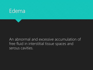
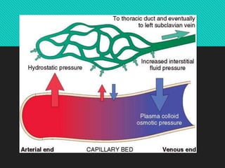
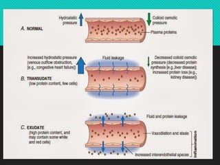
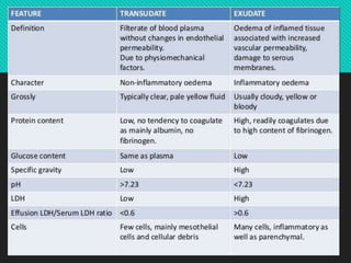
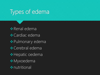
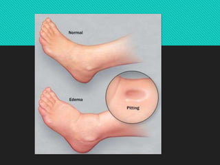
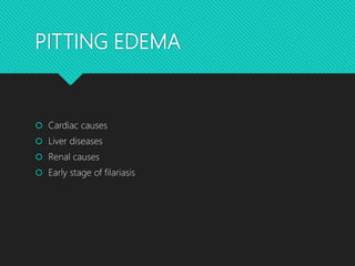
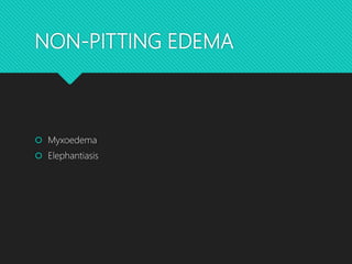
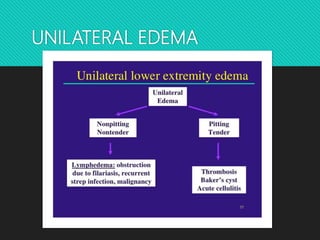
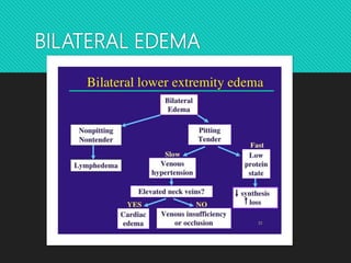
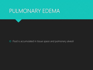
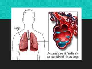
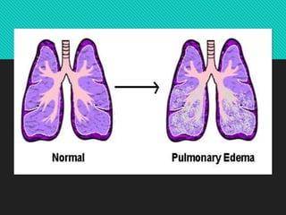
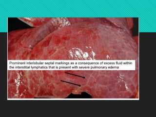
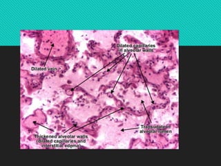
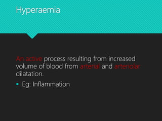
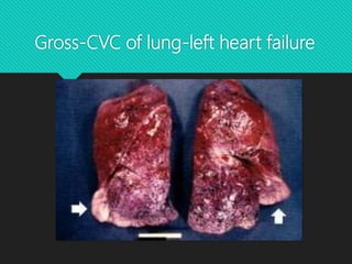
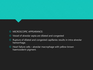
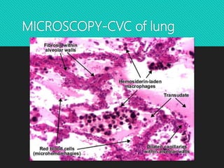
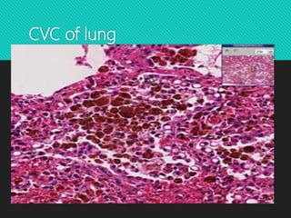
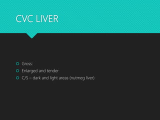
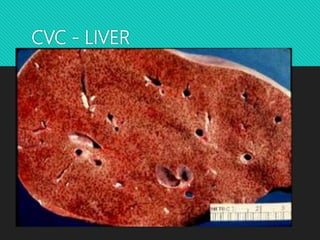
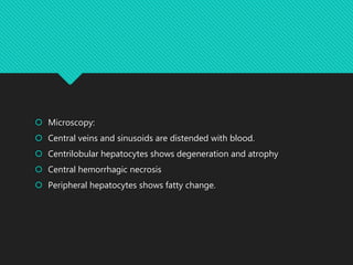
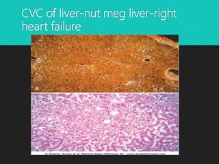
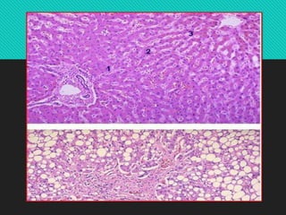
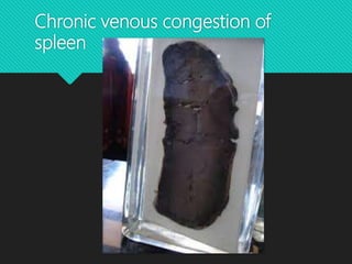
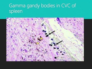
OEDEMA AND
HYPEREMIA
Dr. Janani Mathialagan
Homeostasis
Constancy of the internal environment is
maintained and ensured.
Edema
An abnormal and excessive accumulation of
free fluid in interstitial tissue spaces and
serous cavities.
PATHOGENESIS
1. Increased hydrostatic pressure
2. Decreased oncotic pressure
3. Lymphatic obstruction
4. Tissue factors
5. Increased capillary permeability
6. Sodium and water retention
Types of edema
Renal edema
Cardiac edema
Pulmonary edema
Cerebral edema
Hepatic oedema
Myxoedema
nutritional
TYPES
PITTING
NON- PITTING
PITTING EDEMA
 Cardiac causes
 Liver diseases
 Renal causes
 Early stage of filariasis
NON-PITTING EDEMA
 Myxoedema
 Elephantiasis
UNILATERAL EDEMA
BILATERAL EDEMA
PULMONARY EDEMA
 Fluid is accumulated in tissue space and pulmonary alveoli
Hyperaemia
An active process resulting from increased
volume of blood from arterial and arteriolar
dilatation.
 Eg: Inflammation
Congestion
A passive process resulting from dilatation of
veins and capillaries due to impaired venous
drainage
 Local venous congestion eg-portal venous
obstruction
 Systemic venous congestion eg-left sided
and right sided heart failure
CVC LUNG
GROSS APPEARANCE:
 Dark brown colour, heavy and firm.
 C/S – brown induration seen
Gross-CVC of lung-left heart failure
 MICROSCOPIC APPEARANCE:
 Vessel of alveolar septa are dilated and congested
 Rupture of dilated and congested capillaries results in intra-alveolar
hemorrhage.
 Heart failure cells – alveolar macrophage with yellow-brown
haemosiderin pigment.
MICROSCOPY-CVC of lung
CVC of lung
CVC LIVER
 Gross:
 Enlarged and tender
 C/S – dark and light areas (nutmeg liver)
CVC - LIVER
 Microscopy:
 Central veins and sinusoids are distended with blood.
 Centrilobular hepatocytes shows degeneration and atrophy
 Central hemorrhagic necrosis
 Peripheral hepatocytes shows fatty change.
CVC of liver-nut meg liver-right
heart failure
Chronic venous congestion of
spleen
Gamma gandy bodies in CVC of
spleen

Oedema and hyperemia