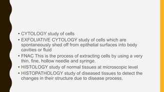
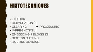
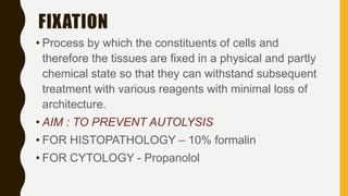
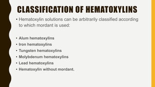
HISTOTECHNIQUES
–
UG PRESENTATION
• CYTOLOGY study of cells
• EXFOLIATIVE CYTOLOGY study of cells which are
spontaneously shed off from epithelial surfaces into body
cavities or fluid
• FNAC This is the process of extracting cells by using a very
thin, fine, hollow needle and syringe.
• HISTOLOGY study of normal tissues at microscopic level
• HISTOPATHOLOGY study of diseased tissues to detect the
changes in their structure due to disease process.
• BIOPSY examination of tissue removed from a living body.
• TYPES:
Endoscopic biopsy
Excision biopsy
Punch biopsy
FNA
Imprint cytology
Exfoliative
HISTOTECHNIQUES
• FIXATION
• DEHYDRATION
• CLEARING PROCESSING
• IMPREGNATION
• EMBEDDING & BLOCKING
• SECTION CUTTING
• ROUTINE STAINING
FIXATION
• Process by which the constituents of cells and
therefore the tissues are fixed in a physical and partly
chemical state so that they can withstand subsequent
treatment with various reagents with minimal loss of
architecture.
• AIM : TO PREVENT AUTOLYSIS
• FOR HISTOPATHOLOGY – 10% formalin
• FOR CYTOLOGY - Propanolol
TYPES OF FIXATIVES:
CHEMICAL
Aldehydes
Protein denaturing agents
• -alcoholic
• -acidic
Oxidising agent – osmium tetroxide
Miscellaneous
BASIC CONCEPTS
• Ratio of tissue with fixative 1 : 10-20
• Time for fixation - 8hrs minimum at room
temperature.
• Tissue thickness should be approximately 4mm
• Fixing solution should never be heated for faster
fixation. (shrinkage and affects staining quality)
EXAMPLE:
• 10% FORMALIN
Formaldehyde 40% - 10ml
Distilled water - 90ml
• If specimen sent to lab in not properly fixed, surgeon
can be charged with medical negligence – omission.
HEMATOXYLIN
AND EOSIN
STAINING
The Hematoxylin and Eosin stain (H&E) is the most
widely used histological stain because :
• comparative simplicity
• Ability to demonstrate clearly an enormous number of different
tissue structures.
• Hematoxylin stains cell nuclei blue black  shows good
intranuclear detail.
• Eosin stains cell cytoplasm and most connective tissue fibers
in varying shades and intensities of pink, orange, and red.
INTRODUCTION
• Outlines tissues and cellular components.
• Identification of tissues.
• Establishes the presence or absence of disease
processes.
PURPOSE OF STAINING
• Most commonly used staining methods are –
• Hematoxylin and Eosin staining in Histopathology
• Gram’s Stain and Ziehl-Neelson staining in Microbiology.
• Romanowsky staining in Hematology.
• Papanicoloau staining in Cytology
COMMON STAINING METHODS
• The word Hematoxylin is derived from old Greek
word Haimato meaning blood & Xylon meaning
wood.
• A natural dye extracted from the core or heartwood
of tree Haematoxylon campechianum.
• The hematoxylin is extracted from logwood with hot
water, and then precipitated out from the aqueous
solution using urea.
• The major oxidization product of hematoxylin is
hematein, a natural dye that is responsible for the
color properties.
HEMATOXYLIN
• Hematoxylin solutions can be arbitrarily classified according
to which mordant is used:
• Alum hematoxylins
• Iron hematoxylins
• Tungsten hematoxylins
• Molybdenum hematoxylins
• Lead hematoxylins
• Hematoxylin without mordant.
CLASSIFICATION OF HEMATOXYLINS
• Xanthine dyes which stains connective tissue and cytoplasm
in varying intensity and shades (red to pink).
• Available in the following types :
 Eosin Y ( Eosin Yellowish, Eosin water soluble) – most widely
available.
 Ethyl Eosin (Eosin S, eosin alcohol soluble).
 Eosin B ( Eosin Bluish, Erythrosine B).
• Ethyl eosin and eosin B are now rarely used, although occasional old
methods specify their use – e.g the Harris stain for Negri bodies.
EOSIN
Eosin Y
• Most commonly used eosin.
• Readily soluble in water.
• Satisfactorily soluble in alcohol.
• Preparation
 Eosin Y, water soluble 5 gm
 Distilled water 1000 ml
 Crystals of Thymol added to inhibit fungal growth.
 Addition of little acetic acid (0.5 -1000 ml stain) sharpens the
staining.
EOSIN
Principle
• Hematoxylin and Eosin are principle stains used for
demonstration of nucleus and cytoplasm.
• Alum acts as a mordant and the hematoxylin containing alum
stains the nucleus light blue which turns red in the presence
of acid.
• The cell differentiation is achieved by treating the tissue with
acid solution.
• The counterstaining is performed using eosin which imparts
pink color to cytoplasm.
THE HEMATOXYLIN AND EOSIN STAINING TECHNIQUE
1. REMOVAL OF WAX.
2. HYDRATION WITH GRADED ALCOHOLS.
3. STAINING.
4. DIFFERENTIATION
5. BLUEING
6. COUNTERSTAIN WITH EOSIN
7. DEHYDRATION THROUGH GRADED ALCOHOL.
8. CLEARING IN XYLENE
9. MOUNTING UNDER A COVER SLIP
THE HEMATOXYLIN AND EOSIN STAINING TECHNIQUE
FOR
PARAFFIN SECTIONS
Results
• Nuclei appear blue/black in color
• Cytoplasm appears in varying shades of pink
• Muscle fibers appear deep pink/red in color
• Red blood cells appear orange/red in color
• Fibrin appears deep pink in color.
THE HEMATOXYLIN AND EOSIN STAINING TECHNIQUE
FOR
PARAFFIN SECTIONS
1. HYDRATION WITH GRADED ALCOHOLS.
2. STAINING.
3. DIFFERENTIATION.
4. BLUEING.
5. COUNTERSTAIN WITH EOSIN.
6. MOUNTING UNDER A COVER SLIP.
THE HEMATOXYLIN AND EOSIN STAINING TECHNIQUE
CYTOLOGY SMEAR STAINING METHOD
1. Freeze suitable tissue block onto a chuck.
2. Cut cryostat sections at 3–6 μm thickness.
3. Fix section in 10% neutral buffered formalin at room temperature for
20 seconds.
4. Rinse in tap water.
5. Stain in double strength Carazzi’s hematoxylin for 1 minute.
6. Wash well in tap water for 10–20 seconds.
7. Stain in 1% aqueous eosin for 10 seconds.
8. Rinse in tap water.
9. Dehydrate, clear, and mount.
RAPID HEMATOXYLIN AND EOSIN STAINING TECHNIQUE
FOR
URGENT FROZEN SECTIONS
Histotechniques for ug

Histotechniques for ug

  • 1.
  • 2.
    • CYTOLOGY studyof cells • EXFOLIATIVE CYTOLOGY study of cells which are spontaneously shed off from epithelial surfaces into body cavities or fluid • FNAC This is the process of extracting cells by using a very thin, fine, hollow needle and syringe. • HISTOLOGY study of normal tissues at microscopic level • HISTOPATHOLOGY study of diseased tissues to detect the changes in their structure due to disease process.
  • 3.
    • BIOPSY examinationof tissue removed from a living body. • TYPES: Endoscopic biopsy Excision biopsy Punch biopsy FNA Imprint cytology Exfoliative
  • 4.
    HISTOTECHNIQUES • FIXATION • DEHYDRATION •CLEARING PROCESSING • IMPREGNATION • EMBEDDING & BLOCKING • SECTION CUTTING • ROUTINE STAINING
  • 5.
    FIXATION • Process bywhich the constituents of cells and therefore the tissues are fixed in a physical and partly chemical state so that they can withstand subsequent treatment with various reagents with minimal loss of architecture. • AIM : TO PREVENT AUTOLYSIS • FOR HISTOPATHOLOGY – 10% formalin • FOR CYTOLOGY - Propanolol
  • 6.
    TYPES OF FIXATIVES: CHEMICAL Aldehydes Proteindenaturing agents • -alcoholic • -acidic Oxidising agent – osmium tetroxide Miscellaneous
  • 7.
    BASIC CONCEPTS • Ratioof tissue with fixative 1 : 10-20 • Time for fixation - 8hrs minimum at room temperature. • Tissue thickness should be approximately 4mm • Fixing solution should never be heated for faster fixation. (shrinkage and affects staining quality)
  • 8.
    EXAMPLE: • 10% FORMALIN Formaldehyde40% - 10ml Distilled water - 90ml
  • 9.
    • If specimensent to lab in not properly fixed, surgeon can be charged with medical negligence – omission.
  • 10.
  • 11.
    The Hematoxylin andEosin stain (H&E) is the most widely used histological stain because : • comparative simplicity • Ability to demonstrate clearly an enormous number of different tissue structures. • Hematoxylin stains cell nuclei blue black  shows good intranuclear detail. • Eosin stains cell cytoplasm and most connective tissue fibers in varying shades and intensities of pink, orange, and red. INTRODUCTION
  • 12.
    • Outlines tissuesand cellular components. • Identification of tissues. • Establishes the presence or absence of disease processes. PURPOSE OF STAINING
  • 13.
    • Most commonlyused staining methods are – • Hematoxylin and Eosin staining in Histopathology • Gram’s Stain and Ziehl-Neelson staining in Microbiology. • Romanowsky staining in Hematology. • Papanicoloau staining in Cytology COMMON STAINING METHODS
  • 14.
    • The wordHematoxylin is derived from old Greek word Haimato meaning blood & Xylon meaning wood. • A natural dye extracted from the core or heartwood of tree Haematoxylon campechianum. • The hematoxylin is extracted from logwood with hot water, and then precipitated out from the aqueous solution using urea. • The major oxidization product of hematoxylin is hematein, a natural dye that is responsible for the color properties. HEMATOXYLIN
  • 15.
    • Hematoxylin solutionscan be arbitrarily classified according to which mordant is used: • Alum hematoxylins • Iron hematoxylins • Tungsten hematoxylins • Molybdenum hematoxylins • Lead hematoxylins • Hematoxylin without mordant. CLASSIFICATION OF HEMATOXYLINS
  • 16.
    • Xanthine dyeswhich stains connective tissue and cytoplasm in varying intensity and shades (red to pink). • Available in the following types :  Eosin Y ( Eosin Yellowish, Eosin water soluble) – most widely available.  Ethyl Eosin (Eosin S, eosin alcohol soluble).  Eosin B ( Eosin Bluish, Erythrosine B). • Ethyl eosin and eosin B are now rarely used, although occasional old methods specify their use – e.g the Harris stain for Negri bodies. EOSIN
  • 17.
    Eosin Y • Mostcommonly used eosin. • Readily soluble in water. • Satisfactorily soluble in alcohol. • Preparation  Eosin Y, water soluble 5 gm  Distilled water 1000 ml  Crystals of Thymol added to inhibit fungal growth.  Addition of little acetic acid (0.5 -1000 ml stain) sharpens the staining. EOSIN
  • 18.
    Principle • Hematoxylin andEosin are principle stains used for demonstration of nucleus and cytoplasm. • Alum acts as a mordant and the hematoxylin containing alum stains the nucleus light blue which turns red in the presence of acid. • The cell differentiation is achieved by treating the tissue with acid solution. • The counterstaining is performed using eosin which imparts pink color to cytoplasm. THE HEMATOXYLIN AND EOSIN STAINING TECHNIQUE
  • 19.
    1. REMOVAL OFWAX. 2. HYDRATION WITH GRADED ALCOHOLS. 3. STAINING. 4. DIFFERENTIATION 5. BLUEING 6. COUNTERSTAIN WITH EOSIN 7. DEHYDRATION THROUGH GRADED ALCOHOL. 8. CLEARING IN XYLENE 9. MOUNTING UNDER A COVER SLIP THE HEMATOXYLIN AND EOSIN STAINING TECHNIQUE FOR PARAFFIN SECTIONS
  • 20.
    Results • Nuclei appearblue/black in color • Cytoplasm appears in varying shades of pink • Muscle fibers appear deep pink/red in color • Red blood cells appear orange/red in color • Fibrin appears deep pink in color. THE HEMATOXYLIN AND EOSIN STAINING TECHNIQUE FOR PARAFFIN SECTIONS
  • 21.
    1. HYDRATION WITHGRADED ALCOHOLS. 2. STAINING. 3. DIFFERENTIATION. 4. BLUEING. 5. COUNTERSTAIN WITH EOSIN. 6. MOUNTING UNDER A COVER SLIP. THE HEMATOXYLIN AND EOSIN STAINING TECHNIQUE CYTOLOGY SMEAR STAINING METHOD
  • 22.
    1. Freeze suitabletissue block onto a chuck. 2. Cut cryostat sections at 3–6 μm thickness. 3. Fix section in 10% neutral buffered formalin at room temperature for 20 seconds. 4. Rinse in tap water. 5. Stain in double strength Carazzi’s hematoxylin for 1 minute. 6. Wash well in tap water for 10–20 seconds. 7. Stain in 1% aqueous eosin for 10 seconds. 8. Rinse in tap water. 9. Dehydrate, clear, and mount. RAPID HEMATOXYLIN AND EOSIN STAINING TECHNIQUE FOR URGENT FROZEN SECTIONS