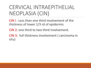
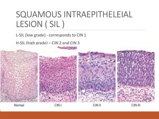
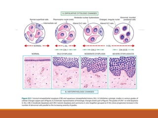
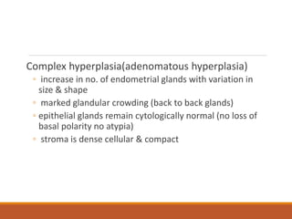
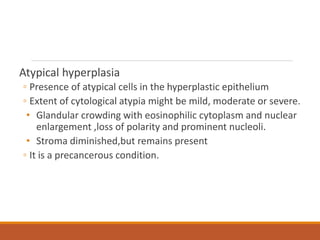
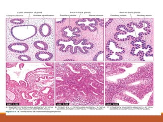
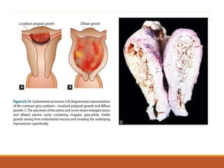
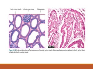
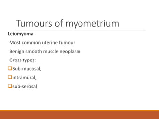
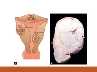
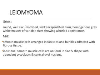
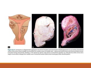
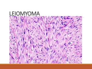
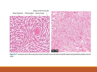
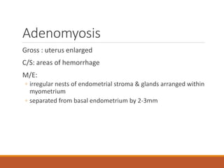
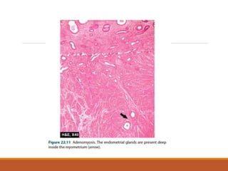
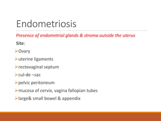
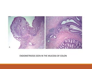
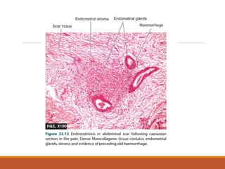
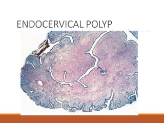
FEMALE GENITAL
TRACT
FGT IS COMPOSED OF
VULVA
VAGINA
CERVIX
UTERUS
FALLOPIAN TUBE
OVARY
CERVIX
OVERVIEW
CIN
SQUAMOUS INTRA-EPITHELIAL LESION
CARCINOMA CERVIX
CERVICAL INTRAEPITHELIAL
NEOPLASIA (CIN)
CIN I : Less than one third involvement of the
thickness of lower 1/3 rd of epidermis
CIN 2: one third to two third involvement.
CIN 3: full thickness involvement ( carcinoma in
situ)
SQUAMOUS INTRAEPITHELEIAL
LESION ( SIL )
L-SIL (low grade) - corresponds to CIN 1
H-SIL (high grade) – CIN 2 and CIN 3
RISK FACTORS
Early onset of sexual activity
Multiple sexual partners
Women with HPV infection : 16, 18, 31, 33, 35
High risk male sexual partner
Multiparous women
Use of OCP
Cigarette smoking
HIV infection
PAP SCREENING:
◦ Annual cervical pap smear in all sexually active women
having any high risk factors.
◦ if negative in 3 consecutive smears, then the frequency
of PAP screening is reduced.
INVASIVE CERVICAL CARCINOMA
Gross:
3 types
fungating,
ulcerative and
infiltrative.
INVASIVE CERVICAL CARCINOMA
Microscopy:
1. Sq cell carcinoma - composed of nests and tongues of
malignant sq epithelium invading the underlying cervical
stroma.
2. Adenocarcinoma – proliferation of glandular endocervical
glands
3. Adenosquamous carcinoma- malignant glandular and sq
epithelium.
Squamous cell carcinoma
moderately differentiated non-
keratinising carcinoma.
UTERUS
OVERVIEW
ENDOMETRIAL HYPERPLASIA
ENDOMETRIAL CARCINOMA
LEIOMYOMA
Endometrial hyperplasia
Characterized by increased proliferation of endometrial
glands relative to stroma resulting in an increased gland to
stroma ratio when compared with normal proliferative
endometrium
Causes:
Prolonged estrogenic stimulation with absence of progestational
activity.
Anovulation,
obesity,
menopause,
PCOD,
functioning granulosa cell tumour,
adrenocortical hyperfunction,
estrogen replacement therapy
Classification of endometrial
hyperplasia
Simple hyperplasia (cystic hyperplasia)
◦ presence of glands of various sizes and irregular shape
with cystic dilatation
◦ mild increase in gland to stroma ratio
Complex hyperplasia(adenomatous hyperplasia)
◦ increase in no. of endometrial glands with variation in
size & shape
◦ marked glandular crowding (back to back glands)
◦ epithelial glands remain cytologically normal (no loss of
basal polarity no atypia)
◦ stroma is dense cellular & compact
Atypical hyperplasia
◦ Presence of atypical cells in the hyperplastic epithelium
◦ Extent of cytological atypia might be mild, moderate or severe.
• Glandular crowding with eosinophilic cytoplasm and nuclear
enlargement ,loss of polarity and prominent nucleoli.
• Stroma diminished,but remains present
◦ It is a precancerous condition.
Tumours of endometrium
Endometrial carcinoma
Most common invasive cancer of FGT
Clinical features is abnormal bleeding
1. Type 1
2. Type 2
TYPE 1 ENDOMETRIAL
CARCINOMA
◦ Most common type (reproductive age group)
◦ Referred as endometrioid carcinoma
◦ Associated with
obesity,
diabetes,
hypertension &
unopposed estrogen stimulation
ASSOCIATED WITH ENDOMETRIAL HYPERPLASIA
TYPE 1 ENDOMETRIAL
CARCINOMA
◦ Gross: 2 patterns –
 localized polypoidal tumour
 diffuse tumour
◦ Extension of growth into
myometrium by direct invasion,
periuterine structures by direct continuity,
lymphatic metastasis,
haematogenous metastasis
TYPE 1 ENDOMETRIAL
CARCINOMA
◦ M/E:
most endometrial carcinomas are endometrioid
adenocarcinomas
Depending on the pattern of glands & cell changes they
are
 well differentiated,
 moderately differentiated or
 poorly differentiated
TYPE 2 ENDOMETRIAL
CARCINOMA
◦ Arise in atrophied endrometrium
◦ Age group 65-75 yrs (post-menopausal age group)
◦ Poorly differentiated tumours
◦ Aggressive tumours
Tumours of myometrium
Leiomyoma
Most common uterine tumour
Benign smooth muscle neoplasm
Gross types:
Sub-mucosal,
intramural,
sub-serosal
LEIOMYOMA
Gross :
round, well circumscribed, well encapsulated, firm, homogenous grey
white masses of variable sizes showing whorled appearance.
M/E:
•smooth muscle cells arranged in fascicles and bundles admixed with
fibrous tissue.
•individual smooth muscle cells are uniform in size & shape with
abundant cytoplasm & central oval nucleus.
LEIOMYOMA
Leiomyosarcoma
Uncommon malignant neoplasm of myometrium
C/F :
uterine enlargement, abnormal uterine bleeding
Gross:
bulky fleshy mass that invade the uterine wall or polypoid mass projecting into
lumen
M/E:
whorled arrangement of spindle cells with large hyperchromatic nuclei(nuclear
atypia),
No. of mitosis per HPF(mitotic index) = 10 or more
Thankyou
Adenomyosis
Presence of endometrial tissue within the myometrium
Clinical features:
◦ menorrhagia,
◦ dysmennorhoea,
◦ pelvic pain
Adenomyosis
Gross : uterus enlarged
C/S: areas of hemorrhage
M/E:
◦ irregular nests of endometrial stroma & glands arranged within
myometrium
◦ separated from basal endometrium by 2-3mm
Endometriosis
Presence of endometrial glands & stroma outside the uterus
Site:
Ovary
uterine ligaments
rectovaginal septum
cul-de –sac
pelvic peritoneum
mucosa of cervix, vagina fallopian tubes
large& small bowel & appendix
Endometriosis
Clinical Features:
infertility,
dysmennorhoea,
pelvic pain,
dyspareunia
Seen only in reproductive years of life
ENDOMETRIOSIS SEEN IN THE MUCOSA OF COLON
Two theories of development of endometriosis
Metastatic theory: retrograde menstruation through fallopian tube
Metaplastic theory: metaplasia of coelomic epithelium
Gross: foci of endometriosis appear blue or brownish black in the sites
mentioned
When disease is extensive organizing hemorrhages causes extensive
fibrous adhesions
Ovaries show numerous cysts filled with dark brown blood called as
chocolate cysts
Microscopy:
Presence of endometrial glands & stroma, areas of hemorrhage,
haemosiderin laden macrophages, surrounding zone of inflammation &
fibrosis
ENDOCERVICAL POLYP
◦ Benign exophytic growths
◦ Gross appearance: Small or sessile or may protrude
throught the external os.
◦ Microscopic appearance:
Soft , mucoid lesions composed of loose
fibromyxomatous stroma ,
mucus secreting endocervical glands and
variable dergee of inflammatory infitrate.
ENDOCERVICAL POLYP

Fgt uterus and cervix

  • 1.
  • 2.
    FGT IS COMPOSEDOF VULVA VAGINA CERVIX UTERUS FALLOPIAN TUBE OVARY
  • 3.
  • 4.
  • 5.
    CERVICAL INTRAEPITHELIAL NEOPLASIA (CIN) CINI : Less than one third involvement of the thickness of lower 1/3 rd of epidermis CIN 2: one third to two third involvement. CIN 3: full thickness involvement ( carcinoma in situ)
  • 6.
    SQUAMOUS INTRAEPITHELEIAL LESION (SIL ) L-SIL (low grade) - corresponds to CIN 1 H-SIL (high grade) – CIN 2 and CIN 3
  • 7.
    RISK FACTORS Early onsetof sexual activity Multiple sexual partners Women with HPV infection : 16, 18, 31, 33, 35 High risk male sexual partner Multiparous women Use of OCP Cigarette smoking HIV infection
  • 8.
    PAP SCREENING: ◦ Annualcervical pap smear in all sexually active women having any high risk factors. ◦ if negative in 3 consecutive smears, then the frequency of PAP screening is reduced.
  • 10.
    INVASIVE CERVICAL CARCINOMA Gross: 3types fungating, ulcerative and infiltrative.
  • 12.
    INVASIVE CERVICAL CARCINOMA Microscopy: 1.Sq cell carcinoma - composed of nests and tongues of malignant sq epithelium invading the underlying cervical stroma. 2. Adenocarcinoma – proliferation of glandular endocervical glands 3. Adenosquamous carcinoma- malignant glandular and sq epithelium.
  • 13.
    Squamous cell carcinoma moderatelydifferentiated non- keratinising carcinoma.
  • 14.
  • 15.
  • 16.
    Endometrial hyperplasia Characterized byincreased proliferation of endometrial glands relative to stroma resulting in an increased gland to stroma ratio when compared with normal proliferative endometrium
  • 17.
    Causes: Prolonged estrogenic stimulationwith absence of progestational activity. Anovulation, obesity, menopause, PCOD, functioning granulosa cell tumour, adrenocortical hyperfunction, estrogen replacement therapy
  • 18.
    Classification of endometrial hyperplasia Simplehyperplasia (cystic hyperplasia) ◦ presence of glands of various sizes and irregular shape with cystic dilatation ◦ mild increase in gland to stroma ratio
  • 19.
    Complex hyperplasia(adenomatous hyperplasia) ◦increase in no. of endometrial glands with variation in size & shape ◦ marked glandular crowding (back to back glands) ◦ epithelial glands remain cytologically normal (no loss of basal polarity no atypia) ◦ stroma is dense cellular & compact
  • 20.
    Atypical hyperplasia ◦ Presenceof atypical cells in the hyperplastic epithelium ◦ Extent of cytological atypia might be mild, moderate or severe. • Glandular crowding with eosinophilic cytoplasm and nuclear enlargement ,loss of polarity and prominent nucleoli. • Stroma diminished,but remains present ◦ It is a precancerous condition.
  • 22.
    Tumours of endometrium Endometrialcarcinoma Most common invasive cancer of FGT Clinical features is abnormal bleeding 1. Type 1 2. Type 2
  • 23.
    TYPE 1 ENDOMETRIAL CARCINOMA ◦Most common type (reproductive age group) ◦ Referred as endometrioid carcinoma ◦ Associated with obesity, diabetes, hypertension & unopposed estrogen stimulation ASSOCIATED WITH ENDOMETRIAL HYPERPLASIA
  • 24.
    TYPE 1 ENDOMETRIAL CARCINOMA ◦Gross: 2 patterns –  localized polypoidal tumour  diffuse tumour ◦ Extension of growth into myometrium by direct invasion, periuterine structures by direct continuity, lymphatic metastasis, haematogenous metastasis
  • 25.
    TYPE 1 ENDOMETRIAL CARCINOMA ◦M/E: most endometrial carcinomas are endometrioid adenocarcinomas Depending on the pattern of glands & cell changes they are  well differentiated,  moderately differentiated or  poorly differentiated
  • 26.
    TYPE 2 ENDOMETRIAL CARCINOMA ◦Arise in atrophied endrometrium ◦ Age group 65-75 yrs (post-menopausal age group) ◦ Poorly differentiated tumours ◦ Aggressive tumours
  • 29.
    Tumours of myometrium Leiomyoma Mostcommon uterine tumour Benign smooth muscle neoplasm Gross types: Sub-mucosal, intramural, sub-serosal
  • 31.
    LEIOMYOMA Gross : round, wellcircumscribed, well encapsulated, firm, homogenous grey white masses of variable sizes showing whorled appearance. M/E: •smooth muscle cells arranged in fascicles and bundles admixed with fibrous tissue. •individual smooth muscle cells are uniform in size & shape with abundant cytoplasm & central oval nucleus.
  • 33.
  • 35.
    Leiomyosarcoma Uncommon malignant neoplasmof myometrium C/F : uterine enlargement, abnormal uterine bleeding Gross: bulky fleshy mass that invade the uterine wall or polypoid mass projecting into lumen M/E: whorled arrangement of spindle cells with large hyperchromatic nuclei(nuclear atypia), No. of mitosis per HPF(mitotic index) = 10 or more
  • 38.
  • 39.
    Adenomyosis Presence of endometrialtissue within the myometrium Clinical features: ◦ menorrhagia, ◦ dysmennorhoea, ◦ pelvic pain
  • 40.
    Adenomyosis Gross : uterusenlarged C/S: areas of hemorrhage M/E: ◦ irregular nests of endometrial stroma & glands arranged within myometrium ◦ separated from basal endometrium by 2-3mm
  • 42.
    Endometriosis Presence of endometrialglands & stroma outside the uterus Site: Ovary uterine ligaments rectovaginal septum cul-de –sac pelvic peritoneum mucosa of cervix, vagina fallopian tubes large& small bowel & appendix
  • 43.
  • 44.
    ENDOMETRIOSIS SEEN INTHE MUCOSA OF COLON
  • 45.
    Two theories ofdevelopment of endometriosis Metastatic theory: retrograde menstruation through fallopian tube Metaplastic theory: metaplasia of coelomic epithelium
  • 46.
    Gross: foci ofendometriosis appear blue or brownish black in the sites mentioned When disease is extensive organizing hemorrhages causes extensive fibrous adhesions Ovaries show numerous cysts filled with dark brown blood called as chocolate cysts
  • 47.
    Microscopy: Presence of endometrialglands & stroma, areas of hemorrhage, haemosiderin laden macrophages, surrounding zone of inflammation & fibrosis
  • 49.
    ENDOCERVICAL POLYP ◦ Benignexophytic growths ◦ Gross appearance: Small or sessile or may protrude throught the external os. ◦ Microscopic appearance: Soft , mucoid lesions composed of loose fibromyxomatous stroma , mucus secreting endocervical glands and variable dergee of inflammatory infitrate.
  • 50.