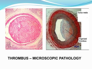
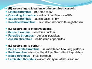
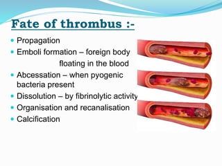
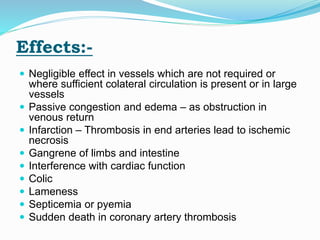
DEFINITION
 THROMBOSIS is the formation of a clotted mass of
blood within the cardiovascular system.
 The clotted mass is called as THROMBUS.
THROMBUS BLOOD CLOT
1- Involves wall of blood
vessels, formed elements
of blood & blood clotting
system.
1- Involves only blood
clotting system.
2- composed of platelets
and fibrin.
2- composed of fibrin only.
3- may be life-threatening. 3- mostly life saving.
THROMBUS – GROSS PATHOLOGY
THROMBUS – MICROSCOPIC PATHOLOGY
Antithrombotic & prothrombotic
property of endothelial cells
AETIOLOGY:-
1- Endothelial injury :-
 Trauma
 Bacteria- Toxins by Erysipelothrix, Streptococcus,
Staphylococcus, Coryanobacterium
 Virus- Hog cholera virus thrombosis in spleen
 Parasites- Strongylus vulgaris in anterior mesenteric
artery of horse
 Tumours invading endothelium
Normal endothelium is thromboresistant, but when
injured the highly thrombogenic subendothelium is
exposed and promotes adhesion of fibrin and
platelets.
2- Abnormal bloodflow:-
 RBC , WBC – Heavier axial stream
Platelets – Lighter plasmatic / laminar stream
 When blood flow slows down, platelets beingd the
outermost in the blood stream, fall out to the periphery
and stick to endothelium by virtue of their adhesive
property.
 Cause of slow bloodflow – Stasis and turbulence
1- chronic venous congestion
2- aged and debilitated animals
3- vericose vein- nasal submucosa in cattle, scrotal
plexus of horse, large veins of broad ligament of bovine
uterus
4- congestive heart failure
3- Hypercoagulability :-
 Increase in level of fibrinogen, prothrombin, factor
VIIa, VIIIa, Xa
 Increse in number of platelets
 Decrease in levels of antithrombin III, protein C and
fibrinolysin
Hepatic disease - formation of prothrombin
Bile deficiency in intestine - less absorption of Vit-K
Pathogenesis :-
 Endothelial injury – platelets adhere to
subendothelial collagen
 Platelets secretes ADP and thromboxane A2
 Plateles expose phospholipid complex – activates
intrinsic coagulation pathway
 Tissue factor from injured endothelium – activates
extrinsic coagulation pathway
 ADP – reversible primary hemostatic plug
ADP, thrombin, TXA2 – irreversible secondary plug
 Fibrin deposition around platelets
Classification :-
(A) According to location-
 1-Cardiac thrombus
Valvular thrombus- in valves – Erysipelothrix infection in
pig
Mural thrombus – in wall of heart – Clostridium infection in
cattle
 2- Arterial thrombus - Strongylus vulgaris in anterior
mesenteric artery of horse
 3- Venous thrombus – more common in human
 4- Capillary thrombus
 5- Lymphatic thrombus
VALVULAR THROMBUS MURAL THROMBUS
 (B) According to location within the blood vessel :-
 Lateral thrombus – one side of BV
 Occluding thrombus – entire circumference of BV
 Saddle thrombus – at bifurcation of BV
 Canalised thrombus – new blood channels through the clot
 (C) According to infective agent :-
 Septic thrombus – contains bacteria
 Parasitic thrombus – contains parasites
 Aseptic thrombus – no bacteria or parasites
 (D) According to colour :-
 Pale or white thrombus – in rapid blood flow, only platelets
 Red thrombus – in slow blood flow, fibrin attach to platelets
 Mixed thrombus – most common
 Laminated thrombus – alternate layers of white and red
WHITE AND RED THROMBI
Fate of thrombus :-
 Propagation
 Emboli formation – foreign body
floating in the blood
 Abcessation – when pyogenic
bacteria present
 Dissolution – by fibrinolytic activity
 Organisation and recanalisation
 Calcification
Effects:-
 Negligible effect in vessels which are not required or
where sufficient colateral circulation is present or in large
vessels
 Passive congestion and edema – as obstruction in
venous return
 Infarction – Thrombosis in end arteries lead to ischemic
necrosis
 Gangrene of limbs and intestine
 Interference with cardiac function
 Colic
 Lameness
 Septicemia or pyemia
 Sudden death in coronary artery thrombosis
Rudolf Virchow
Thrombosis

Thrombosis

  • 2.
    DEFINITION  THROMBOSIS isthe formation of a clotted mass of blood within the cardiovascular system.  The clotted mass is called as THROMBUS. THROMBUS BLOOD CLOT 1- Involves wall of blood vessels, formed elements of blood & blood clotting system. 1- Involves only blood clotting system. 2- composed of platelets and fibrin. 2- composed of fibrin only. 3- may be life-threatening. 3- mostly life saving.
  • 3.
  • 4.
  • 5.
  • 6.
  • 7.
    1- Endothelial injury:-  Trauma  Bacteria- Toxins by Erysipelothrix, Streptococcus, Staphylococcus, Coryanobacterium  Virus- Hog cholera virus thrombosis in spleen  Parasites- Strongylus vulgaris in anterior mesenteric artery of horse  Tumours invading endothelium Normal endothelium is thromboresistant, but when injured the highly thrombogenic subendothelium is exposed and promotes adhesion of fibrin and platelets.
  • 8.
    2- Abnormal bloodflow:- RBC , WBC – Heavier axial stream Platelets – Lighter plasmatic / laminar stream  When blood flow slows down, platelets beingd the outermost in the blood stream, fall out to the periphery and stick to endothelium by virtue of their adhesive property.  Cause of slow bloodflow – Stasis and turbulence 1- chronic venous congestion 2- aged and debilitated animals 3- vericose vein- nasal submucosa in cattle, scrotal plexus of horse, large veins of broad ligament of bovine uterus 4- congestive heart failure
  • 9.
    3- Hypercoagulability :- Increase in level of fibrinogen, prothrombin, factor VIIa, VIIIa, Xa  Increse in number of platelets  Decrease in levels of antithrombin III, protein C and fibrinolysin Hepatic disease - formation of prothrombin Bile deficiency in intestine - less absorption of Vit-K
  • 10.
    Pathogenesis :-  Endothelialinjury – platelets adhere to subendothelial collagen  Platelets secretes ADP and thromboxane A2  Plateles expose phospholipid complex – activates intrinsic coagulation pathway  Tissue factor from injured endothelium – activates extrinsic coagulation pathway  ADP – reversible primary hemostatic plug ADP, thrombin, TXA2 – irreversible secondary plug  Fibrin deposition around platelets
  • 11.
    Classification :- (A) Accordingto location-  1-Cardiac thrombus Valvular thrombus- in valves – Erysipelothrix infection in pig Mural thrombus – in wall of heart – Clostridium infection in cattle  2- Arterial thrombus - Strongylus vulgaris in anterior mesenteric artery of horse  3- Venous thrombus – more common in human  4- Capillary thrombus  5- Lymphatic thrombus
  • 12.
  • 13.
     (B) Accordingto location within the blood vessel :-  Lateral thrombus – one side of BV  Occluding thrombus – entire circumference of BV  Saddle thrombus – at bifurcation of BV  Canalised thrombus – new blood channels through the clot  (C) According to infective agent :-  Septic thrombus – contains bacteria  Parasitic thrombus – contains parasites  Aseptic thrombus – no bacteria or parasites  (D) According to colour :-  Pale or white thrombus – in rapid blood flow, only platelets  Red thrombus – in slow blood flow, fibrin attach to platelets  Mixed thrombus – most common  Laminated thrombus – alternate layers of white and red
  • 14.
  • 15.
    Fate of thrombus:-  Propagation  Emboli formation – foreign body floating in the blood  Abcessation – when pyogenic bacteria present  Dissolution – by fibrinolytic activity  Organisation and recanalisation  Calcification
  • 16.
    Effects:-  Negligible effectin vessels which are not required or where sufficient colateral circulation is present or in large vessels  Passive congestion and edema – as obstruction in venous return  Infarction – Thrombosis in end arteries lead to ischemic necrosis  Gangrene of limbs and intestine  Interference with cardiac function  Colic  Lameness  Septicemia or pyemia  Sudden death in coronary artery thrombosis
  • 17.