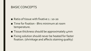
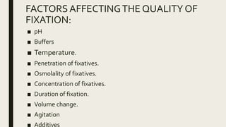
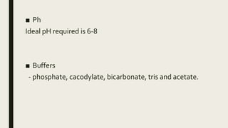
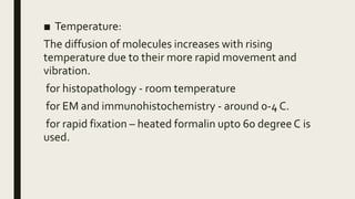
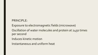
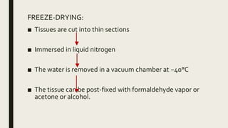
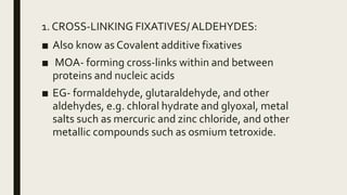
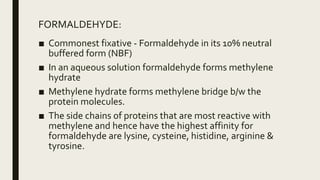
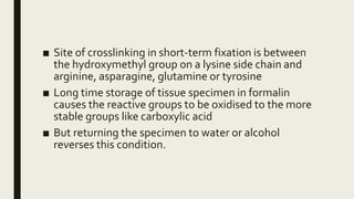
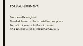
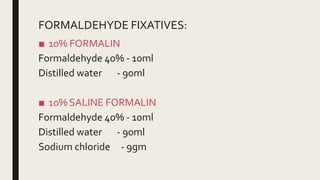
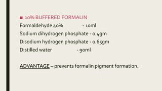
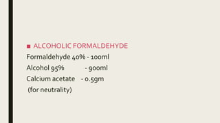
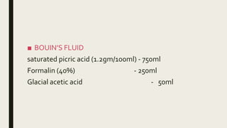
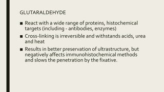
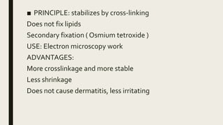
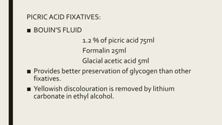
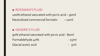
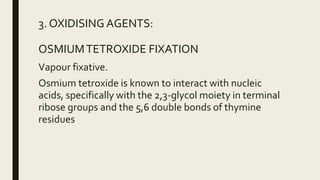
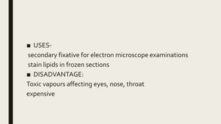
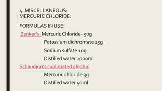
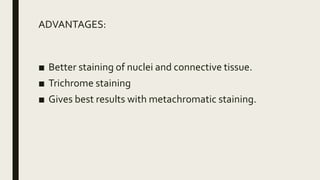
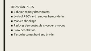
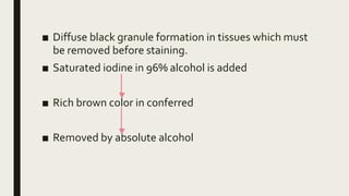
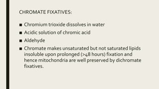
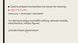
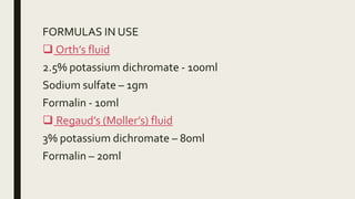
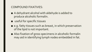
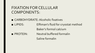
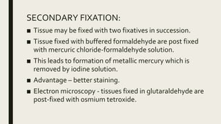
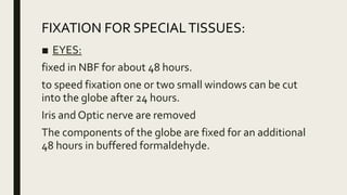
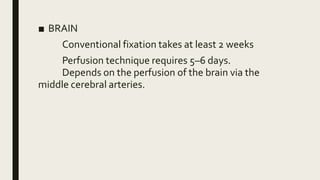
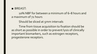
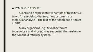
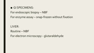
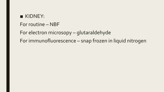
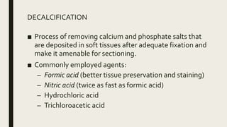
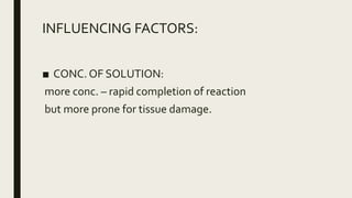
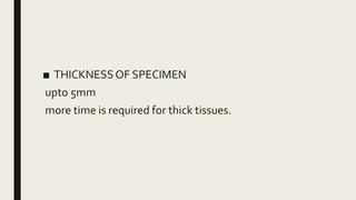
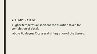
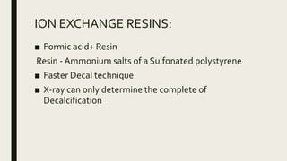
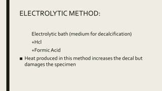
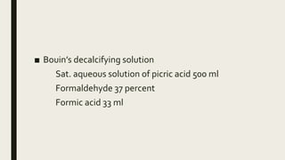
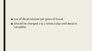
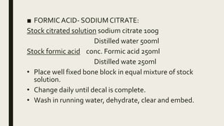
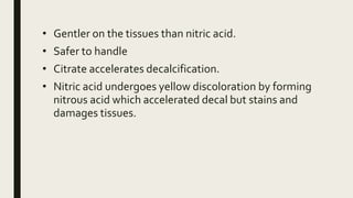
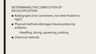
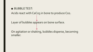
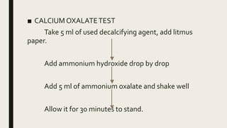
HISTOTECHNIQUES-
FIXATION &
DECALCIFICATION
DR. JANANI MATHIALAGAN
FIXATION:
■ Process by which the constituents of cells and
therefore the tissues are fixed in a physical and partly
chemical state so that they can withstand subsequent
treatment with various reagents with minimal loss of
architecture.
OBJECTIVE OF FIXATION:
■ maintain clear and consistent morphological features
■ to visualize the microanatomy of a tissue with little
disruption of the organization of the tissue
■ must maintain the tissue’s local chemical
composition.
FEATURES OF A FIXATIVE:
■ minimizing the loss or enzymatic destruction of
cellular and extracellular molecules
■ maintaining macromolecular structures
■ protecting tissues from destruction by
microorganisms
■ prevent the subsequent breakdown during longterm
storage
■ high quality and consistent staining with H&E
IDEAL FIXATIVE:
■ penetrate and fix tissues rapidly
■ cost- effective
■ readily disposable or recyclable
■ support long-term tissue storage giving excellent
microtomy of paraffin blocks
■ Prevent autolysis and bacterial decomposition
■ Non-toxic and non-allergenic
■ Fix all components and make them insoluble to
agents used in tissue processing.
BASIC CONCEPTS
■ Ratio of tissue with fixative 1 : 10-20
■ Time for fixation - 8hrs minimum at room
temperature.
■ Tissue thickness should be approximately 4mm
■ Fixing solution should never be heated for faster
fixation. (shrinkage and affects staining quality)
FACTORSAFFECTINGTHE QUALITY OF
FIXATION:
■ pH
■ Buffers
■ Temperature.
■ Penetration of fixatives.
■ Osmolality of fixatives.
■ Concentration of fixatives.
■ Duration of fixation.
■ Volume change.
■ Agitation
■ Additives
■ Ph
Ideal pH required is 6-8
■ Buffers
- phosphate, cacodylate, bicarbonate, tris and acetate.
■ Temperature:
The diffusion of molecules increases with rising
temperature due to their more rapid movement and
vibration.
for histopathology - room temperature
for EM and immunohistochemistry - around 0-4 C.
for rapid fixation – heated formalin upto 60 degreeC is
used.
■ Penetration of fixatives:
done to ensure fixation and hardening.
depends on diffusion
rate of reaction with the tissue.
■ Osmolality of fixatives and ionic composition
hypertonic causes shrinkage
hypotonic causes swelling.
The best morphological results are obtained with
solutions that are slightly hypertonic.
Eg: 0.9% NaCl and 15% calcium chloride
Similarly, various ions (Na+, K+, Ca2+, Mg2+) can
affect cell shape and structure.
The ionic composition of fluids should be as isotonic
as possible to the tissues.
■ Concentration of the fixative:
Concentrations of formalin above 10% causes increased
hardening and gets deposited as white precipitate.
But at 4% provides for greatest solubility.
For electron microscopy – 0.25% glutaraldehyde is used
■ Duration of fixation and size of specimens
depth (d) reached by a fixative is directly proportional to
the square root of duration of fixation (t) and (k) is
coefficient of diffusability
d=k x √t
Prolonged fixation
-Shrinkage and hardening of the tissue.
-Inhibition of enzyme activity .
-Loss of immunological reaction.
■ Volume change:
During fixation tissue commonly changes its volume.
The swelling effect by fixation and the shrinking effect
by dehydration compensate each other.
Usually tissue fixed in formalin at the end of tissue
processing shrinks by 33%.
■ Agitation:
Brisk stirring or disturbance of a liquid.
- increases the speed of penetration of fixatives into the
tissues.
- Slow agitation - No effect on penetration.
- Fast agitation – Damages soft/ friable tissue.
■ Additives
Electrolytes: calcium chloride,
potassium thiocyanate,
ammonium sulfate,
potassium dihydrogen phosphate.
Non-electrolytes: sucrose,
dextran,
and detergent.
TYPES OF FIXATIVES:
■ PHYSICAL
1. Heating
2. Microwaving
3. Freeze-drying
■ CHEMICAL
1. Aldehydes
(formaldehyde
glutaraldehyde)
2. Protein denaturing
agents
-alcoholic
-acidic
3. Oxidising agent –
osmium tetroxide
4. Miscellaneous
HEAT FIXATION:
Eg- Picking up a frozen section on a warm microscope
slide both attaches the section to the slide and partially
fixes it by heat and dehydration.
Mainly used to accelerate other forms of fixation as well
as the steps of tissue processing.
PRINCIPLE:
Rapid freezing of tissues at -160degree C isopentane
immersion - QUENCHING
Removal of water (ice) by sublimation in vacuum at a
higher temperature of -40degreeC in a drying chamber
phosphorus pentoxide
Vacuum embedding with molten wax
10 minutes for completion
■ ADVANTAGES
Little shrinkage of tissue
Enzyme studies
Little chemical alteration of protein
No loss of glycogen
MICROWAVE FIXATION:
■ Microwave heating speeds fixation.
■ from 12 hours to less than 20 minutes.
■ Disadvantage- production of dangerous vapors
resulting in safety problems
■ Recent development- commercial glyoxal-based
fixatives which do not form vapors have been
introduced.
PRINCIPLE:
Exposure to electromagnetic fields (microwave)
Oscillation of water molecules and protein at 2450 times
per second
Induces kinetic motion
Instantaneous and uniform heat
FREEZE-DRYING:
■ Tissues are cut into thin sections
■ Immersed in liquid nitrogen
■ The water is removed in a vacuum chamber at −40°C
■ The tissue can be post-fixed with formaldehyde vapor or
acetone or alcohol.
1. CROSS-LINKING FIXATIVES/ALDEHYDES:
■ Also know as Covalent additive fixatives
■ MOA- forming cross-links within and between
proteins and nucleic acids
■ EG- formaldehyde, glutaraldehyde, and other
aldehydes, e.g. chloral hydrate and glyoxal, metal
salts such as mercuric and zinc chloride, and other
metallic compounds such as osmium tetroxide.
FORMALDEHYDE:
■ Commonest fixative - Formaldehyde in its 10% neutral
buffered form (NBF)
■ In an aqueous solution formaldehyde forms methylene
hydrate
■ Methylene hydrate forms methylene bridge b/w the
protein molecules.
■ The side chains of proteins that are most reactive with
methylene and hence have the highest affinity for
formaldehyde are lysine, cysteine, histidine, arginine &
tyrosine.
■ Site of crosslinking in short-term fixation is between
the hydroxymethyl group on a lysine side chain and
arginine, asparagine, glutamine or tyrosine
■ Long time storage of tissue specimen in formalin
causes the reactive groups to be oxidised to the more
stable groups like carboxylic acid
■ But returning the specimen to water or alcohol
reverses this condition.
FORMALIN PIGMENT:
From laked hemoglobin
Fine dark brown or black crystalline precipitate
Formalin pigment – Artifacts in tissues
TO PREVENT - USE BUFFERED FORMALIN
FORMALDEHYDE FIXATIVES:
■ 10% FORMALIN
Formaldehyde 40% - 10ml
Distilled water - 90ml
■ 10% SALINE FORMALIN
Formaldehyde 40% - 10ml
Distilled water - 90ml
Sodium chloride - 9gm
■ 10% BUFFERED FORMALIN
Formaldehyde 40% - 10ml
Sodium dihydrogen phosphate - 0.4gm
Disodium hydrogen phosphate - 0.65gm
Distilled water - 90ml
ADVANTAGE – prevents formalin pigment formation.
■ ALCOHOLIC FORMALDEHYDE
Formaldehyde 40% - 100ml
Alcohol 95% - 900ml
Calcium acetate - 0.5gm
(for neutrality)
■ BOUIN’S FLUID
saturated picric acid (1.2gm/100ml) - 750ml
Formalin (40%) - 250ml
Glacial acetic acid - 50ml
GLUTARALDEHYDE
■ React with a wide range of proteins, histochemical
targets (including - antibodies, enzymes)
■ Cross-linking is irreversible and withstands acids, urea
and heat
■ Results in better preservation of ultrastructure, but
negatively affects immunohistochemical methods
and slows the penetration by the fixative.
■ PRINCIPLE: stabilizes by cross-linking
Does not fix lipids
Secondary fixation ( Osmium tetroxide )
USE: Electron microscopy work
ADVANTAGES:
More crosslinkage and more stable
Less shrinkage
Does not cause dermatitis, less irritating
■ DISADVANTAGE:
Expensive
Solution is less stable
Slower penetration than formalin
2. COAGULANT FIXATIVE:
■ Disruption of the tertiary structure of proteins
(denaturation, changes their physical properties, causing
insolubility, loss of function)
■ Coagulation of protein (lipoprotein and collagen)
maintains cellular architecture
■ But also causes cytoplasmic flocculation and poor
preservation of mitochondria and secretory granules.
■ So not useful in ultrastructural analysis.
■ Alcoholic coagulant- ethanol, methanol and acetone
■ Acidic coagulant- picric acid and trichloroacetic acid
ALCOHOLIC COAGULANT FIXATIVES
- Used for nucleic acid studies.
- Used as a fixatives for urgent biopsies where alcohol
penetrates the tissue rapidly.
■ CARNOY’S FIXATIVE:
Absolute ethyl alcohol - 60 ml
Chloroform - 30 ml
Glacial acetic acid - 10 ml
■ Advantage-Small tissue fragments (eg-curettings)
For glycogen and nuclear staining.
PICRIC ACID FIXATIVES:
■ BOUIN’S FLUID
1.2 % of picric acid 75ml
Formalin 25ml
Glacial acetic acid 5ml
■ Provides better preservation of glycogen than other
fixatives.
■ Yellowish discolouration is removed by lithium
carbonate in ethyl alcohol.
■ ROSSMAN’S FLUID
100% ethanol saturated with picric acid – 90ml
Neutralized commercial formalin – 10ml
■ GENDRE’S FLUID
90% ethanol saturated with picric acid - 80ml
Formaldehyde 40% - 15ml
Glacial acetic acid - 5ml
3. OXIDISING AGENTS:
OSMIUMTETROXIDE FIXATION
Vapour fixative.
Osmium tetroxide is known to interact with nucleic
acids, specifically with the 2,3-glycol moiety in terminal
ribose groups and the 5,6 double bonds of thymine
residues
■ USES-
secondary fixative for electron microscope examinations
stain lipids in frozen sections
■ DISADVANTAGE:
Toxic vapours affecting eyes, nose, throat
expensive
4. MISCELLANEOUS:
MERCURIC CHLORIDE:
FORMULAS IN USE:
Zenker’s Mercuric Chloride- 50g
Potassium dichromate 25g
Sodium sulfate 10g
Distilled water 1000ml
Schaudinn’s sublimated alcohol
Mercuric chloride 3g
Distilled water 50ml
ADVANTAGES:
■ Better staining of nuclei and connective tissue.
■ Trichrome staining
■ Gives best results with metachromatic staining.
DISADVANTAGES
■ Solution rapidly deteriorates.
■ Lysis of RBC’s and removes hemosiderin.
■ Marked shrinkage
■ Reduces demonstrable glycogen amount
■ slow penetration
■ Tissue becomes hard and brittle
■ Diffuse black granule formation in tissues which must
be removed before staining.
■ Saturated iodine in 96% alcohol is added
■ Rich brown color in conferred
■ Removed by absolute alcohol
CHROMATE FIXATIVES:
■ Chromium trioxide dissolves in water
■ Acidic solution of chromic acid
■ Aldehyde
■ Chromate makes unsaturated but not saturated lipids
insoluble upon prolonged (>48 hours) fixation and
hence mitochondria are well preserved by dichromate
fixatives.
■ Used to prepare neuroendocrine tissues for staining.
■ HELLY’S FLUID
(mercury + chromate + formalin)
For demonstrating chromaffin cells eg-adrenal medulla,
mitochondria, mitotic figures.
provides faster preservation
FORMULAS IN USE
 Orth’s fluid
2.5% potassium dichromate - 100ml
Sodium sulfate – 1gm
Formalin - 10ml
 Regaud’s (Moller’s) fluid
3% potassium dichromate – 80ml
Formalin – 20ml
COMPOUND FIXATIVES:
■ A dehydrant alcohol with aldehyde is added to
produce alcoholic formalin.
■ useful for specific tissues
■ e.g. fatty tissues such as breast, in which preservation
of the lipid is not important.
■ Also fixation of gross specimens in alcoholic formalin
may aid in identifying lymph nodes embedded in fat.
FIXATION FOR CELLULAR
COMPONENTS:
■ CARBOHYDRATE: Alcoholic fixatives
■ LIPIDS: Elftman’s fluid for cryostat method
Baker’s formol calcium
■ PROTEIN: Neutral buffered formalin
Saline formalin
SECONDARY FIXATION:
■ Tissue may be fixed with two fixatives in succession.
■ Tissue fixed with buffered formaldehyde are post fixed
with mercuric chloride-formaldehyde solution.
■ This leads to formation of metallic mercury which is
removed by iodine solution.
■ Advantage – better staining.
■ Electron microscopy - tissues fixed in glutaraldehyde are
post-fixed with osmium tetroxide.
FIXATION FOR SPECIALTISSUES:
■ EYES:
fixed in NBF for about 48 hours.
to speed fixation one or two small windows can be cut
into the globe after 24 hours.
Iris and Optic nerve are removed
The components of the globe are fixed for an additional
48 hours in buffered formaldehyde.
■ BRAIN
Conventional fixation takes at least 2 weeks
Perfusion technique requires 5–6 days.
Depends on the perfusion of the brain via the
middle cerebral arteries.
■ LUNG
Lung biopsies are typically fixed in NBF.
These lungs can be cut within 2 hours
Fixed with NBF through a cannula into the main
bronchi.
■ BREAST:
10% NBF for between a minimum of 6–8 hours and
a maximum of 72 hours
Should be sliced at 5mm intervals
Time from tissue acquisition to fixation should be
as short as possible in order to prevent lysis of clinically
important biomarkers, such as estrogen receptors,
progesterone receptors.
■ LYMPHOIDTISSUE:
Sliced and a representative sample of fresh tissue
taken for special studies (e.g. flow cytometry or
molecular analysis).The rest of the lymph node is fixed
in NBF.
Many organisms (e.g. Mycobacterium
tuberculosis and viruses) may sequester themselves in
the lymphoid reticular system.
■ GI SPECIMENS:
For endoscopic biopsy – NBF
For enzyme assay – snap-frozen without fixation
LIVER:
Routine – NBF
For electron microscopy - glutaraldehyde
■ KIDNEY:
For routine – NBF
For electron microsopy – glutaraldehyde
For immunofluorescence – snap frozen in liquid nitrogen
DECALCIFICATION
DECALCIFICATION
■ Process of removing calcium and phosphate salts that
are deposited in soft tissues after adequate fixation and
make it amenable for sectioning.
■ Commonly employed agents:
– Formic acid (better tissue preservation and staining)
– Nitric acid (twice as fast as formic acid)
– Hydrochloric acid
– Trichloroacetic acid
INFLUENCING FACTORS:
■ CONC. OF SOLUTION:
more conc. – rapid completion of reaction
but more prone for tissue damage.
■ THICKNESS OF SPECIMEN
upto 5mm
more time is required for thick tissues.
■ TEMPERATURE
Higher temperature shortens the duration taken for
completion of decal.
above 60 degree C causes disintegration of the tissues.
BASIC CONCEPTS:
■ SELECTEDTISSUE MUST BE SUSPENDED IN DECAL
FLUIDWITHWAXEDTHREAD ASTHIS ALLOWSTHE
SOLUTIONTO PASSTO ALL SURFACE OFTISSUE
■ THEVOLUME OFTISSUE : DECAL FLUID
1 : 50-100
■ THE PROGRESS OF DECALCIFICATION ISTESTED AT
REGULAR INTERVALS.
■ WHEN DECALCIFICATION IS COMPLETE,TISSUE IS
TRANSFERRED DIRECTLYTO 70% ALCOHOL & GIVEN
SEVERAL CHANGES OVER 8-12 HRS ASTHIS
EFFECTIVELYWASHESTHE ACID AND DEHYDRATES.
■ TISSUE ISTHEN COMPETELY DEHYDRATED AND
PROCESSED ACCORDINGTO REQUIRED EMBEDDING
TECHNIQUE.
METHODS:
■ ACID DECALCIFICATION
■ ION EXCHANGE RESINS
■ ELECTRICAL IONIZATION
■ CHELATING METHOD
■ SURFACE DECALCIFICATIION
ACID DECALCIFICATION
1. NITRIC ACID
2. FORMIC ACID
3. HYDROCHLORIC ACID
4. TCA
ALONGWITH COMBINATIONOF NEUTRALIZERTO
PREVENT EXCESSIVETISSUE SWELLING
Ideal time required is 24-48 hrs.
■ AQUEOUS NITRIC ACID(CLAYDEN,1952)
-NITRICACID 5-10 ml
-DISTILLEDWATER 100 ml
Advantages: Rapid action
Good nuclear staining
Disadvantage:Tissue left for long time causes damage
to cell
■ NITRIC ACID- FORMALDEHYDE
NITRIC ACID 10 ml
FORMALIN 5-10 ml
DISTILLEDWATER 100 ml
Advantage: Urgent biopsy
Good nuclear staining
■ GOODING & STEWART’S FLUID(1932)
FORMIC ACID 5 ml
DISTILLEDWATER 90 ml
FORMALIN(40%) 5 ml
■ TCA: FOR SMALL PIECES OF DELICATETISSUE
■ VON EBNER’S FLUID: (NaCl+ distilled water+
Hcl)..Decal the teeth= 3-5 days
■ CITRATE CITRIC ACID BUFFER
Citric acid monohydrate
Ammonium citrate anhydrous
Zinc sulphate
Chloroform
Advantage - No damage to cell, Good staining
■ PERENYI’S FLUID
Nitric acid
Abs Ethanol
Chromic acid
CHELATINGAGENTS:
EDTA binds with Calcium forming non ionized soluble
complex
Slow process - 2-4 days for 3-4 mm thickness
■ EDTA( Hilleman /Lee):
EDTA + Formalin+ Distilled water
Used in Bone Marrow Biopsy
ION EXCHANGE RESINS:
■ Formic acid+ Resin
Resin - Ammonium salts of a Sulfonated polystyrene
■ Faster Decal technique
■ X-ray can only determine the complete of
Decalcification
ELECTROLYTIC METHOD:
Electrolytic bath (medium for decalcification)
+Hcl
+FormicAcid
■ Heat produced in this method increases the decal but
damages the specimen
■ Bouin’s decalcifying solution
Sat. aqueous solution of picric acid 500 ml
Formaldehyde 37 percent
Formic acid 33 ml
■ 1oz of decal solution per gram of tissue
■ Should be changed 1 to 2 times a day until decal is
complete.
■ FORMIC ACID- SODIUM CITRATE:
Stock citrated solution sodium citrate 100g
Distilled water 500ml
Stock formic acid conc. Formic acid 250ml
Distilled wate 250ml
• Place well fixed bone block in equal mixture of stock
solution.
• Change daily until decal is complete.
• Wash in running water, dehydrate, clear and embed.
• Gentler on the tissues than nitric acid.
• Safer to handle
• Citrate accelerates decalcification.
• Nitric acid undergoes yellow discoloration by forming
nitrous acid which accelerated decal but stains and
damages tissues.
DETERMININGTHE COMPLETIONOF
DECALCIFICATION:
■ Radiography (not convenient, not when fixative is
HgCl)
■ Physical methods (damages tissues producing
artifacts)
-Needling, slicing, squeezing, probing.
■ Chemical methods
■ BUBBLETEST:
Acids react with CaCo3 in bone to produce C02.
Layer of bubbles appears on bone surface.
On agitation or shaking, bubbles disperse, becoming
smaller.
■ CALCIUM OXALATETEST
Take 5 ml of used decalcifying agent, add litmus
paper.
Add ammonium hydroxide drop by drop
Add 5 ml of ammonium oxalate and shake well
Allow it for 30 minutes to stand.
■ If precipitation present after ammonium oxalate
addition (presence of cloudiness), small amount of
calcium is present and decalcification is incomplete.
■ If after 30 minutes of standing fluid gets clear, then
decalcification is complete.
Histotechniques  fixation-&_decalcification

Histotechniques fixation-&_decalcification

  • 1.
  • 2.
    FIXATION: ■ Process bywhich the constituents of cells and therefore the tissues are fixed in a physical and partly chemical state so that they can withstand subsequent treatment with various reagents with minimal loss of architecture.
  • 3.
    OBJECTIVE OF FIXATION: ■maintain clear and consistent morphological features ■ to visualize the microanatomy of a tissue with little disruption of the organization of the tissue ■ must maintain the tissue’s local chemical composition.
  • 4.
    FEATURES OF AFIXATIVE: ■ minimizing the loss or enzymatic destruction of cellular and extracellular molecules ■ maintaining macromolecular structures ■ protecting tissues from destruction by microorganisms ■ prevent the subsequent breakdown during longterm storage ■ high quality and consistent staining with H&E
  • 5.
    IDEAL FIXATIVE: ■ penetrateand fix tissues rapidly ■ cost- effective ■ readily disposable or recyclable ■ support long-term tissue storage giving excellent microtomy of paraffin blocks ■ Prevent autolysis and bacterial decomposition ■ Non-toxic and non-allergenic ■ Fix all components and make them insoluble to agents used in tissue processing.
  • 6.
    BASIC CONCEPTS ■ Ratioof tissue with fixative 1 : 10-20 ■ Time for fixation - 8hrs minimum at room temperature. ■ Tissue thickness should be approximately 4mm ■ Fixing solution should never be heated for faster fixation. (shrinkage and affects staining quality)
  • 7.
    FACTORSAFFECTINGTHE QUALITY OF FIXATION: ■pH ■ Buffers ■ Temperature. ■ Penetration of fixatives. ■ Osmolality of fixatives. ■ Concentration of fixatives. ■ Duration of fixation. ■ Volume change. ■ Agitation ■ Additives
  • 8.
    ■ Ph Ideal pHrequired is 6-8 ■ Buffers - phosphate, cacodylate, bicarbonate, tris and acetate.
  • 9.
    ■ Temperature: The diffusionof molecules increases with rising temperature due to their more rapid movement and vibration. for histopathology - room temperature for EM and immunohistochemistry - around 0-4 C. for rapid fixation – heated formalin upto 60 degreeC is used.
  • 10.
    ■ Penetration offixatives: done to ensure fixation and hardening. depends on diffusion rate of reaction with the tissue.
  • 11.
    ■ Osmolality offixatives and ionic composition hypertonic causes shrinkage hypotonic causes swelling. The best morphological results are obtained with solutions that are slightly hypertonic. Eg: 0.9% NaCl and 15% calcium chloride Similarly, various ions (Na+, K+, Ca2+, Mg2+) can affect cell shape and structure. The ionic composition of fluids should be as isotonic as possible to the tissues.
  • 12.
    ■ Concentration ofthe fixative: Concentrations of formalin above 10% causes increased hardening and gets deposited as white precipitate. But at 4% provides for greatest solubility. For electron microscopy – 0.25% glutaraldehyde is used
  • 13.
    ■ Duration offixation and size of specimens depth (d) reached by a fixative is directly proportional to the square root of duration of fixation (t) and (k) is coefficient of diffusability d=k x √t Prolonged fixation -Shrinkage and hardening of the tissue. -Inhibition of enzyme activity . -Loss of immunological reaction.
  • 14.
    ■ Volume change: Duringfixation tissue commonly changes its volume. The swelling effect by fixation and the shrinking effect by dehydration compensate each other. Usually tissue fixed in formalin at the end of tissue processing shrinks by 33%.
  • 15.
    ■ Agitation: Brisk stirringor disturbance of a liquid. - increases the speed of penetration of fixatives into the tissues. - Slow agitation - No effect on penetration. - Fast agitation – Damages soft/ friable tissue.
  • 16.
    ■ Additives Electrolytes: calciumchloride, potassium thiocyanate, ammonium sulfate, potassium dihydrogen phosphate. Non-electrolytes: sucrose, dextran, and detergent.
  • 17.
    TYPES OF FIXATIVES: ■PHYSICAL 1. Heating 2. Microwaving 3. Freeze-drying ■ CHEMICAL 1. Aldehydes (formaldehyde glutaraldehyde) 2. Protein denaturing agents -alcoholic -acidic 3. Oxidising agent – osmium tetroxide 4. Miscellaneous
  • 18.
    HEAT FIXATION: Eg- Pickingup a frozen section on a warm microscope slide both attaches the section to the slide and partially fixes it by heat and dehydration. Mainly used to accelerate other forms of fixation as well as the steps of tissue processing.
  • 19.
    PRINCIPLE: Rapid freezing oftissues at -160degree C isopentane immersion - QUENCHING Removal of water (ice) by sublimation in vacuum at a higher temperature of -40degreeC in a drying chamber phosphorus pentoxide Vacuum embedding with molten wax 10 minutes for completion
  • 20.
    ■ ADVANTAGES Little shrinkageof tissue Enzyme studies Little chemical alteration of protein No loss of glycogen
  • 21.
    MICROWAVE FIXATION: ■ Microwaveheating speeds fixation. ■ from 12 hours to less than 20 minutes. ■ Disadvantage- production of dangerous vapors resulting in safety problems ■ Recent development- commercial glyoxal-based fixatives which do not form vapors have been introduced.
  • 22.
    PRINCIPLE: Exposure to electromagneticfields (microwave) Oscillation of water molecules and protein at 2450 times per second Induces kinetic motion Instantaneous and uniform heat
  • 23.
    FREEZE-DRYING: ■ Tissues arecut into thin sections ■ Immersed in liquid nitrogen ■ The water is removed in a vacuum chamber at −40°C ■ The tissue can be post-fixed with formaldehyde vapor or acetone or alcohol.
  • 24.
    1. CROSS-LINKING FIXATIVES/ALDEHYDES: ■Also know as Covalent additive fixatives ■ MOA- forming cross-links within and between proteins and nucleic acids ■ EG- formaldehyde, glutaraldehyde, and other aldehydes, e.g. chloral hydrate and glyoxal, metal salts such as mercuric and zinc chloride, and other metallic compounds such as osmium tetroxide.
  • 25.
    FORMALDEHYDE: ■ Commonest fixative- Formaldehyde in its 10% neutral buffered form (NBF) ■ In an aqueous solution formaldehyde forms methylene hydrate ■ Methylene hydrate forms methylene bridge b/w the protein molecules. ■ The side chains of proteins that are most reactive with methylene and hence have the highest affinity for formaldehyde are lysine, cysteine, histidine, arginine & tyrosine.
  • 26.
    ■ Site ofcrosslinking in short-term fixation is between the hydroxymethyl group on a lysine side chain and arginine, asparagine, glutamine or tyrosine ■ Long time storage of tissue specimen in formalin causes the reactive groups to be oxidised to the more stable groups like carboxylic acid ■ But returning the specimen to water or alcohol reverses this condition.
  • 27.
    FORMALIN PIGMENT: From lakedhemoglobin Fine dark brown or black crystalline precipitate Formalin pigment – Artifacts in tissues TO PREVENT - USE BUFFERED FORMALIN
  • 28.
    FORMALDEHYDE FIXATIVES: ■ 10%FORMALIN Formaldehyde 40% - 10ml Distilled water - 90ml ■ 10% SALINE FORMALIN Formaldehyde 40% - 10ml Distilled water - 90ml Sodium chloride - 9gm
  • 29.
    ■ 10% BUFFEREDFORMALIN Formaldehyde 40% - 10ml Sodium dihydrogen phosphate - 0.4gm Disodium hydrogen phosphate - 0.65gm Distilled water - 90ml ADVANTAGE – prevents formalin pigment formation.
  • 30.
    ■ ALCOHOLIC FORMALDEHYDE Formaldehyde40% - 100ml Alcohol 95% - 900ml Calcium acetate - 0.5gm (for neutrality)
  • 31.
    ■ BOUIN’S FLUID saturatedpicric acid (1.2gm/100ml) - 750ml Formalin (40%) - 250ml Glacial acetic acid - 50ml
  • 32.
    GLUTARALDEHYDE ■ React witha wide range of proteins, histochemical targets (including - antibodies, enzymes) ■ Cross-linking is irreversible and withstands acids, urea and heat ■ Results in better preservation of ultrastructure, but negatively affects immunohistochemical methods and slows the penetration by the fixative.
  • 33.
    ■ PRINCIPLE: stabilizesby cross-linking Does not fix lipids Secondary fixation ( Osmium tetroxide ) USE: Electron microscopy work ADVANTAGES: More crosslinkage and more stable Less shrinkage Does not cause dermatitis, less irritating
  • 34.
    ■ DISADVANTAGE: Expensive Solution isless stable Slower penetration than formalin
  • 35.
    2. COAGULANT FIXATIVE: ■Disruption of the tertiary structure of proteins (denaturation, changes their physical properties, causing insolubility, loss of function) ■ Coagulation of protein (lipoprotein and collagen) maintains cellular architecture ■ But also causes cytoplasmic flocculation and poor preservation of mitochondria and secretory granules. ■ So not useful in ultrastructural analysis. ■ Alcoholic coagulant- ethanol, methanol and acetone ■ Acidic coagulant- picric acid and trichloroacetic acid
  • 36.
    ALCOHOLIC COAGULANT FIXATIVES -Used for nucleic acid studies. - Used as a fixatives for urgent biopsies where alcohol penetrates the tissue rapidly.
  • 37.
    ■ CARNOY’S FIXATIVE: Absoluteethyl alcohol - 60 ml Chloroform - 30 ml Glacial acetic acid - 10 ml ■ Advantage-Small tissue fragments (eg-curettings) For glycogen and nuclear staining.
  • 38.
    PICRIC ACID FIXATIVES: ■BOUIN’S FLUID 1.2 % of picric acid 75ml Formalin 25ml Glacial acetic acid 5ml ■ Provides better preservation of glycogen than other fixatives. ■ Yellowish discolouration is removed by lithium carbonate in ethyl alcohol.
  • 39.
    ■ ROSSMAN’S FLUID 100%ethanol saturated with picric acid – 90ml Neutralized commercial formalin – 10ml ■ GENDRE’S FLUID 90% ethanol saturated with picric acid - 80ml Formaldehyde 40% - 15ml Glacial acetic acid - 5ml
  • 40.
    3. OXIDISING AGENTS: OSMIUMTETROXIDEFIXATION Vapour fixative. Osmium tetroxide is known to interact with nucleic acids, specifically with the 2,3-glycol moiety in terminal ribose groups and the 5,6 double bonds of thymine residues
  • 41.
    ■ USES- secondary fixativefor electron microscope examinations stain lipids in frozen sections ■ DISADVANTAGE: Toxic vapours affecting eyes, nose, throat expensive
  • 42.
    4. MISCELLANEOUS: MERCURIC CHLORIDE: FORMULASIN USE: Zenker’s Mercuric Chloride- 50g Potassium dichromate 25g Sodium sulfate 10g Distilled water 1000ml Schaudinn’s sublimated alcohol Mercuric chloride 3g Distilled water 50ml
  • 43.
    ADVANTAGES: ■ Better stainingof nuclei and connective tissue. ■ Trichrome staining ■ Gives best results with metachromatic staining.
  • 44.
    DISADVANTAGES ■ Solution rapidlydeteriorates. ■ Lysis of RBC’s and removes hemosiderin. ■ Marked shrinkage ■ Reduces demonstrable glycogen amount ■ slow penetration ■ Tissue becomes hard and brittle
  • 45.
    ■ Diffuse blackgranule formation in tissues which must be removed before staining. ■ Saturated iodine in 96% alcohol is added ■ Rich brown color in conferred ■ Removed by absolute alcohol
  • 46.
    CHROMATE FIXATIVES: ■ Chromiumtrioxide dissolves in water ■ Acidic solution of chromic acid ■ Aldehyde ■ Chromate makes unsaturated but not saturated lipids insoluble upon prolonged (>48 hours) fixation and hence mitochondria are well preserved by dichromate fixatives.
  • 47.
    ■ Used toprepare neuroendocrine tissues for staining. ■ HELLY’S FLUID (mercury + chromate + formalin) For demonstrating chromaffin cells eg-adrenal medulla, mitochondria, mitotic figures. provides faster preservation
  • 48.
    FORMULAS IN USE Orth’s fluid 2.5% potassium dichromate - 100ml Sodium sulfate – 1gm Formalin - 10ml  Regaud’s (Moller’s) fluid 3% potassium dichromate – 80ml Formalin – 20ml
  • 49.
    COMPOUND FIXATIVES: ■ Adehydrant alcohol with aldehyde is added to produce alcoholic formalin. ■ useful for specific tissues ■ e.g. fatty tissues such as breast, in which preservation of the lipid is not important. ■ Also fixation of gross specimens in alcoholic formalin may aid in identifying lymph nodes embedded in fat.
  • 50.
    FIXATION FOR CELLULAR COMPONENTS: ■CARBOHYDRATE: Alcoholic fixatives ■ LIPIDS: Elftman’s fluid for cryostat method Baker’s formol calcium ■ PROTEIN: Neutral buffered formalin Saline formalin
  • 51.
    SECONDARY FIXATION: ■ Tissuemay be fixed with two fixatives in succession. ■ Tissue fixed with buffered formaldehyde are post fixed with mercuric chloride-formaldehyde solution. ■ This leads to formation of metallic mercury which is removed by iodine solution. ■ Advantage – better staining. ■ Electron microscopy - tissues fixed in glutaraldehyde are post-fixed with osmium tetroxide.
  • 52.
    FIXATION FOR SPECIALTISSUES: ■EYES: fixed in NBF for about 48 hours. to speed fixation one or two small windows can be cut into the globe after 24 hours. Iris and Optic nerve are removed The components of the globe are fixed for an additional 48 hours in buffered formaldehyde.
  • 53.
    ■ BRAIN Conventional fixationtakes at least 2 weeks Perfusion technique requires 5–6 days. Depends on the perfusion of the brain via the middle cerebral arteries.
  • 54.
    ■ LUNG Lung biopsiesare typically fixed in NBF. These lungs can be cut within 2 hours Fixed with NBF through a cannula into the main bronchi.
  • 55.
    ■ BREAST: 10% NBFfor between a minimum of 6–8 hours and a maximum of 72 hours Should be sliced at 5mm intervals Time from tissue acquisition to fixation should be as short as possible in order to prevent lysis of clinically important biomarkers, such as estrogen receptors, progesterone receptors.
  • 56.
    ■ LYMPHOIDTISSUE: Sliced anda representative sample of fresh tissue taken for special studies (e.g. flow cytometry or molecular analysis).The rest of the lymph node is fixed in NBF. Many organisms (e.g. Mycobacterium tuberculosis and viruses) may sequester themselves in the lymphoid reticular system.
  • 57.
    ■ GI SPECIMENS: Forendoscopic biopsy – NBF For enzyme assay – snap-frozen without fixation LIVER: Routine – NBF For electron microscopy - glutaraldehyde
  • 58.
    ■ KIDNEY: For routine– NBF For electron microsopy – glutaraldehyde For immunofluorescence – snap frozen in liquid nitrogen
  • 59.
  • 60.
    DECALCIFICATION ■ Process ofremoving calcium and phosphate salts that are deposited in soft tissues after adequate fixation and make it amenable for sectioning. ■ Commonly employed agents: – Formic acid (better tissue preservation and staining) – Nitric acid (twice as fast as formic acid) – Hydrochloric acid – Trichloroacetic acid
  • 61.
    INFLUENCING FACTORS: ■ CONC.OF SOLUTION: more conc. – rapid completion of reaction but more prone for tissue damage.
  • 62.
    ■ THICKNESS OFSPECIMEN upto 5mm more time is required for thick tissues.
  • 63.
    ■ TEMPERATURE Higher temperatureshortens the duration taken for completion of decal. above 60 degree C causes disintegration of the tissues.
  • 64.
    BASIC CONCEPTS: ■ SELECTEDTISSUEMUST BE SUSPENDED IN DECAL FLUIDWITHWAXEDTHREAD ASTHIS ALLOWSTHE SOLUTIONTO PASSTO ALL SURFACE OFTISSUE ■ THEVOLUME OFTISSUE : DECAL FLUID 1 : 50-100 ■ THE PROGRESS OF DECALCIFICATION ISTESTED AT REGULAR INTERVALS.
  • 65.
    ■ WHEN DECALCIFICATIONIS COMPLETE,TISSUE IS TRANSFERRED DIRECTLYTO 70% ALCOHOL & GIVEN SEVERAL CHANGES OVER 8-12 HRS ASTHIS EFFECTIVELYWASHESTHE ACID AND DEHYDRATES. ■ TISSUE ISTHEN COMPETELY DEHYDRATED AND PROCESSED ACCORDINGTO REQUIRED EMBEDDING TECHNIQUE.
  • 66.
    METHODS: ■ ACID DECALCIFICATION ■ION EXCHANGE RESINS ■ ELECTRICAL IONIZATION ■ CHELATING METHOD ■ SURFACE DECALCIFICATIION
  • 67.
    ACID DECALCIFICATION 1. NITRICACID 2. FORMIC ACID 3. HYDROCHLORIC ACID 4. TCA ALONGWITH COMBINATIONOF NEUTRALIZERTO PREVENT EXCESSIVETISSUE SWELLING Ideal time required is 24-48 hrs.
  • 68.
    ■ AQUEOUS NITRICACID(CLAYDEN,1952) -NITRICACID 5-10 ml -DISTILLEDWATER 100 ml Advantages: Rapid action Good nuclear staining Disadvantage:Tissue left for long time causes damage to cell
  • 69.
    ■ NITRIC ACID-FORMALDEHYDE NITRIC ACID 10 ml FORMALIN 5-10 ml DISTILLEDWATER 100 ml Advantage: Urgent biopsy Good nuclear staining ■ GOODING & STEWART’S FLUID(1932) FORMIC ACID 5 ml DISTILLEDWATER 90 ml FORMALIN(40%) 5 ml
  • 70.
    ■ TCA: FORSMALL PIECES OF DELICATETISSUE ■ VON EBNER’S FLUID: (NaCl+ distilled water+ Hcl)..Decal the teeth= 3-5 days
  • 71.
    ■ CITRATE CITRICACID BUFFER Citric acid monohydrate Ammonium citrate anhydrous Zinc sulphate Chloroform Advantage - No damage to cell, Good staining ■ PERENYI’S FLUID Nitric acid Abs Ethanol Chromic acid
  • 72.
    CHELATINGAGENTS: EDTA binds withCalcium forming non ionized soluble complex Slow process - 2-4 days for 3-4 mm thickness ■ EDTA( Hilleman /Lee): EDTA + Formalin+ Distilled water Used in Bone Marrow Biopsy
  • 73.
    ION EXCHANGE RESINS: ■Formic acid+ Resin Resin - Ammonium salts of a Sulfonated polystyrene ■ Faster Decal technique ■ X-ray can only determine the complete of Decalcification
  • 74.
    ELECTROLYTIC METHOD: Electrolytic bath(medium for decalcification) +Hcl +FormicAcid ■ Heat produced in this method increases the decal but damages the specimen
  • 75.
    ■ Bouin’s decalcifyingsolution Sat. aqueous solution of picric acid 500 ml Formaldehyde 37 percent Formic acid 33 ml
  • 76.
    ■ 1oz ofdecal solution per gram of tissue ■ Should be changed 1 to 2 times a day until decal is complete.
  • 77.
    ■ FORMIC ACID-SODIUM CITRATE: Stock citrated solution sodium citrate 100g Distilled water 500ml Stock formic acid conc. Formic acid 250ml Distilled wate 250ml • Place well fixed bone block in equal mixture of stock solution. • Change daily until decal is complete. • Wash in running water, dehydrate, clear and embed.
  • 78.
    • Gentler onthe tissues than nitric acid. • Safer to handle • Citrate accelerates decalcification. • Nitric acid undergoes yellow discoloration by forming nitrous acid which accelerated decal but stains and damages tissues.
  • 79.
    DETERMININGTHE COMPLETIONOF DECALCIFICATION: ■ Radiography(not convenient, not when fixative is HgCl) ■ Physical methods (damages tissues producing artifacts) -Needling, slicing, squeezing, probing. ■ Chemical methods
  • 80.
    ■ BUBBLETEST: Acids reactwith CaCo3 in bone to produce C02. Layer of bubbles appears on bone surface. On agitation or shaking, bubbles disperse, becoming smaller.
  • 81.
    ■ CALCIUM OXALATETEST Take5 ml of used decalcifying agent, add litmus paper. Add ammonium hydroxide drop by drop Add 5 ml of ammonium oxalate and shake well Allow it for 30 minutes to stand.
  • 82.
    ■ If precipitationpresent after ammonium oxalate addition (presence of cloudiness), small amount of calcium is present and decalcification is incomplete. ■ If after 30 minutes of standing fluid gets clear, then decalcification is complete.