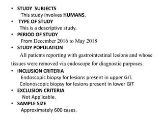
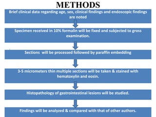
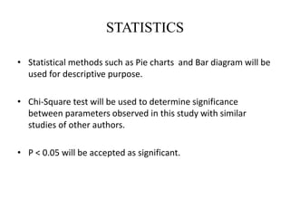
“A CLINICOPATHOLOGICAL STUDY OF
GASTROINTESTINAL ENDOSCOPIC BIOPSIES”
CANDIDATE :
Dr. JANANI MATHILAGAN
1st YEAR POST GRADUATE
DEPARTMENT OF PATHOLOGY
MGMCRI
GUIDE:
Dr. DHANANJAY S. KOTASTHANE MD
PROFESSOR AND HEAD
DEPARTMENT OF PATHOLOGY,
MGMCRI
Co-GUIDE:
DR. TIROU AROUL MS
PROFESSOR
DEPARTMENT OF GENERAL
SURGERY, MGMCRI
Co-GUIDE:
DR.SAJEETH MANIKANDA PRABU DM
ASSISTANT PROFESSOR
DEPARTMENT OF MEDICAL
GASTROENTEROLOGY, MGMCRI
INTRODUCTION
• GI endoscopy in combination with biopsy plays an
important role in the early diagnosis of GI neoplasms.
• Endoscopic screening may detect gastric mucosal lesions
at an early stage so as to prevent progression of lesions
to invasive cancer.
• Biopsy and histological assessment provide a critical
adjunct to endoscopic assessment of the gastrointestinal
tract.
• In diseases such as cancer, coeliac disease and chronic
inflammatory bowel disease, pathological diagnosis
remains the gold standard.
REVIEW OF LITERATURE
• Shanmugasamy K,et al. conducted a study on Clinical correlation of upper
gastrointestinal endoscopic biopsies with histopathological findings and
found there were 24 neoplastic lesions (21%) and 91 non-neoplastic
lesions (79%) were observed out of 115 cases.
• Shefali.H.Karve et al. conducted a clinico-pathological study of colonic
biopsies in which out of 159 colonoscopic biopsies performed, 68 cases
were diagnosed as non-neoplastic lesions, out of which 32 cases of Non-
specific colitis, 11 cases of ulcerative colitis, 5 cases of juvenile polyps, 5
cases of hyperplastic polyps, 4 cases of SRUS, 3 cases of Granulomatous
inflammation, 3 cases of Retention polyp, 2 cases of Crohn’s disease and
one case each of Acute inflammation, Inflammatory polyp and
Endometriosis.
• Rangaswamy R et al conducted a Clinico-colonoscopic and
Histomorphological study on Spectrum of Colonic Diseases and found that
out of 107 biopsies, 76.64% biopsies were non-neoplastic and 23.36%
biopsies were neoplastic lesions.
AIMS & OBJECTIVES
1. To study the histopathological spectrum of endoscopic
gastrointestinal biopsies in MGMCRI, Pondicherry.
2. To correlate histopathological findings with clinical
history and endoscopic findings.
• STUDY SUBJECTS
This study involves HUMANS.
• TYPE OF STUDY
This is a descriptive study.
• PERIOD OF STUDY
From December 2016 to May 2018
• STUDY POPULATION
All patients reporting with gastrointestinal lesions and whose
tissues were removed via endoscope for diagnostic purposes.
• INCLUSION CRITERIA
Endoscopic biopsy for lesions present in upper GIT.
Colonoscopic biopsy for lesions present in lower GIT
• EXCLUSION CRITERIA
Not Applicable.
• SAMPLE SIZE
Approximately 600 cases.
METHODS
`
Brief clinical data regarding age, sex, clinical findings and endoscopic findings
are noted
Specimen received in 10% formalin will be fixed and subjected to gross
examination.
Sections will be processed followed by paraffin embedding
3-5 micrometers thin multiple sections will be taken & stained with
hematoxylin and eosin.
Findings will be analyzed & compared with that of other authors.
Histopathology of gastrointestinal lesions will be studied.
STATISTICS
• Statistical methods such as Pie charts and Bar diagram will be
used for descriptive purpose.
• Chi-Square test will be used to determine significance
between parameters observed in this study with similar
studies of other authors.
• P < 0.05 will be accepted as significant.
ETHICAL ISSUES
The Study Involves:
Collection of biopsy

Pathology janani mathialagan

  • 1.
    “A CLINICOPATHOLOGICAL STUDYOF GASTROINTESTINAL ENDOSCOPIC BIOPSIES” CANDIDATE : Dr. JANANI MATHILAGAN 1st YEAR POST GRADUATE DEPARTMENT OF PATHOLOGY MGMCRI GUIDE: Dr. DHANANJAY S. KOTASTHANE MD PROFESSOR AND HEAD DEPARTMENT OF PATHOLOGY, MGMCRI Co-GUIDE: DR. TIROU AROUL MS PROFESSOR DEPARTMENT OF GENERAL SURGERY, MGMCRI Co-GUIDE: DR.SAJEETH MANIKANDA PRABU DM ASSISTANT PROFESSOR DEPARTMENT OF MEDICAL GASTROENTEROLOGY, MGMCRI
  • 2.
    INTRODUCTION • GI endoscopyin combination with biopsy plays an important role in the early diagnosis of GI neoplasms. • Endoscopic screening may detect gastric mucosal lesions at an early stage so as to prevent progression of lesions to invasive cancer. • Biopsy and histological assessment provide a critical adjunct to endoscopic assessment of the gastrointestinal tract. • In diseases such as cancer, coeliac disease and chronic inflammatory bowel disease, pathological diagnosis remains the gold standard.
  • 3.
    REVIEW OF LITERATURE •Shanmugasamy K,et al. conducted a study on Clinical correlation of upper gastrointestinal endoscopic biopsies with histopathological findings and found there were 24 neoplastic lesions (21%) and 91 non-neoplastic lesions (79%) were observed out of 115 cases. • Shefali.H.Karve et al. conducted a clinico-pathological study of colonic biopsies in which out of 159 colonoscopic biopsies performed, 68 cases were diagnosed as non-neoplastic lesions, out of which 32 cases of Non- specific colitis, 11 cases of ulcerative colitis, 5 cases of juvenile polyps, 5 cases of hyperplastic polyps, 4 cases of SRUS, 3 cases of Granulomatous inflammation, 3 cases of Retention polyp, 2 cases of Crohn’s disease and one case each of Acute inflammation, Inflammatory polyp and Endometriosis. • Rangaswamy R et al conducted a Clinico-colonoscopic and Histomorphological study on Spectrum of Colonic Diseases and found that out of 107 biopsies, 76.64% biopsies were non-neoplastic and 23.36% biopsies were neoplastic lesions.
  • 4.
    AIMS & OBJECTIVES 1.To study the histopathological spectrum of endoscopic gastrointestinal biopsies in MGMCRI, Pondicherry. 2. To correlate histopathological findings with clinical history and endoscopic findings.
  • 5.
    • STUDY SUBJECTS Thisstudy involves HUMANS. • TYPE OF STUDY This is a descriptive study. • PERIOD OF STUDY From December 2016 to May 2018 • STUDY POPULATION All patients reporting with gastrointestinal lesions and whose tissues were removed via endoscope for diagnostic purposes. • INCLUSION CRITERIA Endoscopic biopsy for lesions present in upper GIT. Colonoscopic biopsy for lesions present in lower GIT • EXCLUSION CRITERIA Not Applicable. • SAMPLE SIZE Approximately 600 cases.
  • 6.
    METHODS ` Brief clinical dataregarding age, sex, clinical findings and endoscopic findings are noted Specimen received in 10% formalin will be fixed and subjected to gross examination. Sections will be processed followed by paraffin embedding 3-5 micrometers thin multiple sections will be taken & stained with hematoxylin and eosin. Findings will be analyzed & compared with that of other authors. Histopathology of gastrointestinal lesions will be studied.
  • 7.
    STATISTICS • Statistical methodssuch as Pie charts and Bar diagram will be used for descriptive purpose. • Chi-Square test will be used to determine significance between parameters observed in this study with similar studies of other authors. • P < 0.05 will be accepted as significant.
  • 8.
    ETHICAL ISSUES The StudyInvolves: Collection of biopsy