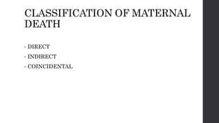
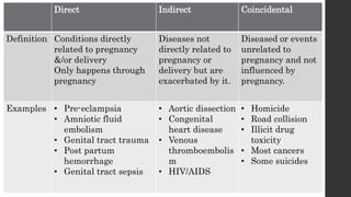
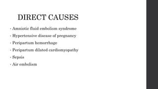
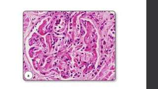
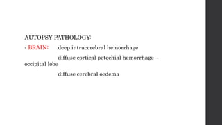
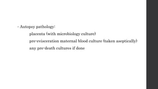
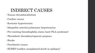
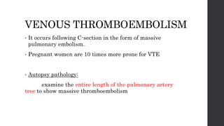
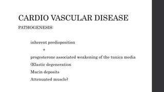
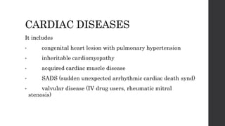
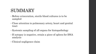
MATERNAL DEATH
AUTOPSY
DR.JANANI MATHIALAGAN
1st year PG - Pathology
OVERVIEW
• Definition
• Classification
• Direct causes
• Indirect causes
• Summary
DEFINITION
• Death of a woman while pregnant or within 42 days of
termination of pregnancy, irrespective of the duration and
site of pregnancy, from any cause related to or aggravated
by pregnancy or its management but not accidental or
incidental causes.
MATERNAL MORTALITY RATIO =
No. of maternal death x 100,000
No. of live births
INDIA:
2007-2009 = 212/100,000 live births
2011-2013 = 167/100,000 live births
CLASSIFICATION OF MATERNAL
DEATH
• DIRECT
• INDIRECT
• COINCIDENTAL
Direct Indirect Coincidental
Definition Conditions directly
related to pregnancy
&/or delivery
Only happens through
pregnancy
Diseases not
directly related to
pregnancy or
delivery but are
exacerbated by it.
Diseased or events
unrelated to
pregnancy and not
influenced by
pregnancy.
Examples • Pre-eclampsia
• Amniotic fluid
embolism
• Genital tract trauma
• Post partum
hemorrhage
• Genital tract sepsis
• Aortic dissection
• Congenital
heart disease
• Venous
thromboembolis
m
• HIV/AIDS
• Homicide
• Road collision
• Illicit drug
toxicity
• Most cancers
• Some suicides
DIRECT CAUSES
• Amniotic fluid embolism syndrome
• Hypertensive disease of pregnancy
• Peripartum hemorrhage
• Peripartum dilated cardiomyopathy
• Sepsis
• Air embolism
AMNIOTIC FLUID EMBOLISM
SYNDROME
• Clinical triad
Hypotension/ cardiac arrest
Pulmonary vasospasm
Coagulopathy with severe bleeding
During or just after labor or caesarean section
Entry of amniotic fluid, fetal hair, amniotic &
fetal squamous cells into maternal circulation
Embolise in the small vessels of the lungs
Triggers acute anaphylactic response
Cardiopulmonary shutdown, clotting cascade,
consumptive coagulopathy.
Important autopsy pathology is in the lungs.
Stains used:
 H & E
 Alcian blue (amniotic acid mucin)
 LP 34 (high molecular wt keratin)
 Endothelial CD31 (diff b/w embolic squames &
sloughed endothelial cells)
• Renal glomeruli:
fibrin thrombi is usually found in capillary lumens
(indicating DIC as a part of AFES)
• Uterus:
mucosal bleeding sites
Amniotic fluid material in mural veins
MEDICO LEGAL ASPECT:
• AFE is inevitably fatal.
• So can be used as a defence against claims of clinical
negligence where there has been fatal peri- or
postpartum hemorrhage.
HYPERTENSIVE DISEASES OF
PREGNANCY
Pre-eclampsia: raised blood pressure,
oedema,
proteinuria.
Predisposing factors
essential hypertension,
renal disease,
obesity
Asssociated with HELLP syndrome (hemolysis, elevated
liver enzymes, low platelet count)
• Eclampsia:
clonic tonic seizures occurring in a patient with pre-
eclampsia.
It has high mortality rate.
Eclampsia induced
endothelial cell damage
Vasogenic oedema
Encephalopathy
HELLP syndrome
Liver failure and
capsular rupture
Intra-abdominal
hemorrhage
Eclampsia
Intracerebral
hemorrhage
death
AUTOPSY PATHOLOGY:
• BRAIN: deep intracerebral hemorrhage
diffuse cortical petechial hemorrhage –
occipital lobe
diffuse cerebral oedema
• KIDNEY: Glomerular endotheliosis
endothelial cells are swollen and appears
bloodless
glomerulus herniates into proximal tubule
endothelial cells vacuolated with lipid
Special stain: Silver – basement membrane thickening and
remodeling resulting in string of beads appearance
• UTERUS AND PLACENTA:
reduced arterial blood supply on the villi, with foci of
infarction.
decidua shows atherosis and fibrinoid necrosis of the
spiral arterioles.
• LIVER:
focal and confluent hemorrhagic necrosis
Microscopically – Periportal fibrin deposition,
hemorrhage,
hepatocyte necrosis
PERI & POSTPARTUM
HEMORRHAGE
CAUSES:
• uterine atony, placenta praevia, retained placenta
• placental abruption
• creta syndromes
• genital tract trauma
• uterine rupture
• abortion
UTERINE ATONY, PLACENTA
PRAEVIA, RETAINED PLACENTA
PLACENTAL ABRUPTION
DEFINITION:
Premature separation of placenta from the walls of
the uterus during pregnancy.
Leaves a clot b/w maternal placental surface and
uterus
CRETA SYNDROMES
Adherent placenta
It can be accreta
increta
percreta
Previous c-
section
Fibrotic scar
Decidua becomes
sub-optimal
Placental villi
attaches directly to
uterine muscle
Adherent placenta
GENITAL TRACT TRAUMA
By large babies or clumsly assisted delivery.
Vagina, cevix, lower uterus can be torn
Entitre genital tract en block dissection is done (from
vagina to uterine fundus)
Fixed
Serially horizontally sliced and sampled to depict tear
dimention and vesses rupture.
UTERINE RUPTURE
• Causes: big baby
small pelvis
prolonged labour
drugs – misoprotol and oxytocics
On examination of uterus, typically lateral rupture is seen.
ABORTION
• Expulsion of products of conception before completion of
28 weeks of gestation.
• Causes of maternal death is due to
ascending genital tract sepsis
uterine hemorrhage
SEPSIS
• Sepsis is commonly due to bacteraemic septic shock
multi-organ failure
DIC
SEPSIS CLASSIFICATION
S.No Category Case definition Agent Pathology
1. Unsafe abortion Illegal termination
of pregnancy
Clostridium
spp.
Mulit organ
failure
2. Ruptured membranes GT infections during
the time of
membrane rupture
E.coli Infected and
inflamed placenta,
cord, membrane,
GT sepsis, MOF
3. Postdelivery Delivery followed by
1-2 days of wellness
followed by GT
infection
Group A
streptococcus
pyogenes
(GAS)
GT sepsis, MOF,
4. Community acquired
sepsis
Membranes intact,
not in labour
GAS,
Pneumococcus
TSS,
MOF
5. Postpartal sepsis related
to birth process, but GT
not involved
C-section wound
infection, infected
spinal anaesthesia
Gram negative
& gram
positive
organisms
Localised sepsis
leading to MOF
• Autopsy pathology:
placenta (with microbiology culture)
pre-evisceration maternal blood culture (taken aseptically)
any pre-death cultures if done
• SEMELWEIS SYNDROME:
INDIRECT CAUSES
• Venous thromboembolism
• Cardiac causes
• Systemic hypertension
• Idiopathic arterial pulmonary hypertension
• Pre-existing thrombophilia states (anti-PLA syndrome)
• Thrombotic thrombocytopaenic purpura
• Stroke
• Psychiatric causes
• SUDEP (sudden unexplained death in epilepsy)
VENOUS THROMBOEMBOLISM
• It occurs following C-section in the form of massive
pulmonary embolism.
• Pregnant women are 10 times more prone for VTE
• Autopsy pathology:
examine the entire length of the pulmonary artery
tree to show massive thromboembolism
CARDIO VASCULAR DISEASE
PATHOGENESIS:
inherent predisposition
+
progesterone associated weakening of the tunica media
(Elastic degeneration
Mucin deposits
Attenuated muscle)
Weakening of the
wall of aorta,
medium and large
arteries
Aneurysm
Dissection
Rupture
CARDIAC DISEASES
It includes
• congenital heart lesion with pulmonary hypertension
• inheritable cardiomyopathy
• acquired cardiac muscle disease
• SADS (sudden unexpected arrhythmic cardiac death synd)
• valvular disease (IV drug users, rheumatic mitral
stenosis)
• Predisposing factors:
lifestyle
obesity
increasing age of pregnant women
In inheritable cardiac conditions (long QT syndrome),
autopsy will be negative and heart morphologically
normal.
Retaining a piece of frozen spleen tissue is done for later
DNA analysis.
PERIPARTUM CARDIOMYOPATHY:
• Heart failure during the last month of pregnancy and
upto 5 months post-delivery with all other causes
excluded.
Aetiology:
oxidative pro-apoptotic stress on myocytes, related
to prolactin.
THROMBOTIC
THROMBOCYTOPAENIC PURPURA
Abnormalities of von
Willebrand factor
Platelet clustering and
adhesion to endothelium
Platele thrombi blocking
small vessels to brain,
kidney, heart
Laboratory data: low platelets
normal clotting factors and fibrin
C/P:
microangiopathic anaemia
renal failure
blockage of arterioles and venules in
myocardium resulting in hemorrhagic infarction and acute heart
failure
PREGNANCY ASSOCIATED
INFECTIONS
• Pregnancy is a relative immunodepressed state
• So
listeriosis,
tuberculosis,
viral infections are more aggressive
EPIDEMIC INFLUENZA
• Type A/ H1N1 influenza
• Mainly affects third trimester
• Results in
influenza pneumonitis,
acute lung injury,
secondary bacterial pneumonia.
HIV
• More prevalent in low-income countries with high HIV
prevalence
• 10 fold increase in maternal mortality
• Death is mostly due to TB or other opportunistic
infections or sepsis.
SUMMARY
• Before evisceration, sterile blood cultures is to be
sampled
• Close attention to pulmonary artery, heart and genital
tract
• Systemic sampling of all organs for histopathology
• If autopsy is negative, retain a piece of spleen for DNA
analysis
• Clinical negligence claim
References:
• Recent advances in histopathology 23 ( chapter 2 )
• WHO website ( maternal mortality )
Thankyou

Maternal death autopsy

  • 1.
  • 2.
    OVERVIEW • Definition • Classification •Direct causes • Indirect causes • Summary
  • 3.
    DEFINITION • Death ofa woman while pregnant or within 42 days of termination of pregnancy, irrespective of the duration and site of pregnancy, from any cause related to or aggravated by pregnancy or its management but not accidental or incidental causes.
  • 4.
    MATERNAL MORTALITY RATIO= No. of maternal death x 100,000 No. of live births INDIA: 2007-2009 = 212/100,000 live births 2011-2013 = 167/100,000 live births
  • 5.
    CLASSIFICATION OF MATERNAL DEATH •DIRECT • INDIRECT • COINCIDENTAL
  • 6.
    Direct Indirect Coincidental DefinitionConditions directly related to pregnancy &/or delivery Only happens through pregnancy Diseases not directly related to pregnancy or delivery but are exacerbated by it. Diseased or events unrelated to pregnancy and not influenced by pregnancy. Examples • Pre-eclampsia • Amniotic fluid embolism • Genital tract trauma • Post partum hemorrhage • Genital tract sepsis • Aortic dissection • Congenital heart disease • Venous thromboembolis m • HIV/AIDS • Homicide • Road collision • Illicit drug toxicity • Most cancers • Some suicides
  • 7.
    DIRECT CAUSES • Amnioticfluid embolism syndrome • Hypertensive disease of pregnancy • Peripartum hemorrhage • Peripartum dilated cardiomyopathy • Sepsis • Air embolism
  • 8.
    AMNIOTIC FLUID EMBOLISM SYNDROME •Clinical triad Hypotension/ cardiac arrest Pulmonary vasospasm Coagulopathy with severe bleeding
  • 9.
    During or justafter labor or caesarean section Entry of amniotic fluid, fetal hair, amniotic & fetal squamous cells into maternal circulation Embolise in the small vessels of the lungs Triggers acute anaphylactic response Cardiopulmonary shutdown, clotting cascade, consumptive coagulopathy.
  • 10.
    Important autopsy pathologyis in the lungs. Stains used:  H & E  Alcian blue (amniotic acid mucin)  LP 34 (high molecular wt keratin)  Endothelial CD31 (diff b/w embolic squames & sloughed endothelial cells)
  • 13.
    • Renal glomeruli: fibrinthrombi is usually found in capillary lumens (indicating DIC as a part of AFES)
  • 15.
    • Uterus: mucosal bleedingsites Amniotic fluid material in mural veins
  • 16.
    MEDICO LEGAL ASPECT: •AFE is inevitably fatal. • So can be used as a defence against claims of clinical negligence where there has been fatal peri- or postpartum hemorrhage.
  • 17.
    HYPERTENSIVE DISEASES OF PREGNANCY Pre-eclampsia:raised blood pressure, oedema, proteinuria. Predisposing factors essential hypertension, renal disease, obesity Asssociated with HELLP syndrome (hemolysis, elevated liver enzymes, low platelet count)
  • 18.
    • Eclampsia: clonic tonicseizures occurring in a patient with pre- eclampsia. It has high mortality rate.
  • 19.
    Eclampsia induced endothelial celldamage Vasogenic oedema Encephalopathy HELLP syndrome Liver failure and capsular rupture Intra-abdominal hemorrhage Eclampsia Intracerebral hemorrhage death
  • 20.
    AUTOPSY PATHOLOGY: • BRAIN:deep intracerebral hemorrhage diffuse cortical petechial hemorrhage – occipital lobe diffuse cerebral oedema
  • 21.
    • KIDNEY: Glomerularendotheliosis endothelial cells are swollen and appears bloodless glomerulus herniates into proximal tubule endothelial cells vacuolated with lipid Special stain: Silver – basement membrane thickening and remodeling resulting in string of beads appearance
  • 23.
    • UTERUS ANDPLACENTA: reduced arterial blood supply on the villi, with foci of infarction. decidua shows atherosis and fibrinoid necrosis of the spiral arterioles.
  • 24.
    • LIVER: focal andconfluent hemorrhagic necrosis Microscopically – Periportal fibrin deposition, hemorrhage, hepatocyte necrosis
  • 25.
    PERI & POSTPARTUM HEMORRHAGE CAUSES: •uterine atony, placenta praevia, retained placenta • placental abruption • creta syndromes • genital tract trauma • uterine rupture • abortion
  • 26.
  • 28.
    PLACENTAL ABRUPTION DEFINITION: Premature separationof placenta from the walls of the uterus during pregnancy. Leaves a clot b/w maternal placental surface and uterus
  • 29.
    CRETA SYNDROMES Adherent placenta Itcan be accreta increta percreta
  • 30.
    Previous c- section Fibrotic scar Deciduabecomes sub-optimal Placental villi attaches directly to uterine muscle Adherent placenta
  • 32.
    GENITAL TRACT TRAUMA Bylarge babies or clumsly assisted delivery. Vagina, cevix, lower uterus can be torn Entitre genital tract en block dissection is done (from vagina to uterine fundus) Fixed Serially horizontally sliced and sampled to depict tear dimention and vesses rupture.
  • 33.
    UTERINE RUPTURE • Causes:big baby small pelvis prolonged labour drugs – misoprotol and oxytocics On examination of uterus, typically lateral rupture is seen.
  • 35.
    ABORTION • Expulsion ofproducts of conception before completion of 28 weeks of gestation. • Causes of maternal death is due to ascending genital tract sepsis uterine hemorrhage
  • 36.
    SEPSIS • Sepsis iscommonly due to bacteraemic septic shock multi-organ failure DIC
  • 37.
    SEPSIS CLASSIFICATION SEPSIS CLASSIFICATION S.NoCategory Case definition Agent Pathology 1. Unsafe abortion Illegal termination of pregnancy Clostridium spp. Mulit organ failure 2. Ruptured membranes GT infections during the time of membrane rupture E.coli Infected and inflamed placenta, cord, membrane, GT sepsis, MOF 3. Postdelivery Delivery followed by 1-2 days of wellness followed by GT infection Group A streptococcus pyogenes (GAS) GT sepsis, MOF, 4. Community acquired sepsis Membranes intact, not in labour GAS, Pneumococcus TSS, MOF 5. Postpartal sepsis related to birth process, but GT not involved C-section wound infection, infected spinal anaesthesia Gram negative & gram positive organisms Localised sepsis leading to MOF
  • 38.
    • Autopsy pathology: placenta(with microbiology culture) pre-evisceration maternal blood culture (taken aseptically) any pre-death cultures if done
  • 39.
  • 40.
    INDIRECT CAUSES • Venousthromboembolism • Cardiac causes • Systemic hypertension • Idiopathic arterial pulmonary hypertension • Pre-existing thrombophilia states (anti-PLA syndrome) • Thrombotic thrombocytopaenic purpura • Stroke • Psychiatric causes • SUDEP (sudden unexplained death in epilepsy)
  • 41.
    VENOUS THROMBOEMBOLISM • Itoccurs following C-section in the form of massive pulmonary embolism. • Pregnant women are 10 times more prone for VTE • Autopsy pathology: examine the entire length of the pulmonary artery tree to show massive thromboembolism
  • 42.
    CARDIO VASCULAR DISEASE PATHOGENESIS: inherentpredisposition + progesterone associated weakening of the tunica media (Elastic degeneration Mucin deposits Attenuated muscle)
  • 43.
    Weakening of the wallof aorta, medium and large arteries Aneurysm Dissection Rupture
  • 44.
    CARDIAC DISEASES It includes •congenital heart lesion with pulmonary hypertension • inheritable cardiomyopathy • acquired cardiac muscle disease • SADS (sudden unexpected arrhythmic cardiac death synd) • valvular disease (IV drug users, rheumatic mitral stenosis)
  • 45.
    • Predisposing factors: lifestyle obesity increasingage of pregnant women In inheritable cardiac conditions (long QT syndrome), autopsy will be negative and heart morphologically normal. Retaining a piece of frozen spleen tissue is done for later DNA analysis.
  • 46.
    PERIPARTUM CARDIOMYOPATHY: • Heartfailure during the last month of pregnancy and upto 5 months post-delivery with all other causes excluded. Aetiology: oxidative pro-apoptotic stress on myocytes, related to prolactin.
  • 47.
    THROMBOTIC THROMBOCYTOPAENIC PURPURA Abnormalities ofvon Willebrand factor Platelet clustering and adhesion to endothelium Platele thrombi blocking small vessels to brain, kidney, heart
  • 48.
    Laboratory data: lowplatelets normal clotting factors and fibrin C/P: microangiopathic anaemia renal failure blockage of arterioles and venules in myocardium resulting in hemorrhagic infarction and acute heart failure
  • 49.
    PREGNANCY ASSOCIATED INFECTIONS • Pregnancyis a relative immunodepressed state • So listeriosis, tuberculosis, viral infections are more aggressive
  • 50.
    EPIDEMIC INFLUENZA • TypeA/ H1N1 influenza • Mainly affects third trimester • Results in influenza pneumonitis, acute lung injury, secondary bacterial pneumonia.
  • 51.
    HIV • More prevalentin low-income countries with high HIV prevalence • 10 fold increase in maternal mortality • Death is mostly due to TB or other opportunistic infections or sepsis.
  • 52.
    SUMMARY • Before evisceration,sterile blood cultures is to be sampled • Close attention to pulmonary artery, heart and genital tract • Systemic sampling of all organs for histopathology • If autopsy is negative, retain a piece of spleen for DNA analysis • Clinical negligence claim
  • 53.
    References: • Recent advancesin histopathology 23 ( chapter 2 ) • WHO website ( maternal mortality )
  • 54.