Download to read offline


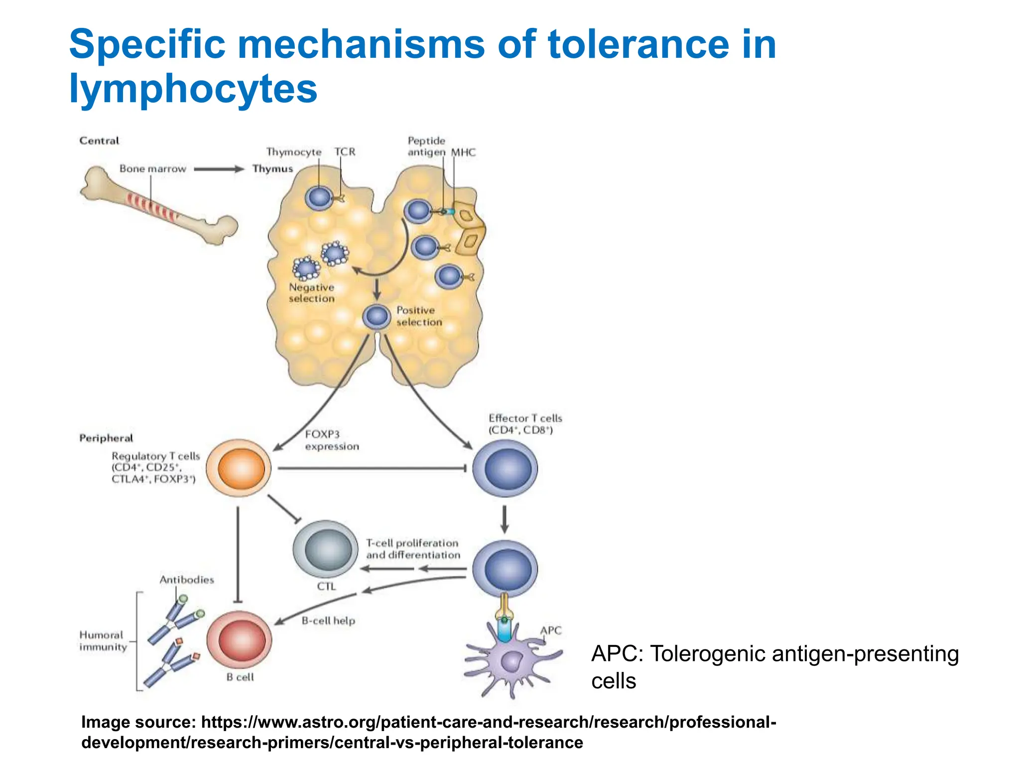


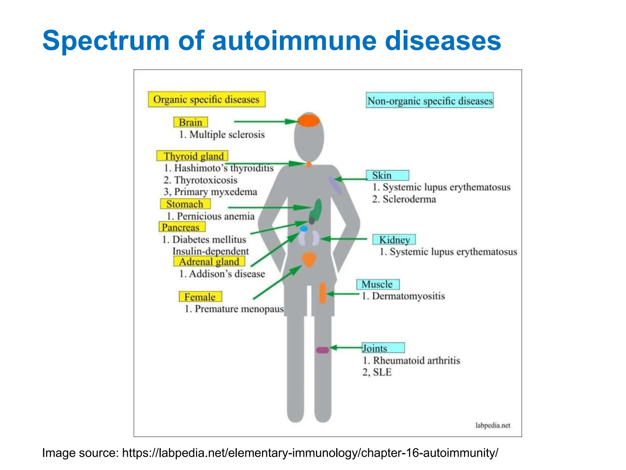
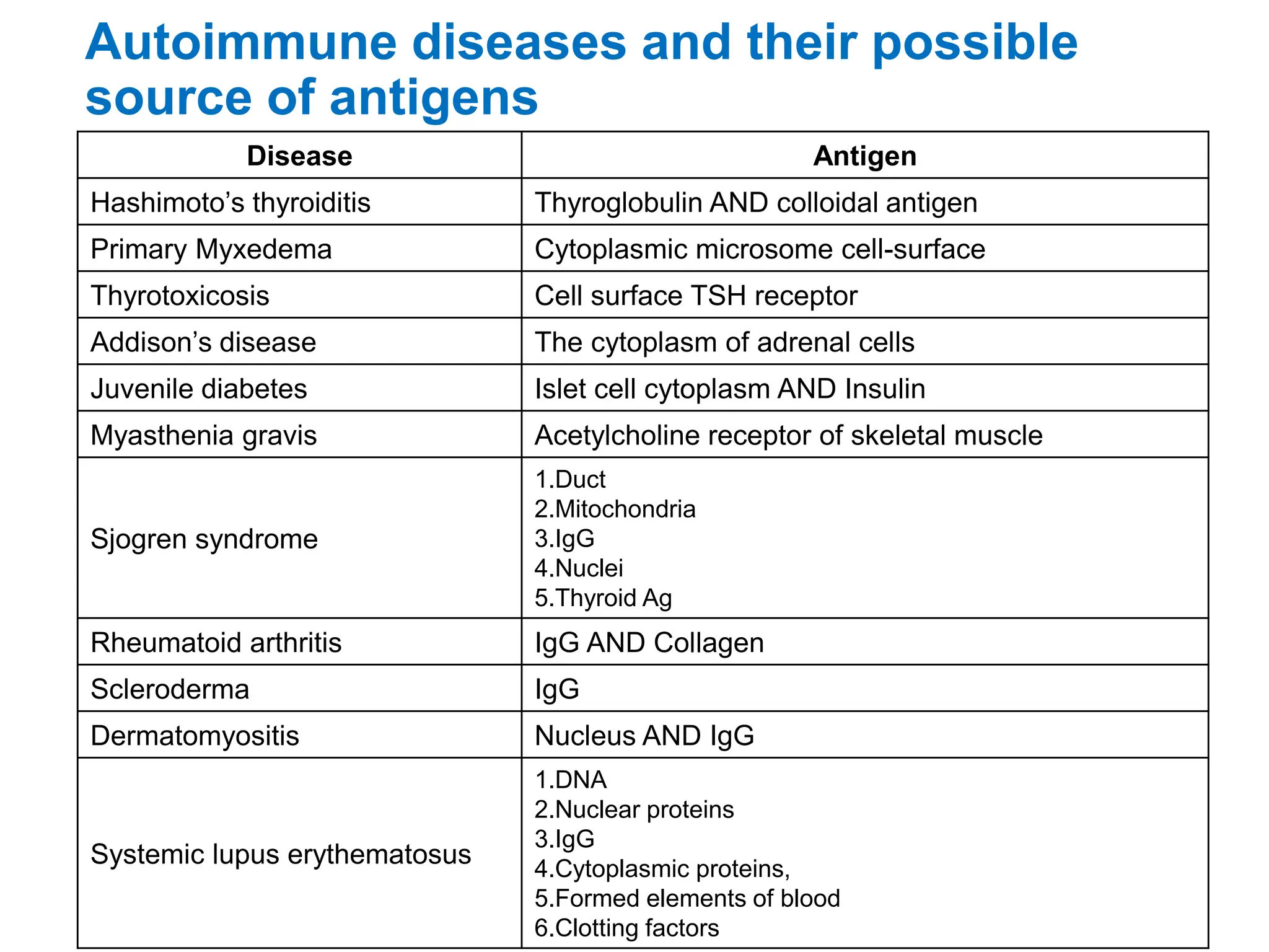

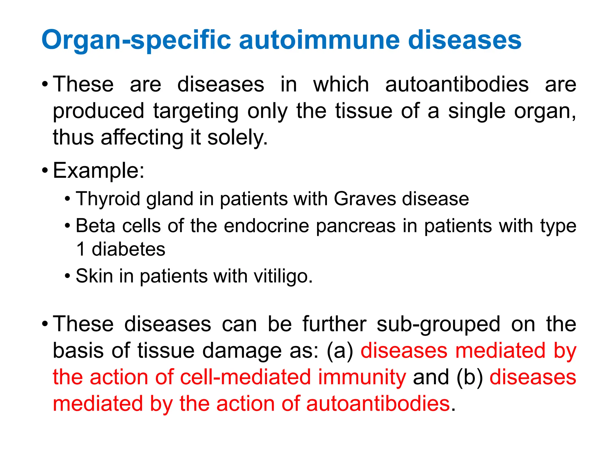


The document discusses immunological tolerance and autoimmunity, outlining the mechanisms by which immune cells develop tolerance to self-antigens and the factors leading to autoimmune diseases. It categorizes autoimmune diseases based on systemic versus organ-specific criteria and describes risk factors such as genetic predisposition, gender, and environmental influences. Additionally, it highlights the importance of therapeutic tolerance induction as a potential treatment for various autoimmune conditions.


