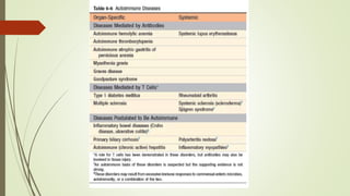
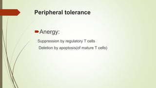
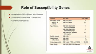
AUTOIMMUNITY
Dr. P. TEJASWI REDDY
MBBS,MD
ASST. PROFESSOR PATHOLOGY
 Introduction
 Examples
 Immunologic tolerance (self tolerance)
 Introduction
• Central tolerance
• Peripheral tolerance
Mechanisms of autoimmunity
 Defective tolerance
 Abnormal display of self antigens
 Inflammation/innate immune response
 Pathogenesis
 Role of Susceptibility Genes
 Role of Infections
 General principles
 Spectrum of autoimmune antibodies
 Etiology and pathogenesis
 Morphology
 Clinical features
 Rheumatoid arthritis
 Sjogren’s syndrome
 Systemic sclerosis
 Immune reactions against self antigens
The presence of an immune reaction specific for some self antigen
or self tissue
 Evidence that such a reaction is not secondary to tissue damage but
is of primary pathogenic significance
 Absence of another well-defined cause of the disease
Introduction
 The antigen receptors of lymphocytes are generated by somatic
recombination of genes in a random fashion
 Hence lymphocytes with receptors capable of recognizing self
antigens are generated constantly, and these cells have to be
eliminated to prevent them from causing harm
 This phenomenon of eliminating self reactive lymphocytes is called
self tolerance
Mechanisms of Central tolerance
 In the central (or generative) lymphoid organs (the thymus for T
cells and the bone marrow for B cells), immature self-reactive T
and B lymphocyte clones that recognize self antigens are killed
 However, not all self antigens may be present in the thymus, and
hence T cells bearing receptors for such autoantigens escape
into the periphery
 There is similar “slippage” in the B-cell system
 Self-reactive lymphocytes that escape negative selection can
inflict tissue injury they are deleted or muzzled in the peripheral
tissue
Negative selection (In T cells)
 In developing T cells, random somatic gene rearrangements
generate diverse TCRs
 TCR generation produces many lymphocytes that express high
affinity receptors for self antigens
 When immature lymphocytes encounter the antigens in the
thymus, many of the cells die by apoptosis
Receptor editing (In B cells)
 Developing B cells strongly recognize self antigens in the
bone marrow
 Many of the cells reactivate the machinery of antigen receptor
gene rearrangement and begin to express new antigen
receptors, not specific for self antigens
 If receptor editing does not occur, the self reactive cells
undergo apoptosis
Peripheral tolerance
Anergy:
Suppression by regulatory T cells
Deletion by apoptosis(of mature T cells)
Anergy
 Lymphocytes that recognize self antigens are rendered functionally
unresponsive by this phenomenon
Activation of antigen-specific T cells requires two signals:
1. Recognition of peptide antigen in association with self MHC
molecules on the surface of APCs
2. A set of costimulatory signals (“second signals”) provided by
molecules such as CD28
 If the antigen is presented to T cells without adequate levels of
costimulators, the cells become anergic
 Because costimulatory molecules are not expressed on dendritic cells
in normal tissues, the encounter between autoreactive T cells and their
specific self antigens displayed by these dendritic cells may lead to
anergy
Clinical application of Anergy
 T cells that recognize self antigens receive an inhibitory signal
from receptors that are structurally homologous to CD28 but
serve the opposite functions
 Two of these inhibitory receptors are
CTLA-4, which (like CD28) binds to B7 molecules &
PD-1 which binds to PDL1
CTLA4
 CTLA-4 has higher affinity for B7 molecules than does CD28
 CTLA-4 may be preferentially engaged when the levels of B7
are low, as when APCs are presenting self antigens
 Conversely, microbial products elicit innate immune reactions,
during which B7 levels on APCs increase and the low-affinity
receptor CD28 is engaged more
Suppression by regulatory T cells
 Regulatory T cells functions to prevent immune reactions
against self antigens
 Develop mainly in the thymus, but they may also be induced
in peripheral lymphoid tissues
 Regulatory T cells are CD4+ cells that express high levels of
CD25, the α chain of the IL-2 receptor, and a transcription
factor of the forkheadfamily, called FOXP3
 Clinical significance
Foxp3 – Prevent immune reactions against fetal antigens that are
inherited from the father and therefore foreign to the mother and
hence acceptance of the fetus
Possible defects in these cells are the basis for recurrent spontaneous
abortions??
 Mutations in FOXP3 result systemic autoimmune disease called
IPEX (an acronym for immune dysregulation, polyendocrinopathy,
enteropathy, X-linked)
 CD25 polymorphisms in the CD25 gene are associated with
multiple sclerosis
Deletion by apoptosis (of mature T cells)
 Two mechanisms of deletion of mature Tcells which encounter
self antigens have been proposed
• Bim pathway
• Fas-Fas ligand system
 Mutations in the FAS (CD95) gene leads to autoimmune
lymphoproliferative syndrome (ALPS)
Immune privileged sites
 Some antigens are hidden (sequestered) from the immune
system, because the tissues in which these antigens are
located do not communicate with the blood and lymph
 As a result, self antigens in these tissues fail to elicit immune
responses and are essentially ignored by the immune system
 Believed to be the case for the testis, eye, and brain
 If the antigens of these tissues are released, for example, as
a consequence of trauma or infection, the result may be an
immune response that leads to prolonged tissue inflammation
and injury.
 This is the postulated mechanism for post-traumatic orchitis
and uveitis
Mechanisms of autoimmunity
 Susceptibility Genes Infections
 Susceptibility genes - contribute to the breakdown of
self-tolerance
 Environmental triggers - such as infections and
tissue damage promote the activation of self-reactive
lymphocytes
Role of Susceptibility Genes
 Association of HLA Alleles with Disease
 Association of Non-MHC Genes with
Autoimmune Diseases
Infections
General Principles
 Autoimmune diseases tend to be chronic Epitope spreading -
in which an immune response against one self antigen
causes tissue damage, releasing other antigens, and resulting
in the activation of lymphocytes by these newly encountered
epitopes
 Manifestations of an autoimmune disease are determined by
the nature of the underlying immune response Whether
caused by antibodies or T cells
Rheumatoid arthritis
 Majority of manifestations of this autoimmune disease are in Joints
 Rheumatoid arthritis is a common autoimmune disease in which the
normal immune response is directed against an individual's own
tissue, including the :
Joints
Tendons
Bones
 Resulting in inflammation and destruction of these tissues with
progressive disability, systemic complications (cardiovascular,
pulmonary ..) and early death.
 Both prevalence and incidence are 2-3 times greater in
women than in men.
 The cause of rheumatoid arthritis is not known: complex
interplay among genotype, environmental triggers.
 Genetic factors: HLA-DR B1 locus alleles that contain a
common amino acid motif (QKRAA) in the HLA-DRB1 region,
termed the shared epitope, confer particular susceptibility
 Inflammatory cells produce pro inflammatory cytokines/ TNF-α, IL-1
that induce the secretion of metalloproteinases; which are known to
cause joint destruction
 T cell activation due to unknown antigens also contributes to the
inflammation in RA
 There is a lack of tolerance to citrullinated proteins and the
appearance of autoantibodies directed against citrullinated proteins
 The classicrheumatoid factor is an
IgM antibody directed against Fc
region of IgG
 Such auto-antibodies bind to normal circulating IgG, forming
IgM-IgG complexes which may be deposited in joints.
 This leads to activation of synovial macrophages
 The macrophages engulf the immune complexes and then
release TNF and other pro-inflammatory cytokines e.g., IL-1
 Diagnosis:
Anti–citrullinated protein/peptides(ACP) antibodies/ anti-CCP
: specific markers Rheumatoid factor
 Medications
NSAIDS (Non-steroidal anti-inflammatory drugs)
Disease-modifying drugs (eg, gold, hydroxychloroquine,
sulfasalazine, penicillamine)
Immunosuppressive therapy:
Corticosteroids
Methotrexate
Surgery
Physical therapy
Sjogren’s syndrome
 Definition
 Pathogenesis
 Clinical features
 Lab diagnosis and Prognosis
 Morphology
 Chronic disease characterized
by
a.dry eyes (keratoconjunctivitis
sicca) b. dry mouth
(xerostomia) resulting from
immunologically mediated
destruction of the lacrimal and
salivary glands
 • Primary form - Sicca
syndrome
 • Secondary form – associated
with other disorders like
• Rheumatoid arthritis
• SLE
• Polymyositis
• Scleroderma,
• Vasculitis
• Mixed connective tissue
disease
• Thyroiditis
 Pathogenesis
Viral infection of the salivary glands Local cell death and
release of tissue self antigens (cytoskeletal protein called α-
fodrin is a candidate autoantigen)
Aberrant T-cell and B-cell activationAntibodies directed against
two ribonucleoprotein antigens, SS-A (Ro) and SS-B (La) can be
detected in as many as 90% of patients Tissue damage, and,
eventually, fibrosis
 Clinical features:
Women between the ages of 50 and 60
Keratoconjunctivitis - blurring of vision, burning, and itching; thick
secretions accumulate in the conjunctival sac
Xerostomia - difficulty in swallowing solid foods, a decrease in the
ability to taste, cracks and fissures in the mouth, and dryness of the buccal
mucosa
Glandular disease
Diffuse pulmonary fibrosis
Peripheral neuropathy
Tubulointerstitial nephritis
Lab diagnosis and prognosis
Biopsy of the lip (to examine minor salivary glands) is essential for
the diagnosis of Sjögren syndrome
• In early stages of the disease, this immune infiltrate consists of a
mixture of polyclonal T and B cells
• However, if the reaction continues unabated there is a strong
tendency over time for individual clones within the population of B cells
to gain a growth advantage, presumably because of the acquisition of
somatic mutations.
• Emergence of a dominant B-cell clone is usually indicative of the
development of a marginal zone lymphoma (also a feature of other
autoimmune disorders like Hashimoto’s)
Morphology
Lacrimal and salivary (both major and minor) glands
Periductal and perivascular lymphocytic infiltration
Lymphoid follicles with germinal centers
Atrophy of the acini, fibrosis, and hyalinization
Replacement of parenchyma with fat
Systemic Sclerosis (Scleroderma)
 Definition
 Pathogenesis
 Clinical features
 Lab Diagnosis
 Morphology
 Systemic sclerosis is characterized by Chronic inflammation thought to be the
result of autoimmunity
 Widespread damage to small blood vessels
 Progressive interstitial and perivascular fibrosis in the skin and organs like
gastrointestinal tract, kidneys, heart, muscles, and
lungs
 Types
• Diffuse scleroderma
widespread skin involvement at onset
rapid progression
Early visceral involvement (renal failure, cardiac failure, pulmonary insufficiency,
or intestinal malabsorption)
• Limited scleroderma
skin involvement is often confined to fingers, forearms, and face
Slow progression
Visceral involvement occurs late
CREST Syndrome
 Some patents with the limited disease also develop a
combination of
• Calcinosis
• Raynaud phenomenon
• Esophageal dysmotility
• Sclerodactyly
• Telangiectasia
Pathogenesis
 Clinical Features
• Female; 50-60 years
• Striking cutaneous changes, notably skin thickening,
Raynaud phenomenon - episodic vasoconstriction of the arteries and
arterioles of the extremities
• Dysphagia attributable to esophageal fibrosis and its resultant hypomotility
• Abdominal pain, intestinal obstruction, or malabsorption syndrome with
weight loss and anemia reflect involvement of the small intestine
• Respiratory difficulties caused by the pulmonary fibrosis may result in
right-sided cardiac dysfunction, and myocardial fibrosis may cause either
arrhythmias or cardiac failure
• Proteinuria or nephrotic syndrome
• Malignant hypertension
Lab Diagnosis
• Two ANAs strongly associated with systemic sclerosis • DNA
topoisomerase I (anti-Scl 70)
• More likely to have pulmonary fibrosis and peripheral vascular disease
• Anticentromere antibody
• Found in 20% to 30% of patients, who tend to have the CREST
syndrome
• Have relatively limited involvement of skin, often confined to fingers,
forearms, and face
Morphology
• Begins in the fingers and distal regions of the upper extremities and
extends proximally to involve the upper arms, shoulders, neck, and face
• Edema and perivascular infiltrates containing CD4+ T cells, together
with swelling and degeneration of collagen fibres, which become
eosinophilic
• Extensive deposition of dense collagen in the dermis with virtual
absence of appendages (e.g., hair follicles) and foci of inflammation
THANK YOU

AUTOIMMUNITY PPT.pptx pathogenesis, tolerance

  • 1.
    AUTOIMMUNITY Dr. P. TEJASWIREDDY MBBS,MD ASST. PROFESSOR PATHOLOGY
  • 2.
     Introduction  Examples Immunologic tolerance (self tolerance)  Introduction • Central tolerance • Peripheral tolerance Mechanisms of autoimmunity  Defective tolerance  Abnormal display of self antigens  Inflammation/innate immune response  Pathogenesis  Role of Susceptibility Genes  Role of Infections
  • 3.
     General principles Spectrum of autoimmune antibodies  Etiology and pathogenesis  Morphology  Clinical features  Rheumatoid arthritis  Sjogren’s syndrome  Systemic sclerosis
  • 4.
     Immune reactionsagainst self antigens The presence of an immune reaction specific for some self antigen or self tissue  Evidence that such a reaction is not secondary to tissue damage but is of primary pathogenic significance  Absence of another well-defined cause of the disease
  • 5.
    Introduction  The antigenreceptors of lymphocytes are generated by somatic recombination of genes in a random fashion  Hence lymphocytes with receptors capable of recognizing self antigens are generated constantly, and these cells have to be eliminated to prevent them from causing harm  This phenomenon of eliminating self reactive lymphocytes is called self tolerance
  • 7.
    Mechanisms of Centraltolerance  In the central (or generative) lymphoid organs (the thymus for T cells and the bone marrow for B cells), immature self-reactive T and B lymphocyte clones that recognize self antigens are killed  However, not all self antigens may be present in the thymus, and hence T cells bearing receptors for such autoantigens escape into the periphery  There is similar “slippage” in the B-cell system  Self-reactive lymphocytes that escape negative selection can inflict tissue injury they are deleted or muzzled in the peripheral tissue
  • 8.
    Negative selection (InT cells)  In developing T cells, random somatic gene rearrangements generate diverse TCRs  TCR generation produces many lymphocytes that express high affinity receptors for self antigens  When immature lymphocytes encounter the antigens in the thymus, many of the cells die by apoptosis
  • 9.
    Receptor editing (InB cells)  Developing B cells strongly recognize self antigens in the bone marrow  Many of the cells reactivate the machinery of antigen receptor gene rearrangement and begin to express new antigen receptors, not specific for self antigens  If receptor editing does not occur, the self reactive cells undergo apoptosis
  • 10.
    Peripheral tolerance Anergy: Suppression byregulatory T cells Deletion by apoptosis(of mature T cells)
  • 11.
    Anergy  Lymphocytes thatrecognize self antigens are rendered functionally unresponsive by this phenomenon Activation of antigen-specific T cells requires two signals: 1. Recognition of peptide antigen in association with self MHC molecules on the surface of APCs 2. A set of costimulatory signals (“second signals”) provided by molecules such as CD28  If the antigen is presented to T cells without adequate levels of costimulators, the cells become anergic  Because costimulatory molecules are not expressed on dendritic cells in normal tissues, the encounter between autoreactive T cells and their specific self antigens displayed by these dendritic cells may lead to anergy
  • 12.
    Clinical application ofAnergy  T cells that recognize self antigens receive an inhibitory signal from receptors that are structurally homologous to CD28 but serve the opposite functions  Two of these inhibitory receptors are CTLA-4, which (like CD28) binds to B7 molecules & PD-1 which binds to PDL1
  • 13.
    CTLA4  CTLA-4 hashigher affinity for B7 molecules than does CD28  CTLA-4 may be preferentially engaged when the levels of B7 are low, as when APCs are presenting self antigens  Conversely, microbial products elicit innate immune reactions, during which B7 levels on APCs increase and the low-affinity receptor CD28 is engaged more
  • 15.
    Suppression by regulatoryT cells  Regulatory T cells functions to prevent immune reactions against self antigens  Develop mainly in the thymus, but they may also be induced in peripheral lymphoid tissues  Regulatory T cells are CD4+ cells that express high levels of CD25, the α chain of the IL-2 receptor, and a transcription factor of the forkheadfamily, called FOXP3
  • 16.
     Clinical significance Foxp3– Prevent immune reactions against fetal antigens that are inherited from the father and therefore foreign to the mother and hence acceptance of the fetus Possible defects in these cells are the basis for recurrent spontaneous abortions??  Mutations in FOXP3 result systemic autoimmune disease called IPEX (an acronym for immune dysregulation, polyendocrinopathy, enteropathy, X-linked)  CD25 polymorphisms in the CD25 gene are associated with multiple sclerosis
  • 17.
    Deletion by apoptosis(of mature T cells)  Two mechanisms of deletion of mature Tcells which encounter self antigens have been proposed • Bim pathway • Fas-Fas ligand system  Mutations in the FAS (CD95) gene leads to autoimmune lymphoproliferative syndrome (ALPS)
  • 19.
    Immune privileged sites Some antigens are hidden (sequestered) from the immune system, because the tissues in which these antigens are located do not communicate with the blood and lymph  As a result, self antigens in these tissues fail to elicit immune responses and are essentially ignored by the immune system  Believed to be the case for the testis, eye, and brain
  • 20.
     If theantigens of these tissues are released, for example, as a consequence of trauma or infection, the result may be an immune response that leads to prolonged tissue inflammation and injury.  This is the postulated mechanism for post-traumatic orchitis and uveitis
  • 21.
    Mechanisms of autoimmunity Susceptibility Genes Infections  Susceptibility genes - contribute to the breakdown of self-tolerance  Environmental triggers - such as infections and tissue damage promote the activation of self-reactive lymphocytes
  • 22.
    Role of SusceptibilityGenes  Association of HLA Alleles with Disease  Association of Non-MHC Genes with Autoimmune Diseases
  • 24.
  • 25.
    General Principles  Autoimmunediseases tend to be chronic Epitope spreading - in which an immune response against one self antigen causes tissue damage, releasing other antigens, and resulting in the activation of lymphocytes by these newly encountered epitopes  Manifestations of an autoimmune disease are determined by the nature of the underlying immune response Whether caused by antibodies or T cells
  • 26.
    Rheumatoid arthritis  Majorityof manifestations of this autoimmune disease are in Joints  Rheumatoid arthritis is a common autoimmune disease in which the normal immune response is directed against an individual's own tissue, including the : Joints Tendons Bones  Resulting in inflammation and destruction of these tissues with progressive disability, systemic complications (cardiovascular, pulmonary ..) and early death.
  • 27.
     Both prevalenceand incidence are 2-3 times greater in women than in men.  The cause of rheumatoid arthritis is not known: complex interplay among genotype, environmental triggers.  Genetic factors: HLA-DR B1 locus alleles that contain a common amino acid motif (QKRAA) in the HLA-DRB1 region, termed the shared epitope, confer particular susceptibility
  • 29.
     Inflammatory cellsproduce pro inflammatory cytokines/ TNF-α, IL-1 that induce the secretion of metalloproteinases; which are known to cause joint destruction  T cell activation due to unknown antigens also contributes to the inflammation in RA  There is a lack of tolerance to citrullinated proteins and the appearance of autoantibodies directed against citrullinated proteins
  • 30.
     The classicrheumatoidfactor is an IgM antibody directed against Fc region of IgG
  • 31.
     Such auto-antibodiesbind to normal circulating IgG, forming IgM-IgG complexes which may be deposited in joints.  This leads to activation of synovial macrophages  The macrophages engulf the immune complexes and then release TNF and other pro-inflammatory cytokines e.g., IL-1
  • 32.
     Diagnosis: Anti–citrullinated protein/peptides(ACP)antibodies/ anti-CCP : specific markers Rheumatoid factor  Medications NSAIDS (Non-steroidal anti-inflammatory drugs) Disease-modifying drugs (eg, gold, hydroxychloroquine, sulfasalazine, penicillamine) Immunosuppressive therapy: Corticosteroids Methotrexate Surgery Physical therapy
  • 33.
    Sjogren’s syndrome  Definition Pathogenesis  Clinical features  Lab diagnosis and Prognosis  Morphology
  • 34.
     Chronic diseasecharacterized by a.dry eyes (keratoconjunctivitis sicca) b. dry mouth (xerostomia) resulting from immunologically mediated destruction of the lacrimal and salivary glands  • Primary form - Sicca syndrome  • Secondary form – associated with other disorders like • Rheumatoid arthritis • SLE • Polymyositis • Scleroderma, • Vasculitis • Mixed connective tissue disease • Thyroiditis
  • 35.
     Pathogenesis Viral infectionof the salivary glands Local cell death and release of tissue self antigens (cytoskeletal protein called α- fodrin is a candidate autoantigen) Aberrant T-cell and B-cell activationAntibodies directed against two ribonucleoprotein antigens, SS-A (Ro) and SS-B (La) can be detected in as many as 90% of patients Tissue damage, and, eventually, fibrosis
  • 36.
     Clinical features: Womenbetween the ages of 50 and 60 Keratoconjunctivitis - blurring of vision, burning, and itching; thick secretions accumulate in the conjunctival sac Xerostomia - difficulty in swallowing solid foods, a decrease in the ability to taste, cracks and fissures in the mouth, and dryness of the buccal mucosa Glandular disease Diffuse pulmonary fibrosis Peripheral neuropathy Tubulointerstitial nephritis
  • 37.
    Lab diagnosis andprognosis Biopsy of the lip (to examine minor salivary glands) is essential for the diagnosis of Sjögren syndrome • In early stages of the disease, this immune infiltrate consists of a mixture of polyclonal T and B cells • However, if the reaction continues unabated there is a strong tendency over time for individual clones within the population of B cells to gain a growth advantage, presumably because of the acquisition of somatic mutations. • Emergence of a dominant B-cell clone is usually indicative of the development of a marginal zone lymphoma (also a feature of other autoimmune disorders like Hashimoto’s)
  • 38.
    Morphology Lacrimal and salivary(both major and minor) glands Periductal and perivascular lymphocytic infiltration Lymphoid follicles with germinal centers Atrophy of the acini, fibrosis, and hyalinization Replacement of parenchyma with fat
  • 40.
    Systemic Sclerosis (Scleroderma) Definition  Pathogenesis  Clinical features  Lab Diagnosis  Morphology
  • 41.
     Systemic sclerosisis characterized by Chronic inflammation thought to be the result of autoimmunity  Widespread damage to small blood vessels  Progressive interstitial and perivascular fibrosis in the skin and organs like gastrointestinal tract, kidneys, heart, muscles, and lungs  Types • Diffuse scleroderma widespread skin involvement at onset rapid progression Early visceral involvement (renal failure, cardiac failure, pulmonary insufficiency, or intestinal malabsorption) • Limited scleroderma skin involvement is often confined to fingers, forearms, and face Slow progression Visceral involvement occurs late
  • 42.
    CREST Syndrome  Somepatents with the limited disease also develop a combination of • Calcinosis • Raynaud phenomenon • Esophageal dysmotility • Sclerodactyly • Telangiectasia
  • 43.
  • 44.
     Clinical Features •Female; 50-60 years • Striking cutaneous changes, notably skin thickening, Raynaud phenomenon - episodic vasoconstriction of the arteries and arterioles of the extremities • Dysphagia attributable to esophageal fibrosis and its resultant hypomotility • Abdominal pain, intestinal obstruction, or malabsorption syndrome with weight loss and anemia reflect involvement of the small intestine • Respiratory difficulties caused by the pulmonary fibrosis may result in right-sided cardiac dysfunction, and myocardial fibrosis may cause either arrhythmias or cardiac failure • Proteinuria or nephrotic syndrome • Malignant hypertension
  • 45.
    Lab Diagnosis • TwoANAs strongly associated with systemic sclerosis • DNA topoisomerase I (anti-Scl 70) • More likely to have pulmonary fibrosis and peripheral vascular disease • Anticentromere antibody • Found in 20% to 30% of patients, who tend to have the CREST syndrome • Have relatively limited involvement of skin, often confined to fingers, forearms, and face
  • 46.
    Morphology • Begins inthe fingers and distal regions of the upper extremities and extends proximally to involve the upper arms, shoulders, neck, and face • Edema and perivascular infiltrates containing CD4+ T cells, together with swelling and degeneration of collagen fibres, which become eosinophilic • Extensive deposition of dense collagen in the dermis with virtual absence of appendages (e.g., hair follicles) and foci of inflammation
  • 47.