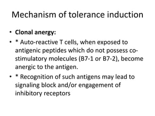
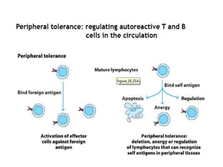
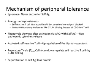
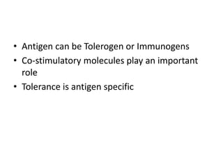
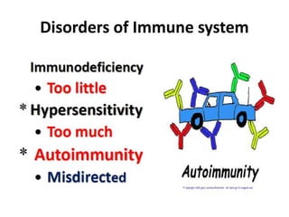
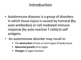
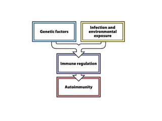
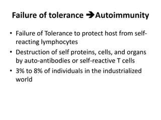
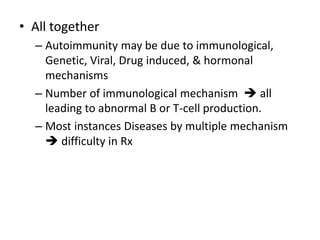
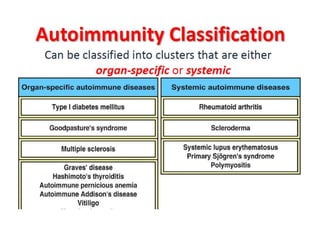
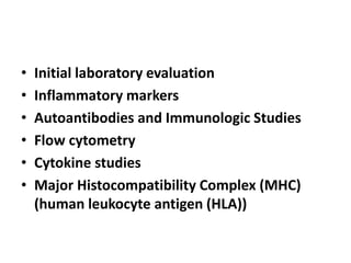
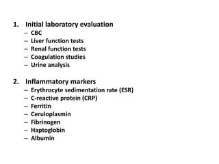
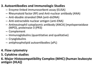
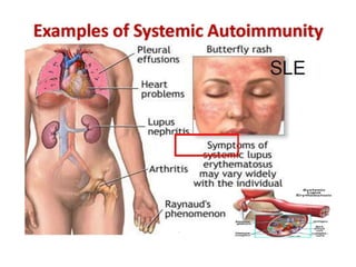
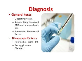
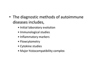
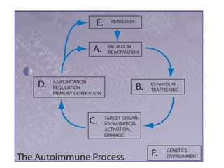
Autoimmunity
Disorders of Immune system
DEFINITION
• Three requirements to call it Autoimmunity :
1. The presence of an immune reaction
specific for some self antigen or self tissue.
2. Evidence that such a reaction is not
secondary to tissue damage but is of
primary pathogenic significance.
3. The absence of another well-defined cause
of the disease.
HISTORY
Julius Donath and Karl Landsteiner (1904)reported
autoantibodies can cause disease by showing that
autoantibodies (‘hemolysins’) caused paroxysmal
cold hemoglobinuria.
Introduction
• Autoimmune diseases is a group of disorders
in which tissue injury is caused by humoral (by
auto-antibodies) or cell mediated immune
response (by auto-reactive T cells) to self
antigens.
• An autoimmune disorder may result in:
 The destruction of one or more types of body tissue
 Abnormal growth of an organ
 Changes in organ function
Causes of Autoimmunity
Tolerance and Autoimmunity
Immune tolerance
• Definition: Immunological tolerance occurs when an
immunocompetent host fails to respond to an
immunogenic challenge with a specific antigen.
• * a state of unresponsiveness specific for a given
antigen
• * It is specific (negative) immune response
• * It is induced by prior exposure to that antigen
• * While the most important form of tolerance is non-
reactivity to self antigens, it is possible to induce
tolerance to non-self antigens. When an antigen
induces tolerance, it is termed tolerogen.
• Immune tolerance
• * Tolerance is different from non-specific
immunosuppression and immunodeficiency. It
is an active antigen-dependent process in
response to the antigen.
• * Like immune response, tolerance is specific
and like immunological memory, it can exist in
T cells, B cells or both and like immunological
memory, tolerance at the T cell level is longer
lasting than tolerance at the B cell level.
TOLERANCE – GENERAL PROPERTIES
• Immature or developing lymphocyte is more
susceptible to tolerance induction than
mature one
• 1.Tolerance to antigens is induced even in
mature lymphocytes under special conditions
• 2.Tolerance of T lymphocytes is a particularly
effective for maintaining long-lived
unresponsiveness to self antigens
Central tolerance: fates of immature
self-reactive lymphocytes
• Induced by antigen in generative lymphoid organs
(thymus for T cells, bone marrow for B cells), and high-
affinity (“strong”) recognition of the antigens
• Immature lymphocytes undergo apoptosis upon
encounter with antigens (negative selection) Eliminates
high-affinity self-reactive (potentially most dangerous)
lymphocytes
• Some self-reactive T cells that encounter self antigens
in the thymus develop into regulatory T cells and
immature B cells in the bone marrow change their
receptors (rendered harmless)
Mechanisms of tolerance induction
• * Clonal deletion – physical elimination of cells
from the repertoire during their lifespan
• * Clonal anergy – downregulating the intrinsic
mechanism of the immune response such as
lack of costimulatory molecules or insufficient
second signal for cell activation
• * Suppression – inhibition of cellular activation
by interaction with other cells:
• (Treg – CD4+CD25+ T lymphocytes)
Mechanism of tolerance induction
• Clonal deletion:
• Functionally immature cells of a clone
encountering antigen undergo a programmed
cell death, as auto-reactive T-cells are
eliminated in the thymus following interaction
with self antigen during their differentiation
(negative selection).
Mechanism of tolerance induction
• Clonal anergy:
• * Auto-reactive T cells, when exposed to
antigenic peptides which do not possess co-
stimulatory molecules (B7-1 or B7-2), become
anergic to the antigen.
• * Recognition of such antigens may lead to
signaling block and/or engagement of
inhibitory receptors
Mechanism of peripheral tolerance
• Ignorance: Never encounter Self Ag
• Anergy: unresponsiveness
• Self reactive T cell interact with APC but co-stimulatory signal blocked
• Immunomodulatory molecules like CTLA4 binding instead of CD 28 on T cell
• Phenotypic skewing: after activation via APC (with Self Ag)---Non
pathogenic cytokines release
• Activated self reactive Tcell---Upregulation of Fas Ligand---apoptosis
• Regulatory T cells (TREG Cells)-can down regulate self reactive T cell (by
IL-10, TGF-b)
• Sequestration of self Ag: lens protein
• Antigen can be Tolerogen or Immunogens
• Co-stimulatory molecules play an important
role
• Tolerance is antigen specific
Immune tolerance: factors that influence
immunity vs. tolerance
• * The stage of differentiation of lymphocytes
at the time of antigen confrontation
– * The site of encounter
• * The nature of cells presenting antigenic
epitopes
• * The number of lymphocytes able to respond
• * Microenvironment of encounter (expression
of cell adhesion molecules, influence of
cytokines etc.)
Immunologically Privileged Sites
• * Sites in the body where foreign antigens or
tissue grafts do not elicit immune responses
• * These antigens do interact with T cells, but
instead of destructive IR they induce tolerance or
a response innocent to the tissue
• Immunosuppressive cytokines such as TGF-beta
seem to be resposible for such unusual response
• * The sites include: brain, eye, testis, uterus
(fetus)
Failure of tolerance Autoimmunity
• Failure of Tolerance to protect host from self-
reacting lymphocytes
• Destruction of self proteins, cells, and organs
by auto-antibodies or self-reactive T cells
• 3% to 8% of individuals in the industrialized
world
Factors responsible for promoting
tolerance
• High dose of antigen
• Persistence of antigen
• Route of administration
• Adjuvents
• Low level of co-stimulators
• Presentation of antigen by
immature/unactivated antigen-presenting
cells (APCs)
• All together
– Autoimmunity may be due to immunological,
Genetic, Viral, Drug induced, & hormonal
mechanisms
– Number of immunological mechanism  all
leading to abnormal B or T-cell production.
– Most instances Diseases by multiple mechanism
 difficulty in Rx
Mechanisms
• Antigenic alteration
• Sequestered Ag
• Cross reacting foreign Ag
• Molecular mimicry
• Polyclonal activation of B cell
• Altered T or B cell function
Antigenic alteration
• Neoantigen formation
• Physical- Irradiation, photosensitivity, cold
allergy
• Chemical- Drug induced anemia, Lecopenia,
thrombocytopenia
• Biological injury-Viral infection (infectious
mononucleosis etc) intracellular pathogen
Sequestered Ag
• Sequestered Ag: Self Ab present in closed
system not accessible to immune system
• Lens protein
• Immunological tolerance not established
during foetal life
• Penetrating injury---leak of lens protein—
immune response—injury to other eye
Sequestered Ag
• Sperm Ag—Puberty
• Mumps---damage to BM seminiferous
tubules—immune response---Orchitis
Cross reacting foreign Ag
• Similary b/w some foreign & Self Ag
• Indivisual Nerve tissue damage-antirabies
immunization of human with neural vaccine of
infected sheep brain
• Streptococcal M protein—heart ms—Heart
damage
• Nephritogenic strain streptococcus-- GN
Molecular mimicry
• Identical peptide sequence in epitopes b/w
microorganism & Self Ag
• HLA B27- Arthritogenic strain of S. flexneri
• Joint membrane- M. tuberculoisis
• Myocardium- Coxsackie B virus
Polyclonal activation of B cell
• Ag--- corresponding B cell activation
• Polyclonal activation of B cells:
– Chemical (2 ME)
– Bacterial product (PPD, LPS)
– Enz (Trypsin)
– Antibotics (Nystatin)
– Infection (Mycoplasma, EBV, Malaria)
Altered T or B cell function
• Enhanced Helper T cell
• ↓↓ Suppressor T cell function
• Defect in thymus
• Stem cell development
• Macrophage function
• Idiotype-antiidiotype network defect
Nature Immunology
• Initial laboratory evaluation
• Inflammatory markers
• Autoantibodies and Immunologic Studies
• Flow cytometry
• Cytokine studies
• Major Histocompatibility Complex (MHC)
(human leukocyte antigen (HLA))
1. Initial laboratory evaluation
– CBC
– Liver function tests
– Renal function tests
– Coagulation studies
– Urine analysis
2. Inflammatory markers
– Erythrocyte sedimentation rate (ESR)
– C-reactive protein (CRP)
– Ferritin
– Ceruloplasmin
– Fibrinogen
– Haptoglobin
– Albumin
3. Autoantibodies and Immunologic Studies
– Enzyme-linked immunosorbent assay (ELISA)
– Rheumatoid factor (RF) and Anti-nuclear antibody (ANA)
– Anti-double stranded DNA (anti-dsDNA)
– Anti-extractable nuclear antigen (anti-ENA)
– Antineutrophil cytoplasmic antibody (ANCA) [myeloperoxidase
(MPO), proteinase-3 (PR3)
– Complement
– Immunoglobulins (quantitative and qualitative)
– Cryoglobulins
– antiphospholipid autoantibodies (aPL)
4. Flow cytometry
5. Cytokine studies
6. Major Histocompatibility Complex (MHC) (human leukocyte
antigen (HLA))
Localised Organ Specific Diseases
Disease
Hashimoto’s Ds Enlargement of thyroid
Hypothyrodism or
Frank myxedma
Glandular structure-replacement
with lymphoid tissue
Ab- to thyoglobulin, acinar
colloid, Microsomal Ag,
thyroid cell surface
components
Thyrotoxicosis
(Grave’s Ds)
↑↑↑ hormone IgG Ab (to thyroid
membrane Ag) act as Long
acting thyroid stimulator
(LATS)
Addison ds Lymphocytic infiltration of
adrenal &
circulating Ab to Zona
glomerulosa
Autoimmune
orchitis
Mumps Lymphocytic infiltration of
Testes &
circulating Ab to sperm &
germinal cell
Disease
Myasthenia gravis Thyorid lymphoid hyperplasia
Numerous germinal center
Ab Ach Receptor on
myoneural junction of
striated Ms---impairment
of Ms Contraction
Autoimmune
Disease of eye
Phacoanaphylaxis- Cataract Sx –
intraocular inflammation
Symathetic ophthamia-
Performating injury to one
eye
Pernicious Anaemia Ab to parietal cell of gastric
mucosa- Achlorhydria, Atrophic
gastritis
Ab to Intrinsic Factor---Vit B12 def
Autoimmune
Disease of Nervous
system
Neuroparalytic accidents
following- neural vaccine—Rabies
GBS- idiopathic polyneuritis
Autoimmune ds of
Skin
Pemphigus vulgaris- Ab to
intracellular adhesion protein
desmoglein
Bullous pemphigoid- Ab
dermoepidermal junction
Ab in Dermatitis
herpetiformis
– Variety of AutoAb
• Nuclei
• Intracytoplasmic cell constituents
– LE cell- Neutrophils containing LE Bodies (large
pale homogenous body) almost filling cytoplasm
– Antinuclear Ab (ANA)—Sensitive but not specific
• Pattern: homogenous, Peripheral, speckled, nucleolar
– Anti DNA Ab- ds
• The diagnostic methods of autoimmune
diseases includes,
• Initial laboratory evolution
• Immunological studies
• Inflammatory markers
• Flowcytometry
• Cytokine studies
• Major histocompatibility complex
• Q: Why are autoimmune diseases challenging to
diagnose?
• A: Diagnosis is challenging for several reasons:
• 1. Patients initially present with nonspecific symptoms such
as fatigue, joint and muscle pain, fever, and/or weight
change.
• 2. Symptoms often flare and remit.
• 3. Patients frequently have more than 1 autoimmune
disease.
• According to a survey by the Autoimmune Diseases
Association, it takes up to 4.6 years and nearly 5 doctors for
a patient to receive a proper autoimmune disease diagnosis
THANK YOU

Autoimmunity

  • 1.
  • 2.
  • 3.
    DEFINITION • Three requirementsto call it Autoimmunity : 1. The presence of an immune reaction specific for some self antigen or self tissue. 2. Evidence that such a reaction is not secondary to tissue damage but is of primary pathogenic significance. 3. The absence of another well-defined cause of the disease.
  • 4.
    HISTORY Julius Donath andKarl Landsteiner (1904)reported autoantibodies can cause disease by showing that autoantibodies (‘hemolysins’) caused paroxysmal cold hemoglobinuria.
  • 5.
    Introduction • Autoimmune diseasesis a group of disorders in which tissue injury is caused by humoral (by auto-antibodies) or cell mediated immune response (by auto-reactive T cells) to self antigens. • An autoimmune disorder may result in:  The destruction of one or more types of body tissue  Abnormal growth of an organ  Changes in organ function
  • 7.
  • 12.
  • 13.
    Immune tolerance • Definition:Immunological tolerance occurs when an immunocompetent host fails to respond to an immunogenic challenge with a specific antigen. • * a state of unresponsiveness specific for a given antigen • * It is specific (negative) immune response • * It is induced by prior exposure to that antigen • * While the most important form of tolerance is non- reactivity to self antigens, it is possible to induce tolerance to non-self antigens. When an antigen induces tolerance, it is termed tolerogen.
  • 14.
    • Immune tolerance •* Tolerance is different from non-specific immunosuppression and immunodeficiency. It is an active antigen-dependent process in response to the antigen. • * Like immune response, tolerance is specific and like immunological memory, it can exist in T cells, B cells or both and like immunological memory, tolerance at the T cell level is longer lasting than tolerance at the B cell level.
  • 15.
    TOLERANCE – GENERALPROPERTIES • Immature or developing lymphocyte is more susceptible to tolerance induction than mature one • 1.Tolerance to antigens is induced even in mature lymphocytes under special conditions • 2.Tolerance of T lymphocytes is a particularly effective for maintaining long-lived unresponsiveness to self antigens
  • 17.
    Central tolerance: fatesof immature self-reactive lymphocytes • Induced by antigen in generative lymphoid organs (thymus for T cells, bone marrow for B cells), and high- affinity (“strong”) recognition of the antigens • Immature lymphocytes undergo apoptosis upon encounter with antigens (negative selection) Eliminates high-affinity self-reactive (potentially most dangerous) lymphocytes • Some self-reactive T cells that encounter self antigens in the thymus develop into regulatory T cells and immature B cells in the bone marrow change their receptors (rendered harmless)
  • 19.
    Mechanisms of toleranceinduction • * Clonal deletion – physical elimination of cells from the repertoire during their lifespan • * Clonal anergy – downregulating the intrinsic mechanism of the immune response such as lack of costimulatory molecules or insufficient second signal for cell activation • * Suppression – inhibition of cellular activation by interaction with other cells: • (Treg – CD4+CD25+ T lymphocytes)
  • 20.
    Mechanism of toleranceinduction • Clonal deletion: • Functionally immature cells of a clone encountering antigen undergo a programmed cell death, as auto-reactive T-cells are eliminated in the thymus following interaction with self antigen during their differentiation (negative selection).
  • 21.
    Mechanism of toleranceinduction • Clonal anergy: • * Auto-reactive T cells, when exposed to antigenic peptides which do not possess co- stimulatory molecules (B7-1 or B7-2), become anergic to the antigen. • * Recognition of such antigens may lead to signaling block and/or engagement of inhibitory receptors
  • 24.
    Mechanism of peripheraltolerance • Ignorance: Never encounter Self Ag • Anergy: unresponsiveness • Self reactive T cell interact with APC but co-stimulatory signal blocked • Immunomodulatory molecules like CTLA4 binding instead of CD 28 on T cell • Phenotypic skewing: after activation via APC (with Self Ag)---Non pathogenic cytokines release • Activated self reactive Tcell---Upregulation of Fas Ligand---apoptosis • Regulatory T cells (TREG Cells)-can down regulate self reactive T cell (by IL-10, TGF-b) • Sequestration of self Ag: lens protein
  • 25.
    • Antigen canbe Tolerogen or Immunogens • Co-stimulatory molecules play an important role • Tolerance is antigen specific
  • 26.
    Immune tolerance: factorsthat influence immunity vs. tolerance • * The stage of differentiation of lymphocytes at the time of antigen confrontation – * The site of encounter • * The nature of cells presenting antigenic epitopes • * The number of lymphocytes able to respond • * Microenvironment of encounter (expression of cell adhesion molecules, influence of cytokines etc.)
  • 27.
    Immunologically Privileged Sites •* Sites in the body where foreign antigens or tissue grafts do not elicit immune responses • * These antigens do interact with T cells, but instead of destructive IR they induce tolerance or a response innocent to the tissue • Immunosuppressive cytokines such as TGF-beta seem to be resposible for such unusual response • * The sites include: brain, eye, testis, uterus (fetus)
  • 28.
    Failure of toleranceAutoimmunity • Failure of Tolerance to protect host from self- reacting lymphocytes • Destruction of self proteins, cells, and organs by auto-antibodies or self-reactive T cells • 3% to 8% of individuals in the industrialized world
  • 29.
    Factors responsible forpromoting tolerance • High dose of antigen • Persistence of antigen • Route of administration • Adjuvents • Low level of co-stimulators • Presentation of antigen by immature/unactivated antigen-presenting cells (APCs)
  • 31.
    • All together –Autoimmunity may be due to immunological, Genetic, Viral, Drug induced, & hormonal mechanisms – Number of immunological mechanism  all leading to abnormal B or T-cell production. – Most instances Diseases by multiple mechanism  difficulty in Rx
  • 33.
    Mechanisms • Antigenic alteration •Sequestered Ag • Cross reacting foreign Ag • Molecular mimicry • Polyclonal activation of B cell • Altered T or B cell function
  • 34.
    Antigenic alteration • Neoantigenformation • Physical- Irradiation, photosensitivity, cold allergy • Chemical- Drug induced anemia, Lecopenia, thrombocytopenia • Biological injury-Viral infection (infectious mononucleosis etc) intracellular pathogen
  • 35.
    Sequestered Ag • SequesteredAg: Self Ab present in closed system not accessible to immune system • Lens protein • Immunological tolerance not established during foetal life • Penetrating injury---leak of lens protein— immune response—injury to other eye
  • 36.
    Sequestered Ag • SpermAg—Puberty • Mumps---damage to BM seminiferous tubules—immune response---Orchitis
  • 37.
    Cross reacting foreignAg • Similary b/w some foreign & Self Ag • Indivisual Nerve tissue damage-antirabies immunization of human with neural vaccine of infected sheep brain • Streptococcal M protein—heart ms—Heart damage • Nephritogenic strain streptococcus-- GN
  • 38.
    Molecular mimicry • Identicalpeptide sequence in epitopes b/w microorganism & Self Ag • HLA B27- Arthritogenic strain of S. flexneri • Joint membrane- M. tuberculoisis • Myocardium- Coxsackie B virus
  • 40.
    Polyclonal activation ofB cell • Ag--- corresponding B cell activation • Polyclonal activation of B cells: – Chemical (2 ME) – Bacterial product (PPD, LPS) – Enz (Trypsin) – Antibotics (Nystatin) – Infection (Mycoplasma, EBV, Malaria)
  • 41.
    Altered T orB cell function • Enhanced Helper T cell • ↓↓ Suppressor T cell function • Defect in thymus • Stem cell development • Macrophage function • Idiotype-antiidiotype network defect
  • 46.
  • 48.
    • Initial laboratoryevaluation • Inflammatory markers • Autoantibodies and Immunologic Studies • Flow cytometry • Cytokine studies • Major Histocompatibility Complex (MHC) (human leukocyte antigen (HLA))
  • 49.
    1. Initial laboratoryevaluation – CBC – Liver function tests – Renal function tests – Coagulation studies – Urine analysis 2. Inflammatory markers – Erythrocyte sedimentation rate (ESR) – C-reactive protein (CRP) – Ferritin – Ceruloplasmin – Fibrinogen – Haptoglobin – Albumin
  • 50.
    3. Autoantibodies andImmunologic Studies – Enzyme-linked immunosorbent assay (ELISA) – Rheumatoid factor (RF) and Anti-nuclear antibody (ANA) – Anti-double stranded DNA (anti-dsDNA) – Anti-extractable nuclear antigen (anti-ENA) – Antineutrophil cytoplasmic antibody (ANCA) [myeloperoxidase (MPO), proteinase-3 (PR3) – Complement – Immunoglobulins (quantitative and qualitative) – Cryoglobulins – antiphospholipid autoantibodies (aPL) 4. Flow cytometry 5. Cytokine studies 6. Major Histocompatibility Complex (MHC) (human leukocyte antigen (HLA))
  • 51.
  • 52.
    Disease Hashimoto’s Ds Enlargementof thyroid Hypothyrodism or Frank myxedma Glandular structure-replacement with lymphoid tissue Ab- to thyoglobulin, acinar colloid, Microsomal Ag, thyroid cell surface components Thyrotoxicosis (Grave’s Ds) ↑↑↑ hormone IgG Ab (to thyroid membrane Ag) act as Long acting thyroid stimulator (LATS) Addison ds Lymphocytic infiltration of adrenal & circulating Ab to Zona glomerulosa Autoimmune orchitis Mumps Lymphocytic infiltration of Testes & circulating Ab to sperm & germinal cell
  • 53.
    Disease Myasthenia gravis Thyoridlymphoid hyperplasia Numerous germinal center Ab Ach Receptor on myoneural junction of striated Ms---impairment of Ms Contraction Autoimmune Disease of eye Phacoanaphylaxis- Cataract Sx – intraocular inflammation Symathetic ophthamia- Performating injury to one eye Pernicious Anaemia Ab to parietal cell of gastric mucosa- Achlorhydria, Atrophic gastritis Ab to Intrinsic Factor---Vit B12 def Autoimmune Disease of Nervous system Neuroparalytic accidents following- neural vaccine—Rabies GBS- idiopathic polyneuritis Autoimmune ds of Skin Pemphigus vulgaris- Ab to intracellular adhesion protein desmoglein Bullous pemphigoid- Ab dermoepidermal junction Ab in Dermatitis herpetiformis
  • 56.
    – Variety ofAutoAb • Nuclei • Intracytoplasmic cell constituents – LE cell- Neutrophils containing LE Bodies (large pale homogenous body) almost filling cytoplasm – Antinuclear Ab (ANA)—Sensitive but not specific • Pattern: homogenous, Peripheral, speckled, nucleolar – Anti DNA Ab- ds
  • 65.
    • The diagnosticmethods of autoimmune diseases includes, • Initial laboratory evolution • Immunological studies • Inflammatory markers • Flowcytometry • Cytokine studies • Major histocompatibility complex
  • 69.
    • Q: Whyare autoimmune diseases challenging to diagnose? • A: Diagnosis is challenging for several reasons: • 1. Patients initially present with nonspecific symptoms such as fatigue, joint and muscle pain, fever, and/or weight change. • 2. Symptoms often flare and remit. • 3. Patients frequently have more than 1 autoimmune disease. • According to a survey by the Autoimmune Diseases Association, it takes up to 4.6 years and nearly 5 doctors for a patient to receive a proper autoimmune disease diagnosis
  • 70.