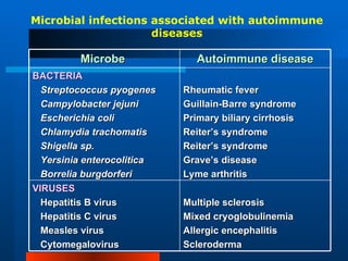
AUTOIMMUNITY FE A. BARTOLOME, MD, FPASMAP  Department of Microbiology  Our Lady of Fatima University
IMMUNOLOGICAL TOLERANCE State in which the individual is incapable of developing an immune response to a specific antigen Self-tolerance     lack of responsiveness to an individual’s antigens Central tolerance & peripheral tolerance
CENTRAL TOLERANCE Clonal deletion of self-reactive T and B lymphocytes during their maturation in the central lymphoid organs
CENTRAL TOLERANCE T cells T lymphocytes that bear high-affinity receptors for self-antigens are  negatively selected  or deleted    undergo  apoptosis occur during fetal development
CENTRAL TOLERANCE B cells Also undergo clonal deletion Developing B cells encounter a membrane-bound antigen within the bone marrow    B cells undergo apoptosis Occur throughout life
PERIPHERAL TOLERANCE Self-reactive T cells that escape  intrathymic negative selection are  deleted or muzzled in the peripheral tissues
PERIPHERAL TOLERANCE MECHANISMS: Clonal deletion by activation-induced cell death Clonal anergy Peripheral suppression by T cells
PERIPHERAL TOLERANCE Clonal deletion by activation-induced cell death Apoptotic death of activated T cells by the  Fas-FasL system Self-antigens abundant in peripheral tissue (e.g. collagen, thyroglobulin)    repeated & persistent stimulation of self-antigen-specific T cells    activation of Fas-mediated apoptosis
Clonal deletion in thymus A A A  Cell death B B B Antigen  Two immature T cells (A and B) with different antigen receptors Binding of self antigen to T-cell A in thymus but not to T-cell B Death of self-reacting T-cell A; survival of T-cell B that reacts against foreign antigen
 
PERIPHERAL TOLERANCE Clonal anergy Prolonged or irreversible functional inactivation of lymphocytes Induced by encounter with antigens T cells –  due to absence of co-stimulatory molecules on APCs, such as B7-1 & B7-2 B cells –  due to lack of T cell help for antibody synthesis (T cell anergy or down-regulation of surface IgM)
PERIPHERAL TOLERANCE APC Helper T cell Class II MHC Antigen  TCR B7 protein CD28 protein CD4 protein APC Helper T cell Class II MHC Antigen  TCR CD28 protein CD4 protein B7 protein on APC interacts with CD28 on helper T cells. Full activation of helper T cells occur. B7 protein on APC is not produced. CD28 on helper T cell does not give a co-stimulatory signal. Anergy occurs.
 
PERIPHERAL TOLERANCE Peripheral suppression by T cells Suppressor T cells – with ability to down-regulate the function of other autoreactive T cells Shift immune response from T H 1 to T H 2 T H 2 generally immunosuppressive    down-modulate T H 1 response
 
Factors Affecting Artificially-Induced Tolerance Form, dose & route of administration Very simple molecules induce tolerance more readily than a complex one Very high or very low doses of an antigen may result in tolerance instead of an immune response Purified polysaccharides or amino acid co-polymers injected in very large doses result in “ immune paralysis ”
Factors Affecting Artificially-Induced Tolerance Immunologic “maturity” of the host E.g. neonates    immunologically immature    do not respond well to foreign antigens Chimerism Tolerance induced by inoculation of allogeneic cells into hosts that lack immune competence Antibodies to CD4 and CD8 Tolerance of transplanted tissues by inoculating graft recipient with monoclonal antibodies against CD4 and CD8
Factors Affecting Artificially-Induced Tolerance Clonal exhaustion Repeated antigenic challenge Stimulate B and T cell to differentiate into short-lived end cells
Factors Affecting Artificially-Induced Tolerance Clonal anergy induced by anti-idiotypic antibodies & antagonistic peptides Antibody combining site (idiotype) act as antigen    induce formation of anti-idiotypic antibodies    cross-link on B cells    prevent interaction with Ag Antagonistic peptides    fit into Ag-binding site of MHC    no activation of T cells
Other aspects of induction/maintenance of tolerance T cells become tolerant more readily and remain tolerant longer than B cells. Administration of a cross-reacting antigen tends to terminate tolerance. Administration of immunosuppressive drugs enhances tolerance. Tolerance is maintained best if the antigen to which the immune system is tolerant continues to be present.
AUTOIMMUNITY Immune reaction against  self-antigens Requirements: Presence of an autoimmune reaction Clinical or experimental evidence that reaction is not secondary to tissue damage but is of primary pathogenetic significance Absence of another well-defined cause of the disease
AUTOIMMUNITY Most important step in production of autoimmune disease:  activation of self-reactive CD4 T cells Most are antibody-mediated
AUTOIMMUNITY GENETIC FACTORS (+) genetic predisposition Strong association with HLA specificities, especially class II genes Class I MHC-related: ankylosing spondylitis & Reiter’s syndrome; more common in men Class II MHC-related: RA, Grave’s disease, SLE; more common in women
AUTOIMMUNITY HORMONAL FACTORS Approximately 90% occur in women Estrogen   can alter the B-cell repertoire and enhance formation of antibody to DNA
AUTOIMMUNITY ENVIRONMENTAL FACTORS Exposure to an environmental agent can trigger a cross-reacting immune response against some component of normal tissue Example:  S. pyogenes  & rheumatic fever
AUTOIMMUNITY: Mechanisms Defects in clonal deletion mechanisms Thymic defects that lead to proliferation of self-reactive T cells Failure of central tolerance
AUTOIMMUNITY: Mechanisms Polyclonal lymphocyte activation Microorganism-derived mitogens stimulate lymphocytes Microbial products (e.g. LPS)    act as  superantigens     activate a large pool of T and B cells
AUTOIMMUNITY: Mechanisms Molecular mimicry Microbial antigens with similar structure to self-antigens    activate autoreactive T cells Cross-reactivity-induced immune response Example:  M protein of  S. pyogenes  and myosin of cardiac muscle
AUTOIMMUNITY: Mechanisms Release of sequestered antigens Immunologically privileged sites ( brain, ant. eye chamber, ovary, placenta, testis, pregnant uterus )    not exposed to immune system Damage    release of antigens    elicit immune response
AUTOIMMUNITY: Mechanisms Defects in the regulation of TH 1  and TH 2  cells Impaired T suppressor cell immunoregulation
Microbial infections associated with autoimmune diseases Microbe  Autoimmune disease BACTERIA Streptococcus pyogenes Campylobacter jejuni Escherichia coli Chlamydia trachomatis Shigella sp. Yersinia enterocolitica Borrelia burgdorferi Rheumatic fever Guillain-Barre syndrome Primary biliary cirrhosis Reiter’s syndrome Reiter’s syndrome Grave’s disease Lyme arthritis VIRUSES Hepatitis B virus Hepatitis C virus Measles virus Cytomegalovirus Multiple sclerosis Mixed cryoglobulinemia Allergic encephalitis Scleroderma
ORGAN-SPECIFIC AUTOIMMUNE DISEASES Type of Immune Response Autoimmune Disease Target of Immune Response Antibody to receptors Myasthenia gravis Grave’s disease Acetylcholine receptor TSH receptor Antibody to cell components other than receptors Pernicious anemia Goodpasture’s synd. IDDM Addison’s disease Male infertility Pemphigus Hashimoto’s Primary myxedema Intrinsic factor and parietal cells BM of kidney & lung Islet cell Adrenal cortex Sperm  Desmoglein in tight junctions of skin Thyroglobulin Thyroid peroxidase
NON-ORGAN SPECIFIC AUTOIMMUNE DISEASES Type of Immune Response Autoimmune Disease Target of Immune Response Antibody to cell components other than receptors Rheumatoid arthritis SLE Sjogren’s syndrome (Sicca syndrome) Guillain-Barre synd. IgG in joints dsDNA, histones RNP antigens (SS-A/Ro and SS-B/La) Myelin protein


Autoimmunity

  • 1.
    AUTOIMMUNITY FE A.BARTOLOME, MD, FPASMAP Department of Microbiology Our Lady of Fatima University
  • 2.
    IMMUNOLOGICAL TOLERANCE Statein which the individual is incapable of developing an immune response to a specific antigen Self-tolerance  lack of responsiveness to an individual’s antigens Central tolerance & peripheral tolerance
  • 3.
    CENTRAL TOLERANCE Clonaldeletion of self-reactive T and B lymphocytes during their maturation in the central lymphoid organs
  • 4.
    CENTRAL TOLERANCE Tcells T lymphocytes that bear high-affinity receptors for self-antigens are negatively selected or deleted  undergo apoptosis occur during fetal development
  • 5.
    CENTRAL TOLERANCE Bcells Also undergo clonal deletion Developing B cells encounter a membrane-bound antigen within the bone marrow  B cells undergo apoptosis Occur throughout life
  • 6.
    PERIPHERAL TOLERANCE Self-reactiveT cells that escape intrathymic negative selection are deleted or muzzled in the peripheral tissues
  • 7.
    PERIPHERAL TOLERANCE MECHANISMS:Clonal deletion by activation-induced cell death Clonal anergy Peripheral suppression by T cells
  • 8.
    PERIPHERAL TOLERANCE Clonaldeletion by activation-induced cell death Apoptotic death of activated T cells by the Fas-FasL system Self-antigens abundant in peripheral tissue (e.g. collagen, thyroglobulin)  repeated & persistent stimulation of self-antigen-specific T cells  activation of Fas-mediated apoptosis
  • 9.
    Clonal deletion inthymus A A A Cell death B B B Antigen Two immature T cells (A and B) with different antigen receptors Binding of self antigen to T-cell A in thymus but not to T-cell B Death of self-reacting T-cell A; survival of T-cell B that reacts against foreign antigen
  • 10.
  • 11.
    PERIPHERAL TOLERANCE Clonalanergy Prolonged or irreversible functional inactivation of lymphocytes Induced by encounter with antigens T cells – due to absence of co-stimulatory molecules on APCs, such as B7-1 & B7-2 B cells – due to lack of T cell help for antibody synthesis (T cell anergy or down-regulation of surface IgM)
  • 12.
    PERIPHERAL TOLERANCE APCHelper T cell Class II MHC Antigen TCR B7 protein CD28 protein CD4 protein APC Helper T cell Class II MHC Antigen TCR CD28 protein CD4 protein B7 protein on APC interacts with CD28 on helper T cells. Full activation of helper T cells occur. B7 protein on APC is not produced. CD28 on helper T cell does not give a co-stimulatory signal. Anergy occurs.
  • 13.
  • 14.
    PERIPHERAL TOLERANCE Peripheralsuppression by T cells Suppressor T cells – with ability to down-regulate the function of other autoreactive T cells Shift immune response from T H 1 to T H 2 T H 2 generally immunosuppressive  down-modulate T H 1 response
  • 15.
  • 16.
    Factors Affecting Artificially-InducedTolerance Form, dose & route of administration Very simple molecules induce tolerance more readily than a complex one Very high or very low doses of an antigen may result in tolerance instead of an immune response Purified polysaccharides or amino acid co-polymers injected in very large doses result in “ immune paralysis ”
  • 17.
    Factors Affecting Artificially-InducedTolerance Immunologic “maturity” of the host E.g. neonates  immunologically immature  do not respond well to foreign antigens Chimerism Tolerance induced by inoculation of allogeneic cells into hosts that lack immune competence Antibodies to CD4 and CD8 Tolerance of transplanted tissues by inoculating graft recipient with monoclonal antibodies against CD4 and CD8
  • 18.
    Factors Affecting Artificially-InducedTolerance Clonal exhaustion Repeated antigenic challenge Stimulate B and T cell to differentiate into short-lived end cells
  • 19.
    Factors Affecting Artificially-InducedTolerance Clonal anergy induced by anti-idiotypic antibodies & antagonistic peptides Antibody combining site (idiotype) act as antigen  induce formation of anti-idiotypic antibodies  cross-link on B cells  prevent interaction with Ag Antagonistic peptides  fit into Ag-binding site of MHC  no activation of T cells
  • 20.
    Other aspects ofinduction/maintenance of tolerance T cells become tolerant more readily and remain tolerant longer than B cells. Administration of a cross-reacting antigen tends to terminate tolerance. Administration of immunosuppressive drugs enhances tolerance. Tolerance is maintained best if the antigen to which the immune system is tolerant continues to be present.
  • 21.
    AUTOIMMUNITY Immune reactionagainst self-antigens Requirements: Presence of an autoimmune reaction Clinical or experimental evidence that reaction is not secondary to tissue damage but is of primary pathogenetic significance Absence of another well-defined cause of the disease
  • 22.
    AUTOIMMUNITY Most importantstep in production of autoimmune disease: activation of self-reactive CD4 T cells Most are antibody-mediated
  • 23.
    AUTOIMMUNITY GENETIC FACTORS(+) genetic predisposition Strong association with HLA specificities, especially class II genes Class I MHC-related: ankylosing spondylitis & Reiter’s syndrome; more common in men Class II MHC-related: RA, Grave’s disease, SLE; more common in women
  • 24.
    AUTOIMMUNITY HORMONAL FACTORSApproximately 90% occur in women Estrogen  can alter the B-cell repertoire and enhance formation of antibody to DNA
  • 25.
    AUTOIMMUNITY ENVIRONMENTAL FACTORSExposure to an environmental agent can trigger a cross-reacting immune response against some component of normal tissue Example: S. pyogenes & rheumatic fever
  • 26.
    AUTOIMMUNITY: Mechanisms Defectsin clonal deletion mechanisms Thymic defects that lead to proliferation of self-reactive T cells Failure of central tolerance
  • 27.
    AUTOIMMUNITY: Mechanisms Polyclonallymphocyte activation Microorganism-derived mitogens stimulate lymphocytes Microbial products (e.g. LPS)  act as superantigens  activate a large pool of T and B cells
  • 28.
    AUTOIMMUNITY: Mechanisms Molecularmimicry Microbial antigens with similar structure to self-antigens  activate autoreactive T cells Cross-reactivity-induced immune response Example: M protein of S. pyogenes and myosin of cardiac muscle
  • 29.
    AUTOIMMUNITY: Mechanisms Releaseof sequestered antigens Immunologically privileged sites ( brain, ant. eye chamber, ovary, placenta, testis, pregnant uterus )  not exposed to immune system Damage  release of antigens  elicit immune response
  • 30.
    AUTOIMMUNITY: Mechanisms Defectsin the regulation of TH 1 and TH 2 cells Impaired T suppressor cell immunoregulation
  • 31.
    Microbial infections associatedwith autoimmune diseases Microbe Autoimmune disease BACTERIA Streptococcus pyogenes Campylobacter jejuni Escherichia coli Chlamydia trachomatis Shigella sp. Yersinia enterocolitica Borrelia burgdorferi Rheumatic fever Guillain-Barre syndrome Primary biliary cirrhosis Reiter’s syndrome Reiter’s syndrome Grave’s disease Lyme arthritis VIRUSES Hepatitis B virus Hepatitis C virus Measles virus Cytomegalovirus Multiple sclerosis Mixed cryoglobulinemia Allergic encephalitis Scleroderma
  • 32.
    ORGAN-SPECIFIC AUTOIMMUNE DISEASESType of Immune Response Autoimmune Disease Target of Immune Response Antibody to receptors Myasthenia gravis Grave’s disease Acetylcholine receptor TSH receptor Antibody to cell components other than receptors Pernicious anemia Goodpasture’s synd. IDDM Addison’s disease Male infertility Pemphigus Hashimoto’s Primary myxedema Intrinsic factor and parietal cells BM of kidney & lung Islet cell Adrenal cortex Sperm Desmoglein in tight junctions of skin Thyroglobulin Thyroid peroxidase
  • 33.
    NON-ORGAN SPECIFIC AUTOIMMUNEDISEASES Type of Immune Response Autoimmune Disease Target of Immune Response Antibody to cell components other than receptors Rheumatoid arthritis SLE Sjogren’s syndrome (Sicca syndrome) Guillain-Barre synd. IgG in joints dsDNA, histones RNP antigens (SS-A/Ro and SS-B/La) Myelin protein
  • 34.
  • 35.