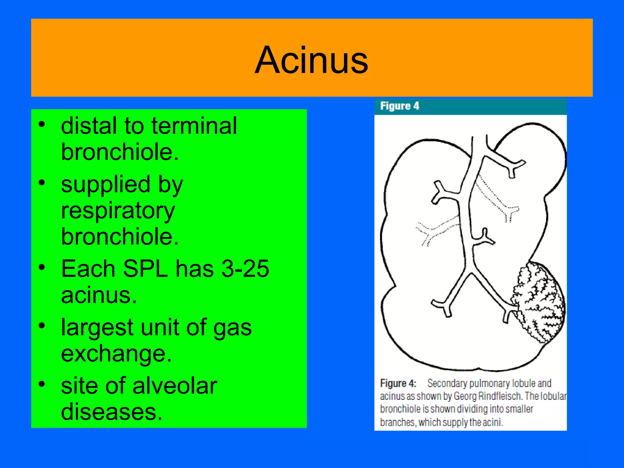
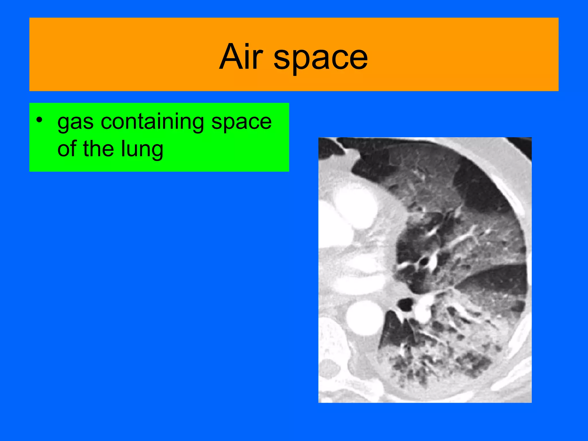
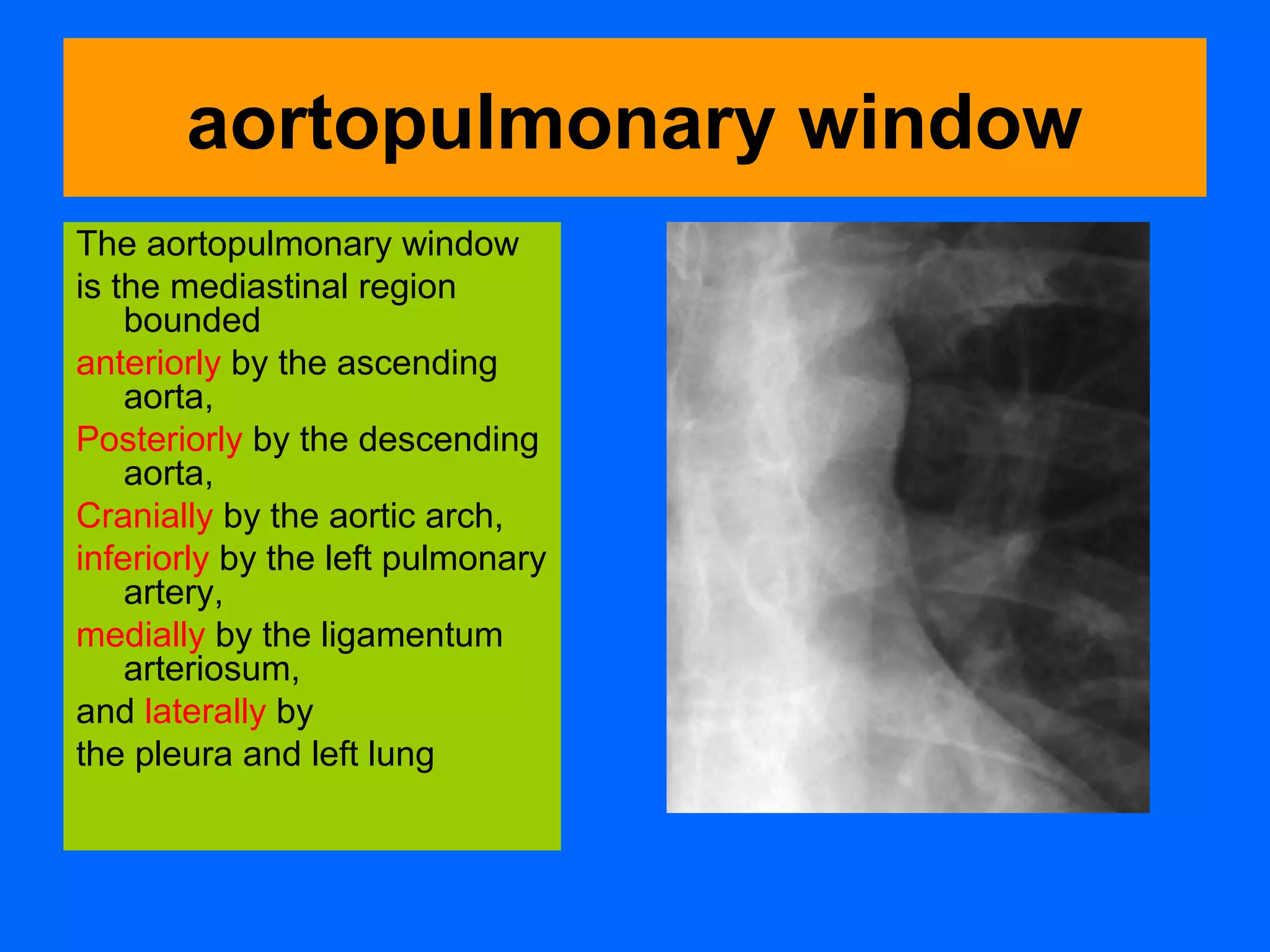
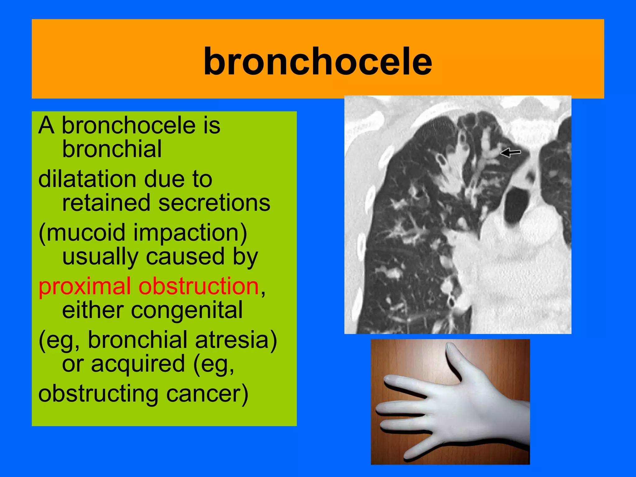
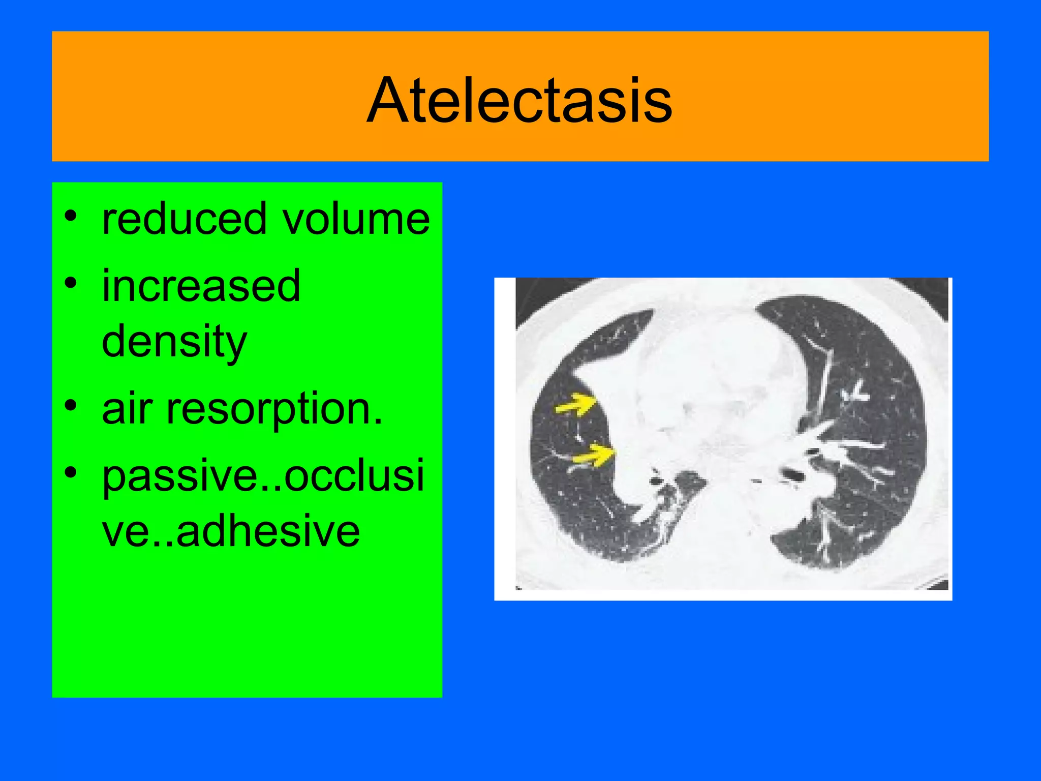
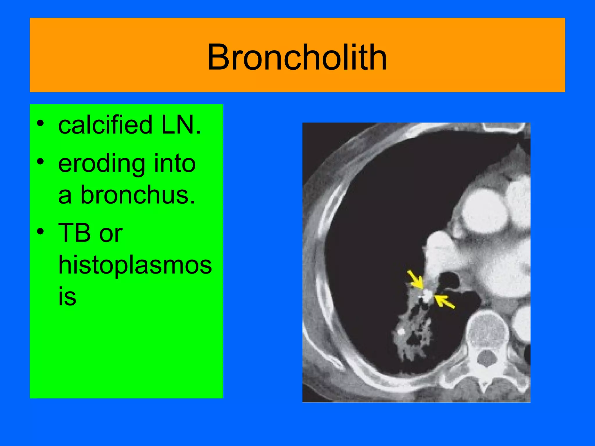
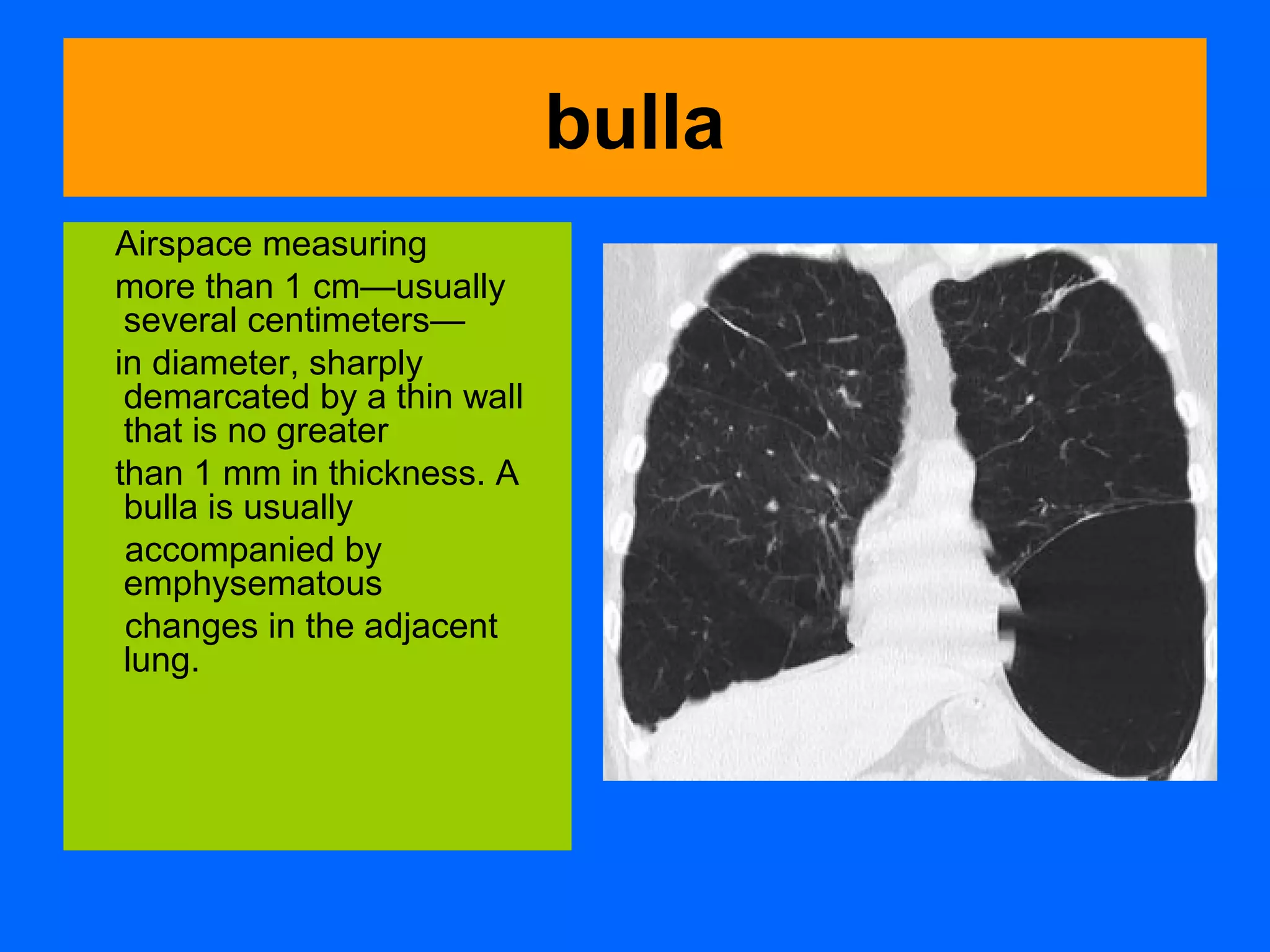
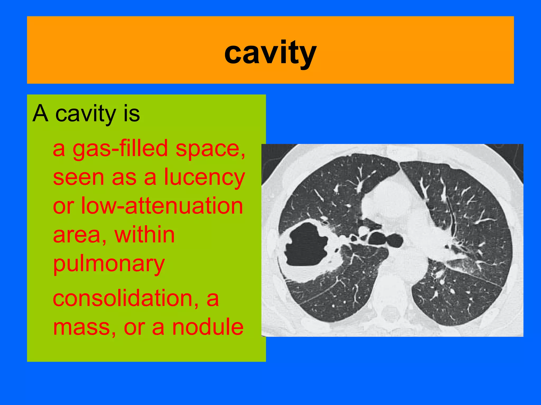
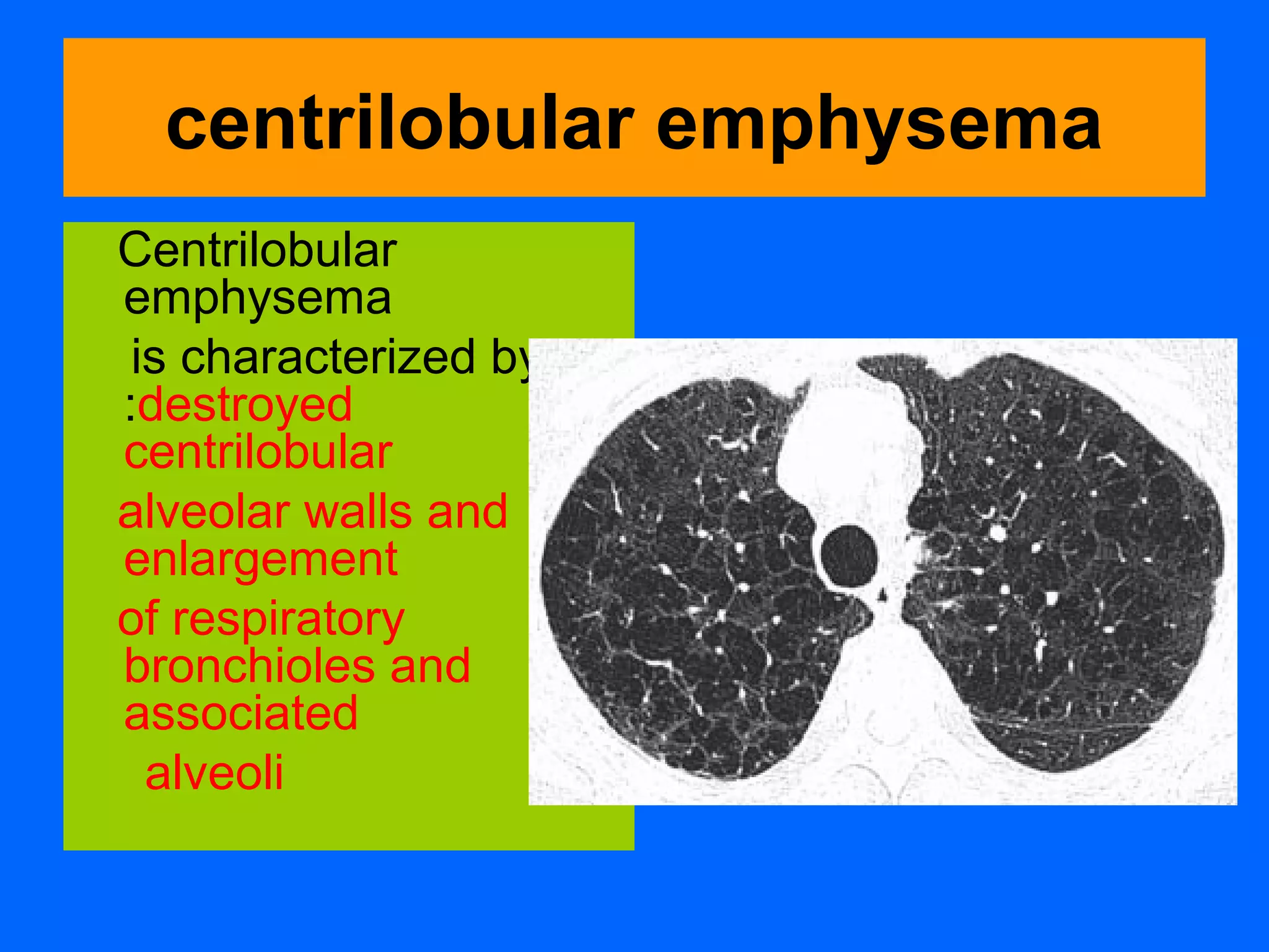
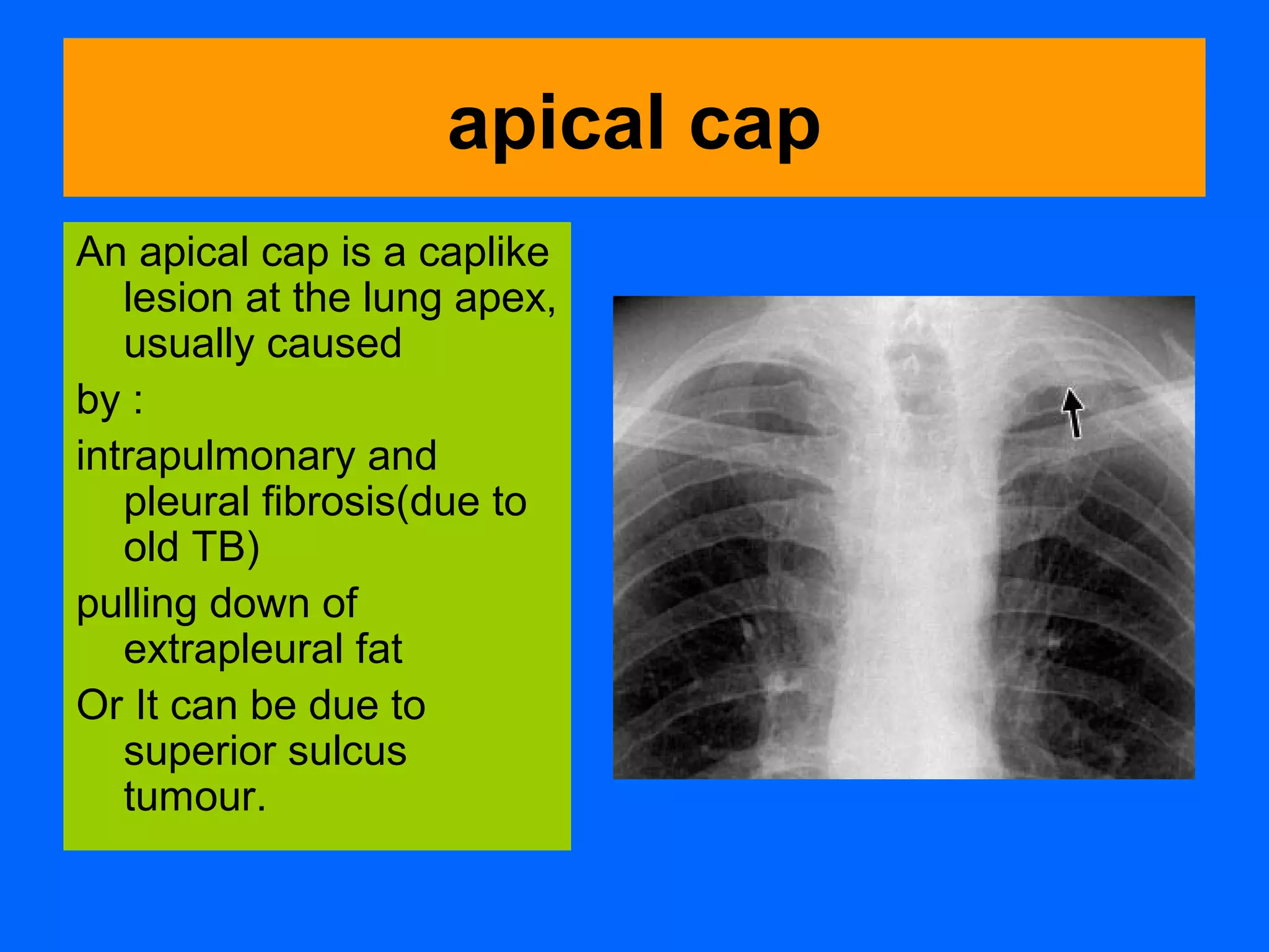
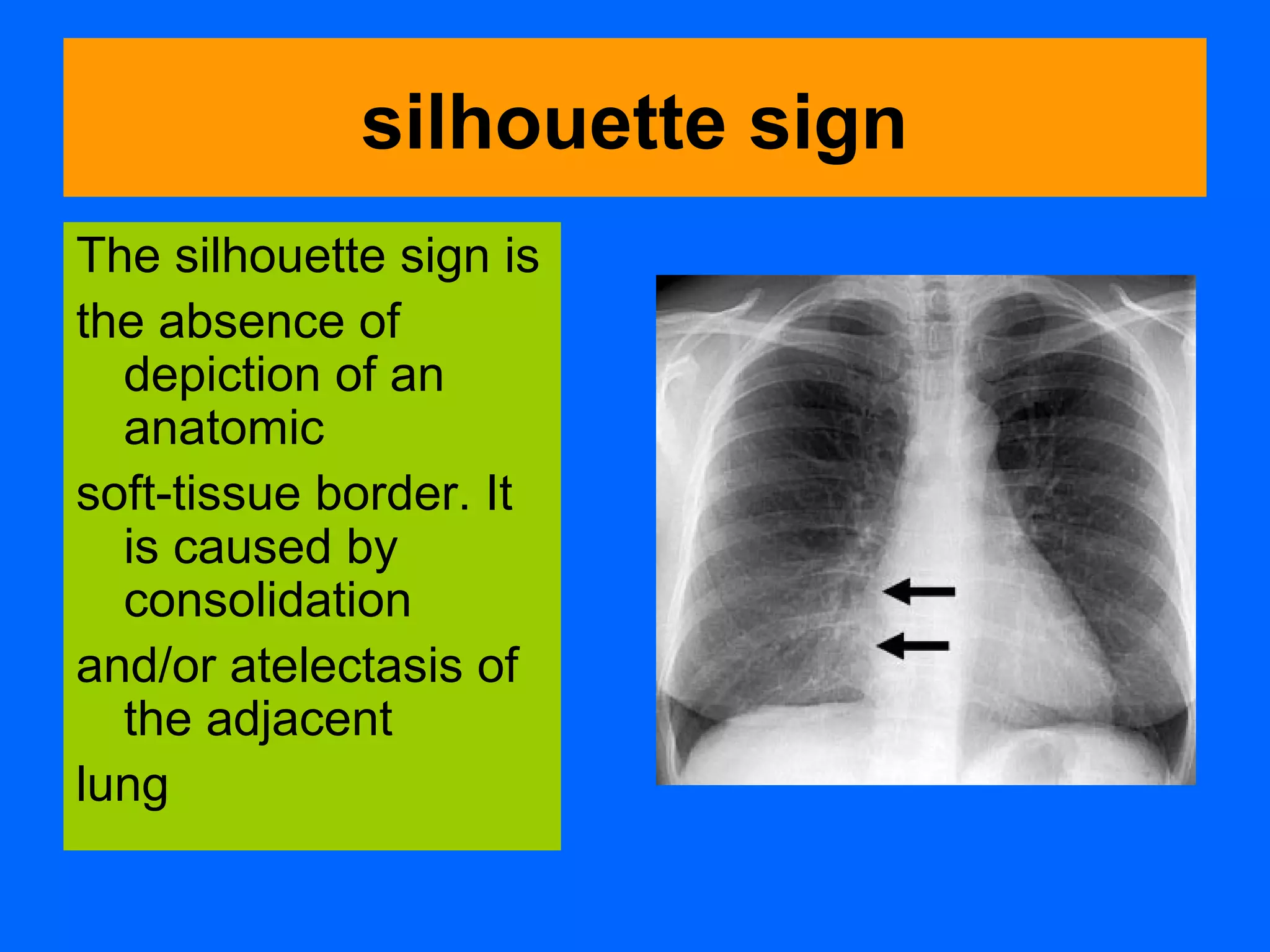
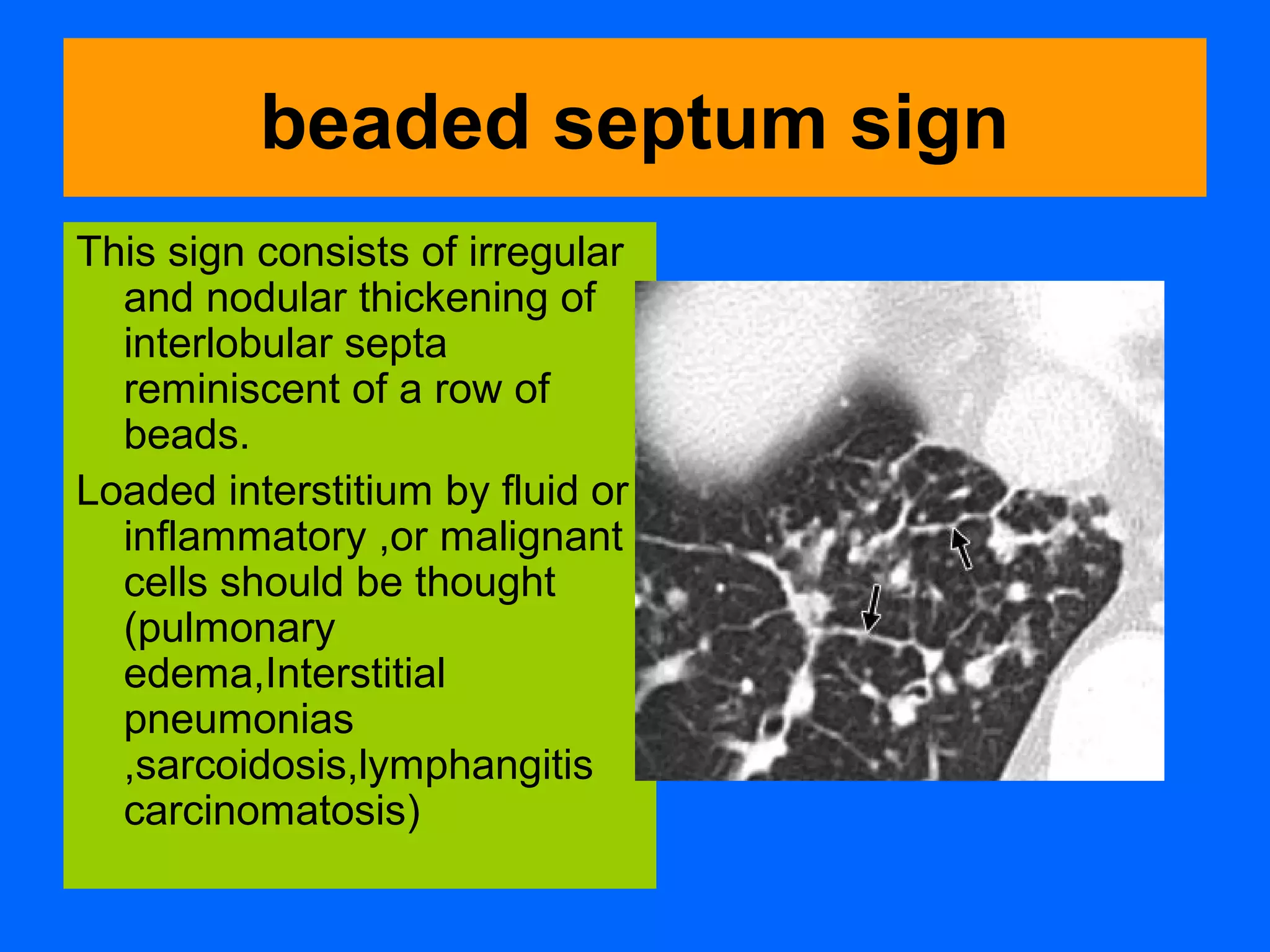
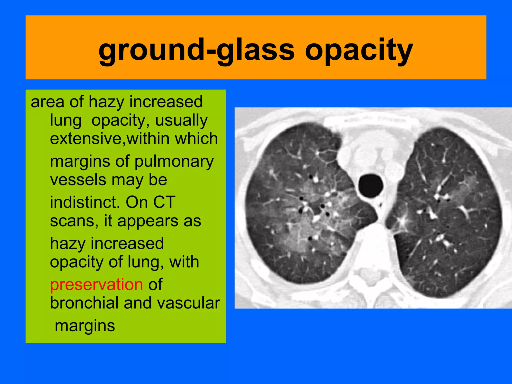
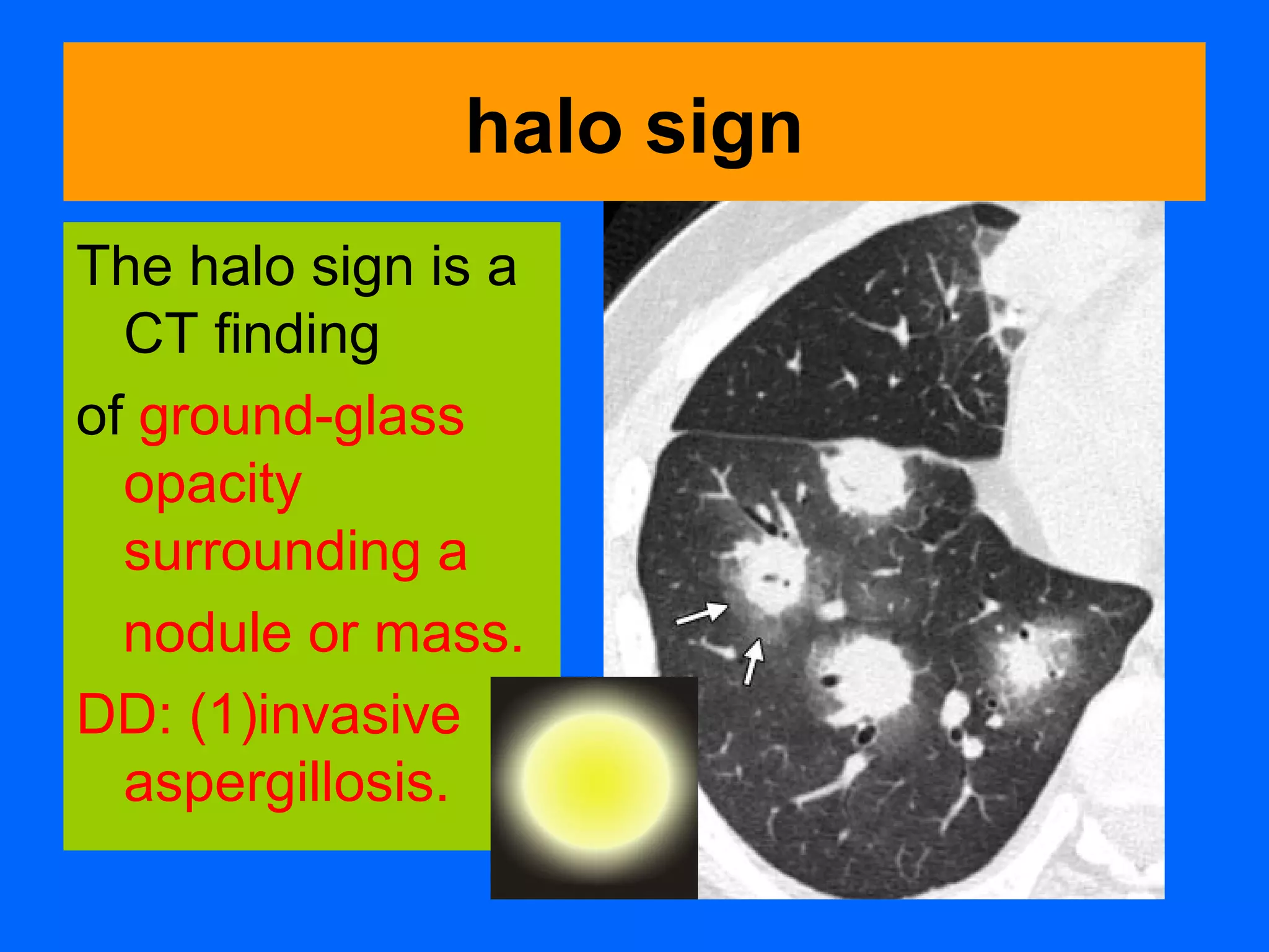
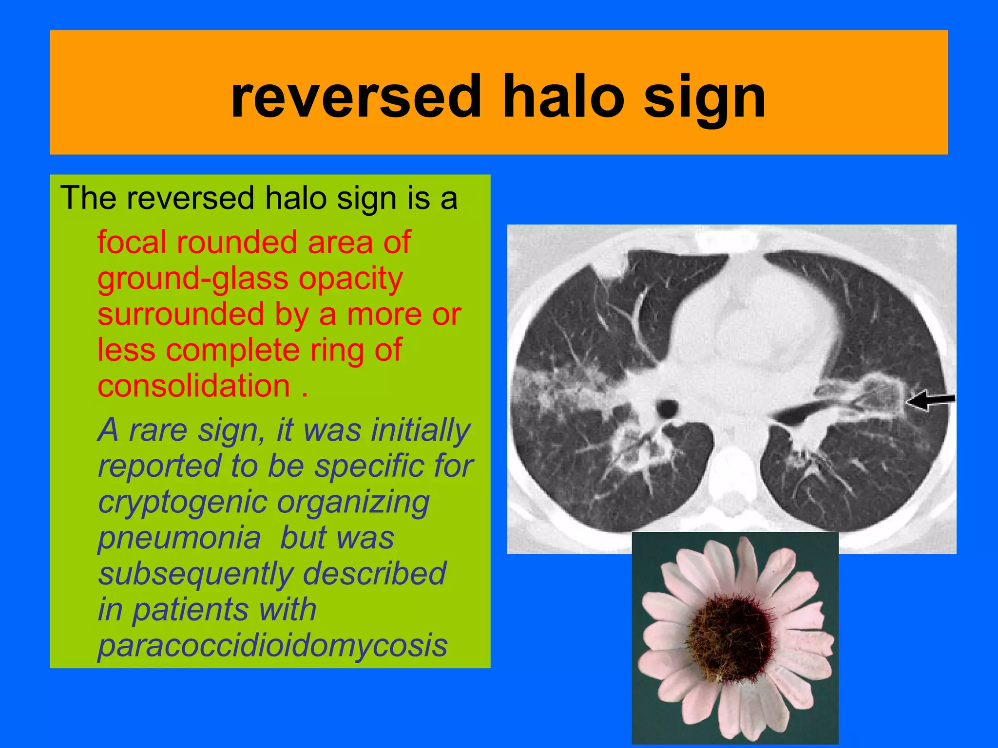
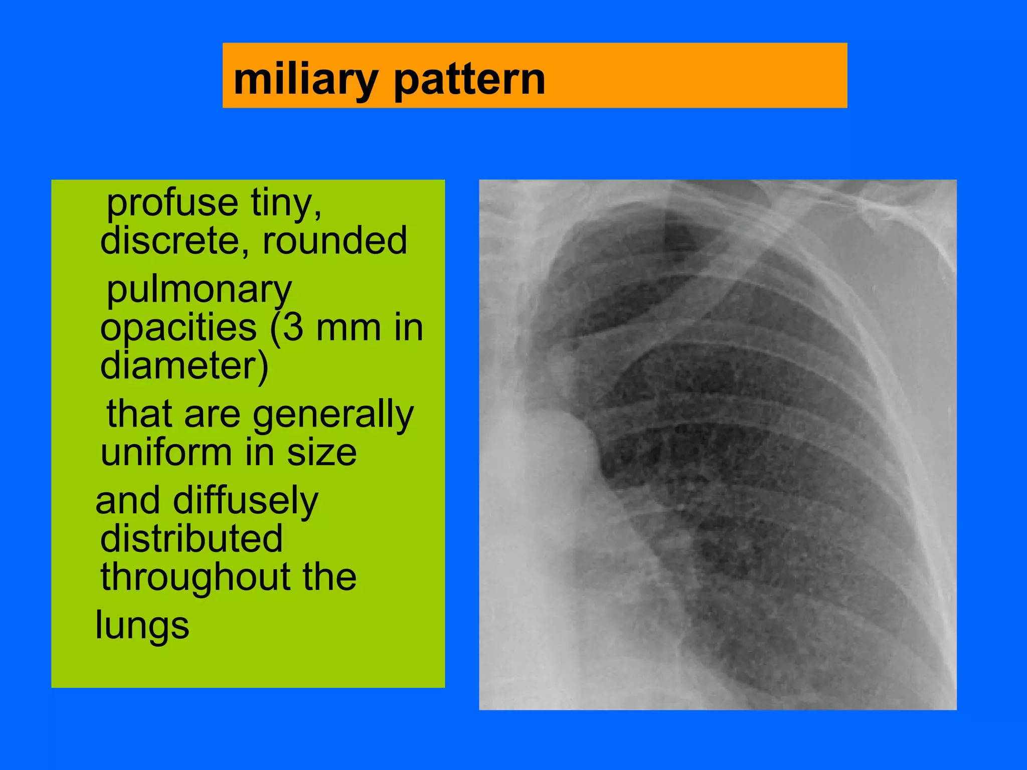
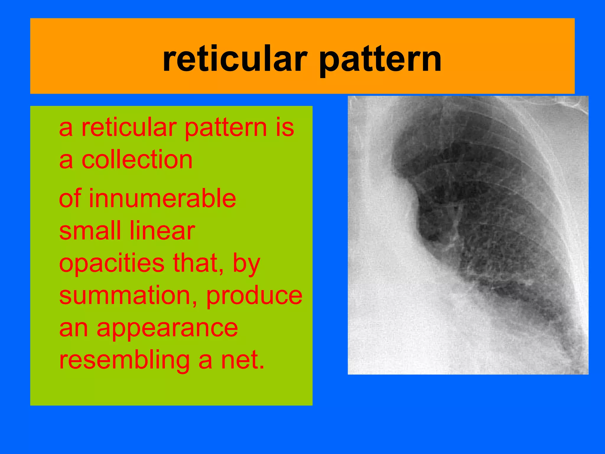
Basic terms inThoracic
Imaging
Dr/Ahmed Bahnassy
Consultant Radiologist
MBCHB-MD-FRCR.
Members of the Fleischner Society
compiled a glossary of terms for thoracic
imaging that replaces previous glossaries
published in 1984 and 1996 for thoracic
radiography and computed tomography
(CT), respectively.
Acinus
• distal to terminal
bronchiole.
• supplied by
respiratory
bronchiole.
• Each SPL has 3-25
acinus.
• largest unit of gas
exchange.
• site of alveolar
diseases.
Air space
• gas containing space
of the lung
aortopulmonary window
The aortopulmonary window
is the mediastinal region
bounded
anteriorly by the ascending
aorta,
Posteriorly by the descending
aorta,
Cranially by the aortic arch,
inferiorly by the left pulmonary
artery,
medially by the ligamentum
arteriosum,
and laterally by
the pleura and left lung
bronchiectasis
Bronchiectasis is
irreversible
localized or diffuse
bronchial dilatation,
usually resulting from
chronic infection,
proximal airway
obstruction, or
congenital bronchial
abnormality
signet ring sign
This finding is
composed of a ring-
shaped opacity
representing a dilated
bronchus in cross
section and a smaller
adjacent opacity
representing its
pulmonary artery, with
the combination
resembling a signet (or
pearl) ring
Bronchiectasis
bronchiolectasis
Bronchiolectasis is
defined as
dilatation of
bronchioles. It is
caused by
inflammatory
airways disease
(potentially
reversible) .
bronchocele
A bronchocele is
bronchial
dilatation due to
retained secretions
(mucoid impaction)
usually caused by
proximal obstruction,
either congenital
(eg, bronchial atresia)
or acquired (eg,
obstructing cancer)
bronchocentric
This descriptor is applied to
disease that is conspicuously
centered on macroscopic
bronchovascular bundles
Examples of diseases with
a bronchocentric distribution
include :
sarcoidosis , Kaposi
sarcoma ,
and organizing pneumonia .
Architectal distorsion
Parenchymal band
Atelectasis
• reduced volume
• increased
density
• air resorption.
• passive..occlusi
ve..adhesive
Broncholith
• calcified LN.
• eroding into
a bronchus.
• TB or
histoplasmos
is
bulla
Airspace measuring
more than 1 cm—usually
several centimeters—
in diameter, sharply
demarcated by a thin wall
that is no greater
than 1 mm in thickness. A
bulla is usually
accompanied by
emphysematous
changes in the adjacent
lung.
cavity
A cavity is
a gas-filled space,
seen as a lucency
or low-attenuation
area, within
pulmonary
consolidation, a
mass, or a nodule
centrilobular emphysema
Centrilobular
emphysema
is characterized by
:destroyed
centrilobular
alveolar walls and
enlargement
of respiratory
bronchioles and
associated
alveoli
cyst
A cyst is any
round
circumscribed
space that is
surrounded by an
epithelial or
fibrous wall of
variable thickness
mycetoma
A mycetoma is a discrete
mass of intertwined
hyphae, usually of
an Aspergillus species,
matted together
by mucus, fibrin, and
cellular debris colonizing
a cavity, usually from
prior fibrocavitary
disease (eg,
tuberculosis or
sarcoidosis)
tree-in-bud pattern
The tree-in-bud pattern
represents
centrilobular branching
structures that
resemble a budding
tree.
The pattern reflects a
spectrum of endo-
and
peribronchiolar
disorders, including
mucoid impaction,
inflammation, and/or
fibrosis
air crescent
An air crescent is a
collection of air in a
crescentic shape that
separates the wall of a
cavity from an inner
mass.
It can represent
aspergilloma on top of
chronic cavity,partially
ruptured hydatid cyst,or
fungus ball in cavitary
bronchiectasis.
apical cap
An apical cap is a caplike
lesion at the lung apex,
usually caused
by :
intrapulmonary and
pleural fibrosis(due to
old TB)
pulling down of
extrapleural fat
Or It can be due to
superior sulcus
tumour.
silhouette sign
The silhouette sign is
the absence of
depiction of an
anatomic
soft-tissue border. It
is caused by
consolidation
and/or atelectasis of
the adjacent
lung
beaded septum sign
This sign consists of irregular
and nodular thickening of
interlobular septa
reminiscent of a row of
beads.
Loaded interstitium by fluid or
inflammatory ,or malignant
cells should be thought
(pulmonary
edema,Interstitial
pneumonias
,sarcoidosis,lymphangitis
carcinomatosis)
ground-glass opacity
area of hazy increased
lung opacity, usually
extensive,within which
margins of pulmonary
vessels may be
indistinct. On CT
scans, it appears as
hazy increased
opacity of lung, with
preservation of
bronchial and vascular
margins
halo sign
The halo sign is a
CT finding
of ground-glass
opacity
surrounding a
nodule or mass.
DD: (1)invasive
aspergillosis.
reversed halo sign
The reversed halo sign is a
focal rounded area of
ground-glass opacity
surrounded by a more or
less complete ring of
consolidation .
A rare sign, it was initially
reported to be specific for
cryptogenic organizing
pneumonia but was
subsequently described
in patients with
paracoccidioidomycosis
honeycombing
Honeycombing represents
destroyed and fibrotic lung
tissue containing numerous
cystic airspaces with thick
fibrous walls,
representing the late stage of
various lung diseases.
The cysts range in size from a
few millimeters to several
centimeters in diameter,
have variable wall thickness, and
are lined by metaplastic
bronchiolar epithelium
profuse tiny,
discrete, rounded
pulmonary
opacities (3 mm in
diameter)
that are generally
uniform in size
and diffusely
distributed
throughout the
lungs
miliary pattern
reticular pattern
a reticular pattern is
a collection
of innumerable
small linear
opacities that, by
summation, produce
an appearance
resembling a net.
reticulonodular pattern
A combined
reticular and nodular
pattern, the
reticulonodular pattern is
usually the result
of the summation of
points of intersection
of innumerable lines,
creating
the effect on chest
radiographs of
superimposed
micronodules
nodule
The chest
radiographic
appearance of a
nodule is
a rounded
opacity, well or
poorly defined,
measuring up to
3 cm in diameter
crazy-paving pattern
This pattern appears
as thickened
interlobular septa and
intralobular
lines superimposed on
a background of
ground-glass opacity ,
resembling irregularly
shaped paving stones.
DD:Alveolar
proteinosis..what
else ?
consolidation
Consolidation refers
to
An exudate or other
product of disease
that replaces
alveolar air,
rendering the lung
solid (as in infective
pneumonia).
Pleural plaque
• fibrohyaline acellular
lesion
• from parietal pleura.
• 15 years post
asbestosis.
• TB?
penumopericardium
pneumomediastinum
pneumothorax
Thoracic imaging terminology

Thoracic imaging terminology

  • 1.
    Basic terms inThoracic Imaging Dr/AhmedBahnassy Consultant Radiologist MBCHB-MD-FRCR.
  • 2.
    Members of theFleischner Society compiled a glossary of terms for thoracic imaging that replaces previous glossaries published in 1984 and 1996 for thoracic radiography and computed tomography (CT), respectively.
  • 3.
    Acinus • distal toterminal bronchiole. • supplied by respiratory bronchiole. • Each SPL has 3-25 acinus. • largest unit of gas exchange. • site of alveolar diseases.
  • 4.
    Air space • gascontaining space of the lung
  • 5.
    aortopulmonary window The aortopulmonarywindow is the mediastinal region bounded anteriorly by the ascending aorta, Posteriorly by the descending aorta, Cranially by the aortic arch, inferiorly by the left pulmonary artery, medially by the ligamentum arteriosum, and laterally by the pleura and left lung
  • 6.
    bronchiectasis Bronchiectasis is irreversible localized ordiffuse bronchial dilatation, usually resulting from chronic infection, proximal airway obstruction, or congenital bronchial abnormality
  • 7.
    signet ring sign Thisfinding is composed of a ring- shaped opacity representing a dilated bronchus in cross section and a smaller adjacent opacity representing its pulmonary artery, with the combination resembling a signet (or pearl) ring
  • 8.
  • 9.
    bronchiolectasis Bronchiolectasis is defined as dilatationof bronchioles. It is caused by inflammatory airways disease (potentially reversible) .
  • 10.
    bronchocele A bronchocele is bronchial dilatationdue to retained secretions (mucoid impaction) usually caused by proximal obstruction, either congenital (eg, bronchial atresia) or acquired (eg, obstructing cancer)
  • 11.
    bronchocentric This descriptor isapplied to disease that is conspicuously centered on macroscopic bronchovascular bundles Examples of diseases with a bronchocentric distribution include : sarcoidosis , Kaposi sarcoma , and organizing pneumonia .
  • 12.
  • 13.
  • 14.
    Atelectasis • reduced volume •increased density • air resorption. • passive..occlusi ve..adhesive
  • 15.
    Broncholith • calcified LN. •eroding into a bronchus. • TB or histoplasmos is
  • 16.
    bulla Airspace measuring more than1 cm—usually several centimeters— in diameter, sharply demarcated by a thin wall that is no greater than 1 mm in thickness. A bulla is usually accompanied by emphysematous changes in the adjacent lung.
  • 17.
    cavity A cavity is agas-filled space, seen as a lucency or low-attenuation area, within pulmonary consolidation, a mass, or a nodule
  • 18.
    centrilobular emphysema Centrilobular emphysema is characterizedby :destroyed centrilobular alveolar walls and enlargement of respiratory bronchioles and associated alveoli
  • 19.
    cyst A cyst isany round circumscribed space that is surrounded by an epithelial or fibrous wall of variable thickness
  • 20.
    mycetoma A mycetoma isa discrete mass of intertwined hyphae, usually of an Aspergillus species, matted together by mucus, fibrin, and cellular debris colonizing a cavity, usually from prior fibrocavitary disease (eg, tuberculosis or sarcoidosis)
  • 21.
    tree-in-bud pattern The tree-in-budpattern represents centrilobular branching structures that resemble a budding tree. The pattern reflects a spectrum of endo- and peribronchiolar disorders, including mucoid impaction, inflammation, and/or fibrosis
  • 22.
    air crescent An aircrescent is a collection of air in a crescentic shape that separates the wall of a cavity from an inner mass. It can represent aspergilloma on top of chronic cavity,partially ruptured hydatid cyst,or fungus ball in cavitary bronchiectasis.
  • 23.
    apical cap An apicalcap is a caplike lesion at the lung apex, usually caused by : intrapulmonary and pleural fibrosis(due to old TB) pulling down of extrapleural fat Or It can be due to superior sulcus tumour.
  • 24.
    silhouette sign The silhouettesign is the absence of depiction of an anatomic soft-tissue border. It is caused by consolidation and/or atelectasis of the adjacent lung
  • 25.
    beaded septum sign Thissign consists of irregular and nodular thickening of interlobular septa reminiscent of a row of beads. Loaded interstitium by fluid or inflammatory ,or malignant cells should be thought (pulmonary edema,Interstitial pneumonias ,sarcoidosis,lymphangitis carcinomatosis)
  • 26.
    ground-glass opacity area ofhazy increased lung opacity, usually extensive,within which margins of pulmonary vessels may be indistinct. On CT scans, it appears as hazy increased opacity of lung, with preservation of bronchial and vascular margins
  • 27.
    halo sign The halosign is a CT finding of ground-glass opacity surrounding a nodule or mass. DD: (1)invasive aspergillosis.
  • 28.
    reversed halo sign Thereversed halo sign is a focal rounded area of ground-glass opacity surrounded by a more or less complete ring of consolidation . A rare sign, it was initially reported to be specific for cryptogenic organizing pneumonia but was subsequently described in patients with paracoccidioidomycosis
  • 29.
    honeycombing Honeycombing represents destroyed andfibrotic lung tissue containing numerous cystic airspaces with thick fibrous walls, representing the late stage of various lung diseases. The cysts range in size from a few millimeters to several centimeters in diameter, have variable wall thickness, and are lined by metaplastic bronchiolar epithelium
  • 30.
    profuse tiny, discrete, rounded pulmonary opacities(3 mm in diameter) that are generally uniform in size and diffusely distributed throughout the lungs miliary pattern
  • 31.
    reticular pattern a reticularpattern is a collection of innumerable small linear opacities that, by summation, produce an appearance resembling a net.
  • 32.
    reticulonodular pattern A combined reticularand nodular pattern, the reticulonodular pattern is usually the result of the summation of points of intersection of innumerable lines, creating the effect on chest radiographs of superimposed micronodules
  • 33.
    nodule The chest radiographic appearance ofa nodule is a rounded opacity, well or poorly defined, measuring up to 3 cm in diameter
  • 34.
    crazy-paving pattern This patternappears as thickened interlobular septa and intralobular lines superimposed on a background of ground-glass opacity , resembling irregularly shaped paving stones. DD:Alveolar proteinosis..what else ?
  • 35.
    consolidation Consolidation refers to An exudateor other product of disease that replaces alveolar air, rendering the lung solid (as in infective pneumonia).
  • 36.
    Pleural plaque • fibrohyalineacellular lesion • from parietal pleura. • 15 years post asbestosis. • TB?
  • 37.
  • 38.
  • 39.