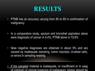
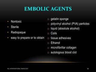
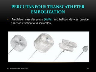
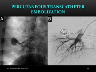
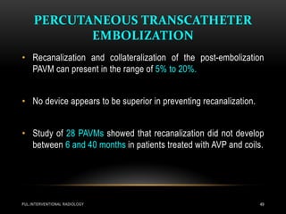
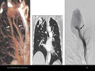
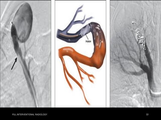
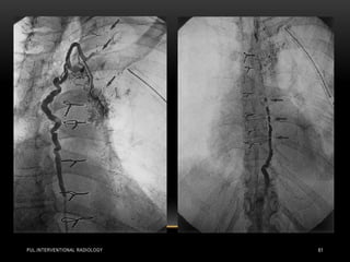
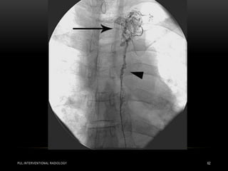
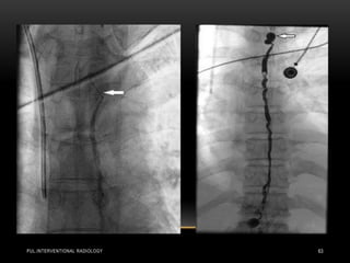
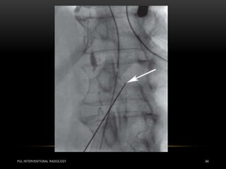
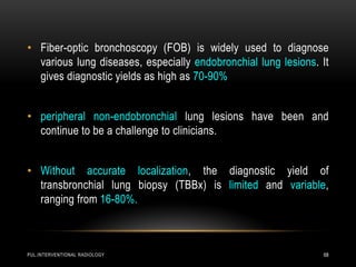
By
Mahmoud E. Abo El-Magd
Assistant lecturer of pulmonary and critical
care medicine
PULMONARY
INTERVENTIONAL
RADIOLOGY
INTRODUCTION
PUL.INTERVENTIONAL RADIOLOGY 2
• Minimally invasive
• Image guided procedures
• Less complications
• No need for general anesthesia
• No surgical interference
• Patients with co-morbidities
• Patients refused surgery
PUL.INTERVENTIONAL RADIOLOGY 3
PERCUTANEOUS TRANSTHORACIC
NEEDLE BIOPSY
PUL.INTERVENTIONAL RADIOLOGY 4
• Percutaneous lung biopsy was first described by Leyden in 1883
to establish the infectious nature of lung disease.
• Actually percutaneous tissue sampling of a pulmonary, pleural or
mediastinal lesion is indicated, when histological diagnosis will
further influence diagnostic strategy and therapeutica options, or
modify tumor staging and the sequence of cancer treatment.
VALUE OF PTNB
PUL.INTERVENTIONAL RADIOLOGY 5
• Cost efficiency by shortening hospital stay .
• Management was altered in 51% of patients.
• Surgery was avoided in 83% of biopsies that altered patient
treatment.
INDICATIONS
PUL.INTERVENTIONAL RADIOLOGY 6
• pulmonary nodule(s) without specific diagnostic criteria on CT
ascertaining benignity .
• pulmonary nodule(s) or mass suggestive of malignancy, when
surgery will be postponed, or replaced by chemotherapy and/or
radiotherapy.
• pulmonary nodule(s) in a patient with a history of
extrapulmonary primary malignancy and in clinical remission or
presenting several primary malignancies .
PUL.INTERVENTIONAL RADIOLOGY 7
• Residual non-regressive lesion following radiotherapy or
chemotherapy .
• Mediastinal mass or adenopathy. PTNB is a useful alternative to
mediastinoscopy or mediastinostomy for tissue diagnosis of
enlarged hilar or mediastinal lymph nodes.
• All areas of the mediastinum are accessible to PTNB. The
technique is faster, better tolerated and less expensive .
PUL.INTERVENTIONAL RADIOLOGY 8
• Chronic diffuse pulmonary infiltrate in selected cases .
• Intracavitary injection for treatment of aspergilloma .
• Percutaneous brachytherapy of pulmonary malignancy
CONTRA-INDICATIONS
PUL.INTERVENTIONAL RADIOLOGY 9
• Vascular structures such as aneurysm or pulmonary
arteriovenous malformation.
• Hydatid cyst and mediastinal pheochromocytoma are absolute
contra-indications of PTNB.
PUL.INTERVENTIONAL RADIOLOGY 10
Relative contra-indications:
• puncture of both lungs during the same day.
• puncture of only one functional lung.
• chronic respiratory insufficiency.
• pulmonary arterial hypertension.
• cardiac insufficiency, recent myocardial infarct.
• severe emphysema and bullae near the pulmonary lesion.
• A coagulation defect .
• Cough, dyspnea and uncooperative patient.
• Mechanical ventilation .
TECHNIQUE
PUL.INTERVENTIONAL RADIOLOGY 11
• The needle path should avoid traversing intercostal vessels (i.e.
needle entry is optimally defined at mid intercostal space).
• Central tumor necrosis is recognized on the basis of central low
densities after i. v. contrast injection.
• The biopsy needle should be directed to the viable tumor
component situated at the periphery of the lesion.
TECHNIQUE - cont
PUL.INTERVENTIONAL RADIOLOGY 12
• Mediastinal and hilar vascular structures surrounding a lesion
are recognized with contrast enhanced CT.
• Real time-CT (fluoro-CT or continuous CT) is now becoming
widely available .
• US can be used as a guidance for puncture of pleural or
subpleural lesions
RESULTS
PUL.INTERVENTIONAL RADIOLOGY 13
• PTNB has an accuracy varying from 80 to 95 in confirmation of
malignancy
• In a comparative study, sputum and bronchial aspiration alone
were diagnostic of cancer in 5.4%, PTNB alone in 72.6%
• false negative diagnoses are obtained in about 5% and are
caused by inadequate sampling, tumor necrosis, crushed cells,
or errors in sampling reading.
• If the sampled material is inadequate, or insufficient or in case
COMPLICATIONS
PUL.INTERVENTIONAL RADIOLOGY 14
• Pneumothorax is the most frequent complication following
PTNB, with an incidence of 8-60%.
• Less than 5% of patients have persistent clinical symptoms and
require aspiration or drainage.
PUL.INTERVENTIONAL RADIOLOGY 15
Factors influencing pneumothorax are :
• Age.
• Co-operation.
• respiratory function.
• biopsy technique.
• experience of the operator.
• duration of the procedure.
• number of needle passes.
• diameter and flexibility of the
needle.
• Emphysema.
• difficult localization of the target.
• depth and diameter of the target.
• mechanical ventilation.
• cavitary lesion.
PUL.INTERVENTIONAL RADIOLOGY 16
• Special care should be taken in patients who underwent thoracic
surgical intervention with a potential communication between
both pleural spaces, increasing the risk of bilateral
pneumothorax.
• Best prevention of complications is to perform the procedure
rapidly and to reduce the number of passes by inspecting the
quality of smears or fragments after each puncture.
PUL.INTERVENTIONAL RADIOLOGY 17
• Pneumothorax is further prevented by the roll-over technique,
which consists of turning the patient following biopsy for 15-30
minutes on the side which had been punctured.
• All patients are maintained in the recumbant position for 4
hours after biopsy; then an erect chest radiograph is obtained.
If no pneumothorax is present, compliant patients can be
discharged.
PUL.INTERVENTIONAL RADIOLOGY 18
• biopsy is completed before the pneumothorax is treated. In the
literature, small bore catheter drainage of iatrogenic
pneumothorax is successful in 75-97 % .
PUL.INTERVENTIONAL RADIOLOGY 19
• To reduce the risk of pneumothorax or pulmonary hemorrhage
during mediastinal biopsy, injection of small amounts of saline
with a small gauge needle can be used to distend the extra-
pleural tissue, which allows access to most mediastinal lesions,
using an exclusive extrapleural pathway without transgression
of the visceral pleura .
• Similarly, extrapleural injection of saline displaces the pleura,
allowing for a safe transpleural access to subpleural pulmonary
lesions
PUL.INTERVENTIONAL RADIOLOGY 20
• Haemoptysis or hemorrhage is encountered in less than 10% of
PTNB, most of which are not life threatening
• A limited perinodular alveolar haemorrhage is commonly
observed on CT and can obscure the nodule, rendering further
punctures impossible
PUL.INTERVENTIONAL RADIOLOGY 21
Other complications :
• mediastinal emphysema.
• thoracic wall haematoma.
• Haemothorax.
• cardiac tamponade.
• Empyema.
• bronchopleural fistula.
• lung torsion and implantation metastasis along the needle tract.
• air embolius and sudden death are rarely observed .
• Mortality rate of PTNB is estimated at 0.02 % .
• thoracic pain.
• pleural effusion.
• superinfection.
PUL.INTERVENTIONAL RADIOLOGY 22
PUL.INTERVENTIONAL RADIOLOGY 23
• Thoracic fluid collections of various origin, located in the pleura,
pericardium, lung or mediastinum can be aspirated or drained
percutaneously with imaging guidance.
• Percutaneous therapy is an important refinement compared to
blind bed side technique and compares favourably with surgery.
• Diagnostic fluid aspiration includes cytological, bacteriological
and chemical analyses.
PUL.INTERVENTIONAL RADIOLOGY 24
• Fluoroscopy, US and CT can be used as guidance modalities,
either alone or in combination.
• The choice of imaging technique depends on technical
characteristics, availability, location of the collection, and
operator expertise.
• Loculated pleural effusions may be treated by thoracocentesis,
thoracoscopy, surgically or CT-guided thoracostomy tube
drainage, open drainage, or thoracotomy and pleural
decortication. Imaging guidance best insures proper catheter
positioning.
GUIDANCE MODALITIES -
ULTRASONOGRAPHY (US)
PUL.INTERVENTIONAL RADIOLOGY 25
• US is particularly indicated to guide bedside percutaneous
aspiration and catheter drainage of pericardial and pleural fluid,
even of reduced amount .
• Pulmonary abscesses that are located in a subpleural location
can also be included.
• Percutaneous approach is performed in the position that
optimally displays access to the fluid collection.
COMPUTED TOMOGRAPHY
(CT)
PUL.INTERVENTIONAL RADIOLOGY 26
• CT offers exquisite anatomical display of all thoracic structures
and allows percutaneous access to all spaces with equal ease
• The optimal percutaneous entry point to a pulmonary abscess is
defined according to its pleural contact, location of fissures and
vascular structures.
• CT is the optimal modality to confirm pneumothorax or
pneumomediastinum following a percutaneous procedure.
PUL.INTERVENTIONAL RADIOLOGY 27
• CT is particularly useful for the percutaneous approach to the
mediastinum, and for pulmonary abscesses with a reduced
pleural contact.
PUL.INTERVENTIONAL RADIOLOGY 28
• Pleural empyema
• Malignant Pleural Effusion
• Post-traumatic Haemothorax
• Gazeous Collections
• Pulmonary Abscess
• Mediastinal collections
• Pericardial Fluid Collection
• Percutaneous Drainage of Tension Pneumomediastinum
COMPLICATIONS OF PERCUTANEOUS
CATHETER DRAINAGE
PUL.INTERVENTIONAL RADIOLOGY 29
• Complications occur in approximately 5% and are mainly due to
inadequate catheter insertion technique
• Injury to an intercostal artery can produce a haemothorax or a
haematoma of the thoracic wall.
• Life threatening haemorrhage is rare, but can be observed
following rupture of the wall of a pulmonary abscess, erosion of
a branch of the pulmonary artery or transfixation of an internal
mammary or mediastinal vessel by the catheter.
PUL.INTERVENTIONAL RADIOLOGY 30
• When normal lung is crossed with a large bore catheter using
the trocar technique, a pulmonary infarction can result.
• Haemoptysis can occur during catheter insertion. Pneumothorax
is a potentially frequent complication.
• Other side-effects include subcutaneous emphysema, local skin
infection at the entry point of the catheter and discomfort during
breathing, rib erosion, catheter leakage, bending, breakage and
obstruction.
PUL.INTERVENTIONAL RADIOLOGY 31
PUL.INTERVENTIONAL RADIOLOGY 32
• Although IV TPA is indicated for treatment of acute massive PE,
many patients cannot receive systemic thrombolysis due to
contraindications.
• The rate of major hemorrhage from systemic thrombolytic
administration is approximately 20%, including a 3%–5% risk of
hemorrhagic stroke .
• In a meta-analysis of 594 patients with acute massive
PE treated with modern CDT, clinical success was achieved
in 86.5%.
PUL.INTERVENTIONAL RADIOLOGY 33
PUL.INTERVENTIONAL RADIOLOGY 34
PUL.INTERVENTIONAL RADIOLOGY 35
• Pulmonary angiography in a 57-year-old woman in shock from
acute bilateral massive PE. Initial right (a) and left
(b) pulmonary angiograms show near-complete obstruction.
Pulmonary artery pressure was 73/18 mm Hg. Final right (c) and
left
(d) images after suction thrombectomy and catheter-directed
thrombolytic agent injection into each main descending
pulmonary
artery. Pulmonary artery pressure was reduced to 36/16 mm Hg.
(Images courtesy of Daniel Y. Sze.)
PUL.INTERVENTIONAL RADIOLOGY 36
• Biopsy – guided biopsies
• Embolization
• Vascular embolization
• Chemo-embolization
• Radio-embolization
• Radiofrequency ablation
• cryoablation
PUL.INTERVENTIONAL RADIOLOGY 37
PUL.INTERVENTIONAL RADIOLOGY 38
 definitive treatment of benign lesions.
 reducing the risk of bleeding prior to biopsy or surgery.
 palliation of pain, bleeding, fever, and hypercalcemia-like symptoms
in inoperable tumors.
 preventing further dissemination of a tumor.
 increasing the response to chemotherapy and radiotherapy.
 retention of selectively delivered anti-mitotic agents or monoclonal
antibodies deep into the tumor substance.
PUL.INTERVENTIONAL RADIOLOGY 39
EMBOLIC AGENTS
• Nontoxic
• Sterile
• Radiopaque
• easy to prepare or to obtain
 gelatin sponge
 polyvinyl alcohol (PVA) particles
 liquid (absolute alcohol)
 Coils
 tissue adhesives
 Ethanol
 microfibrillar collagen
 autologous blood clot
PUL.INTERVENTIONAL RADIOLOGY 40
ADVANTAGES
• Minimal incisions/scarring
• Shorter hospital stay/outpatient
• Reduced recovery time
• Anesthetic
• Lower risk of complications
• Treatment option in poor/risk prone patients
PUL.INTERVENTIONAL RADIOLOGY 41
PUL.INTERVENTIONAL RADIOLOGY 42
DIAGNOSTIC TESTING – pulmonary angiography
PUL.INTERVENTIONAL RADIOLOGY 43
• Pulmonary angiography generally is no longer necessary as a
diagnostic procedure alone.
• It is reserved for therapeutic purposes after a diagnosis has
been established.
• It remains the gold standard for inconclusive cases.
TREATMENT AND MANAGEMENT
PERCUTANEOUS TRANSCATHETER
EMBOLIZATION
PUL.INTERVENTIONAL RADIOLOGY 44
• Percutaneous TCE is the gold standard for the treatment of
PAVM because it is effective in reducing the risk of paradoxical
embolism and other complications associated with PAVM.
• less invasive and easy to repeat .
• disadvantages include collateralization and revascularization
over time.
PERCUTANEOUS TRANSCATHETER
EMBOLIZATION
PUL.INTERVENTIONAL RADIOLOGY 45
• Major indications for treatment are :-
- prevention of neurologic complications, including stroke and
cerebral abscess from paradoxical embolism.
- improvement in exercise tolerance .
- reduction in migraine prevalence .
- prevention of lung hemorrhage.
PERCUTANEOUS TRANSCATHETER
EMBOLIZATION
PUL.INTERVENTIONAL RADIOLOGY 46
• During embolization, the supplying artery immediately preceding
the PAVM is the target to occlude the feeding vessel just
proximal to the aneurysmal sac .
• The deployed coils are designed to coil within the vessel lumen
and carry micro fibers that activate platelets to generate an
occluding platelet plug.
PERCUTANEOUS TRANSCATHETER
EMBOLIZATION
PUL.INTERVENTIONAL RADIOLOGY 47
• Amplatzer vascular plugs (AVPs) and balloon devices provide
direct obstruction to vascular flow.
PERCUTANEOUS TRANSCATHETER
EMBOLIZATION
PUL.INTERVENTIONAL RADIOLOGY 48
PERCUTANEOUS TRANSCATHETER
EMBOLIZATION
PUL.INTERVENTIONAL RADIOLOGY 49
• Recanalization and collateralization of the post-embolization
PAVM can present in the range of 5% to 20%.
• No device appears to be superior in preventing recanalization.
• Study of 28 PAVMs showed that recanalization did not develop
between 6 and 40 months in patients treated with AVP and coils.
PUL.INTERVENTIONAL RADIOLOGY 50
PUL.INTERVENTIONAL RADIOLOGY 51
PUL.INTERVENTIONAL RADIOLOGY 52
PUL.INTERVENTIONAL RADIOLOGY 53
PUL.INTERVENTIONAL RADIOLOGY 54
PUL.INTERVENTIONAL RADIOLOGY 55
PUL.INTERVENTIONAL RADIOLOGY 56
PUL.INTERVENTIONAL RADIOLOGY 57
PUL.INTERVENTIONAL RADIOLOGY 58
PUL.INTERVENTIONAL RADIOLOGY 59
INTERVENTIONAL RADIOLOGY
PUL.INTERVENTIONAL RADIOLOGY 60
• Today there are several radiological treatments that can be used
in both traumatic and non-traumatic chylothorax .
• Much more experience is available for percutaneous
embolization of the thoracic duct , which can be performed as
an alternative to thoracic duct ligation and can be performed in
both adults and children .
• Embolization has a much higher success rate: if the thoracic
duct can be intubated successfully, the procedure is successful
in well over 90% of cases
PUL.INTERVENTIONAL RADIOLOGY 61
PUL.INTERVENTIONAL RADIOLOGY 62
PUL.INTERVENTIONAL RADIOLOGY 63
PUL.INTERVENTIONAL RADIOLOGY 64
PUL.INTERVENTIONAL RADIOLOGY 65
PUL.INTERVENTIONAL RADIOLOGY 66
PUL.INTERVENTIONAL RADIOLOGY 67
PUL.INTERVENTIONAL RADIOLOGY 68
• Fiber-optic bronchoscopy (FOB) is widely used to diagnose
various lung diseases, especially endobronchial lung lesions. It
gives diagnostic yields as high as 70-90%
• peripheral non-endobronchial lung lesions have been and
continue to be a challenge to clinicians.
• Without accurate localization, the diagnostic yield of
transbronchial lung biopsy (TBBx) is limited and variable,
ranging from 16-80%.
PUL.INTERVENTIONAL RADIOLOGY 69
• For nonvisible or peripheral lesions, conventional techniques
such as selective bronchial aspiration or broncho-alveolar
lavage have a diagnostic yield of only 48%.
• The addition of other diagnostic procedures such as
transbronchial needle aspiration or bronchoscopic lung biopsy
(BLB) increases diagnostic yield to more than 70% when used
in combination or when guided by fluoroscopy.
PUL.INTERVENTIONAL RADIOLOGY 70
• Fluoroscopy reduces the risk of pneumothorax during TBBx
• FOB FB extraction fluoroscopic guided.
PUL.INTERVENTIONAL RADIOLOGY 71
PUL.INTERVENTIONAL RADIOLOGY 72
• Most patients either have too alimited pulmonary function for
surgical resection or are unable to tolerate surgery because of
other comorbid medical conditions, especially patients with poor
cardiopulmonary functions.
• Image-guided percutaneous thermal ablation therapies
are minimally invasive interventional techniques
PUL.INTERVENTIONAL RADIOLOGY 73
• Among these techniques, radiofrequency ablation (RFA) has
now attained consideration for therapy of primary and secondary
lung tumors .
• Others include cryoablation , laser , and more recently
microwave .
• Thermal ablative techniques produce irreversible tumor tissue
destruction through application of either hot or cold thermal
energy.
PUL.INTERVENTIONAL RADIOLOGY 74
• Planning, monitoring, targeting, and controlling this
modality are performed with the help of different imaging
modalities, including ultrasound, X-ray, computed tomography
(CT), and magnetic resonance tomography.
PUL.INTERVENTIONAL RADIOLOGY 75

Pulmonary interventional radiology techniques

  • 1.
    By Mahmoud E. AboEl-Magd Assistant lecturer of pulmonary and critical care medicine PULMONARY INTERVENTIONAL RADIOLOGY
  • 2.
    INTRODUCTION PUL.INTERVENTIONAL RADIOLOGY 2 •Minimally invasive • Image guided procedures • Less complications • No need for general anesthesia • No surgical interference • Patients with co-morbidities • Patients refused surgery
  • 3.
  • 4.
    PERCUTANEOUS TRANSTHORACIC NEEDLE BIOPSY PUL.INTERVENTIONALRADIOLOGY 4 • Percutaneous lung biopsy was first described by Leyden in 1883 to establish the infectious nature of lung disease. • Actually percutaneous tissue sampling of a pulmonary, pleural or mediastinal lesion is indicated, when histological diagnosis will further influence diagnostic strategy and therapeutica options, or modify tumor staging and the sequence of cancer treatment.
  • 5.
    VALUE OF PTNB PUL.INTERVENTIONALRADIOLOGY 5 • Cost efficiency by shortening hospital stay . • Management was altered in 51% of patients. • Surgery was avoided in 83% of biopsies that altered patient treatment.
  • 6.
    INDICATIONS PUL.INTERVENTIONAL RADIOLOGY 6 •pulmonary nodule(s) without specific diagnostic criteria on CT ascertaining benignity . • pulmonary nodule(s) or mass suggestive of malignancy, when surgery will be postponed, or replaced by chemotherapy and/or radiotherapy. • pulmonary nodule(s) in a patient with a history of extrapulmonary primary malignancy and in clinical remission or presenting several primary malignancies .
  • 7.
    PUL.INTERVENTIONAL RADIOLOGY 7 •Residual non-regressive lesion following radiotherapy or chemotherapy . • Mediastinal mass or adenopathy. PTNB is a useful alternative to mediastinoscopy or mediastinostomy for tissue diagnosis of enlarged hilar or mediastinal lymph nodes. • All areas of the mediastinum are accessible to PTNB. The technique is faster, better tolerated and less expensive .
  • 8.
    PUL.INTERVENTIONAL RADIOLOGY 8 •Chronic diffuse pulmonary infiltrate in selected cases . • Intracavitary injection for treatment of aspergilloma . • Percutaneous brachytherapy of pulmonary malignancy
  • 9.
    CONTRA-INDICATIONS PUL.INTERVENTIONAL RADIOLOGY 9 •Vascular structures such as aneurysm or pulmonary arteriovenous malformation. • Hydatid cyst and mediastinal pheochromocytoma are absolute contra-indications of PTNB.
  • 10.
    PUL.INTERVENTIONAL RADIOLOGY 10 Relativecontra-indications: • puncture of both lungs during the same day. • puncture of only one functional lung. • chronic respiratory insufficiency. • pulmonary arterial hypertension. • cardiac insufficiency, recent myocardial infarct. • severe emphysema and bullae near the pulmonary lesion. • A coagulation defect . • Cough, dyspnea and uncooperative patient. • Mechanical ventilation .
  • 11.
    TECHNIQUE PUL.INTERVENTIONAL RADIOLOGY 11 •The needle path should avoid traversing intercostal vessels (i.e. needle entry is optimally defined at mid intercostal space). • Central tumor necrosis is recognized on the basis of central low densities after i. v. contrast injection. • The biopsy needle should be directed to the viable tumor component situated at the periphery of the lesion.
  • 12.
    TECHNIQUE - cont PUL.INTERVENTIONALRADIOLOGY 12 • Mediastinal and hilar vascular structures surrounding a lesion are recognized with contrast enhanced CT. • Real time-CT (fluoro-CT or continuous CT) is now becoming widely available . • US can be used as a guidance for puncture of pleural or subpleural lesions
  • 13.
    RESULTS PUL.INTERVENTIONAL RADIOLOGY 13 •PTNB has an accuracy varying from 80 to 95 in confirmation of malignancy • In a comparative study, sputum and bronchial aspiration alone were diagnostic of cancer in 5.4%, PTNB alone in 72.6% • false negative diagnoses are obtained in about 5% and are caused by inadequate sampling, tumor necrosis, crushed cells, or errors in sampling reading. • If the sampled material is inadequate, or insufficient or in case
  • 14.
    COMPLICATIONS PUL.INTERVENTIONAL RADIOLOGY 14 •Pneumothorax is the most frequent complication following PTNB, with an incidence of 8-60%. • Less than 5% of patients have persistent clinical symptoms and require aspiration or drainage.
  • 15.
    PUL.INTERVENTIONAL RADIOLOGY 15 Factorsinfluencing pneumothorax are : • Age. • Co-operation. • respiratory function. • biopsy technique. • experience of the operator. • duration of the procedure. • number of needle passes. • diameter and flexibility of the needle. • Emphysema. • difficult localization of the target. • depth and diameter of the target. • mechanical ventilation. • cavitary lesion.
  • 16.
    PUL.INTERVENTIONAL RADIOLOGY 16 •Special care should be taken in patients who underwent thoracic surgical intervention with a potential communication between both pleural spaces, increasing the risk of bilateral pneumothorax. • Best prevention of complications is to perform the procedure rapidly and to reduce the number of passes by inspecting the quality of smears or fragments after each puncture.
  • 17.
    PUL.INTERVENTIONAL RADIOLOGY 17 •Pneumothorax is further prevented by the roll-over technique, which consists of turning the patient following biopsy for 15-30 minutes on the side which had been punctured. • All patients are maintained in the recumbant position for 4 hours after biopsy; then an erect chest radiograph is obtained. If no pneumothorax is present, compliant patients can be discharged.
  • 18.
    PUL.INTERVENTIONAL RADIOLOGY 18 •biopsy is completed before the pneumothorax is treated. In the literature, small bore catheter drainage of iatrogenic pneumothorax is successful in 75-97 % .
  • 19.
    PUL.INTERVENTIONAL RADIOLOGY 19 •To reduce the risk of pneumothorax or pulmonary hemorrhage during mediastinal biopsy, injection of small amounts of saline with a small gauge needle can be used to distend the extra- pleural tissue, which allows access to most mediastinal lesions, using an exclusive extrapleural pathway without transgression of the visceral pleura . • Similarly, extrapleural injection of saline displaces the pleura, allowing for a safe transpleural access to subpleural pulmonary lesions
  • 20.
    PUL.INTERVENTIONAL RADIOLOGY 20 •Haemoptysis or hemorrhage is encountered in less than 10% of PTNB, most of which are not life threatening • A limited perinodular alveolar haemorrhage is commonly observed on CT and can obscure the nodule, rendering further punctures impossible
  • 21.
    PUL.INTERVENTIONAL RADIOLOGY 21 Othercomplications : • mediastinal emphysema. • thoracic wall haematoma. • Haemothorax. • cardiac tamponade. • Empyema. • bronchopleural fistula. • lung torsion and implantation metastasis along the needle tract. • air embolius and sudden death are rarely observed . • Mortality rate of PTNB is estimated at 0.02 % . • thoracic pain. • pleural effusion. • superinfection.
  • 22.
  • 23.
    PUL.INTERVENTIONAL RADIOLOGY 23 •Thoracic fluid collections of various origin, located in the pleura, pericardium, lung or mediastinum can be aspirated or drained percutaneously with imaging guidance. • Percutaneous therapy is an important refinement compared to blind bed side technique and compares favourably with surgery. • Diagnostic fluid aspiration includes cytological, bacteriological and chemical analyses.
  • 24.
    PUL.INTERVENTIONAL RADIOLOGY 24 •Fluoroscopy, US and CT can be used as guidance modalities, either alone or in combination. • The choice of imaging technique depends on technical characteristics, availability, location of the collection, and operator expertise. • Loculated pleural effusions may be treated by thoracocentesis, thoracoscopy, surgically or CT-guided thoracostomy tube drainage, open drainage, or thoracotomy and pleural decortication. Imaging guidance best insures proper catheter positioning.
  • 25.
    GUIDANCE MODALITIES - ULTRASONOGRAPHY(US) PUL.INTERVENTIONAL RADIOLOGY 25 • US is particularly indicated to guide bedside percutaneous aspiration and catheter drainage of pericardial and pleural fluid, even of reduced amount . • Pulmonary abscesses that are located in a subpleural location can also be included. • Percutaneous approach is performed in the position that optimally displays access to the fluid collection.
  • 26.
    COMPUTED TOMOGRAPHY (CT) PUL.INTERVENTIONAL RADIOLOGY26 • CT offers exquisite anatomical display of all thoracic structures and allows percutaneous access to all spaces with equal ease • The optimal percutaneous entry point to a pulmonary abscess is defined according to its pleural contact, location of fissures and vascular structures. • CT is the optimal modality to confirm pneumothorax or pneumomediastinum following a percutaneous procedure.
  • 27.
    PUL.INTERVENTIONAL RADIOLOGY 27 •CT is particularly useful for the percutaneous approach to the mediastinum, and for pulmonary abscesses with a reduced pleural contact.
  • 28.
    PUL.INTERVENTIONAL RADIOLOGY 28 •Pleural empyema • Malignant Pleural Effusion • Post-traumatic Haemothorax • Gazeous Collections • Pulmonary Abscess • Mediastinal collections • Pericardial Fluid Collection • Percutaneous Drainage of Tension Pneumomediastinum
  • 29.
    COMPLICATIONS OF PERCUTANEOUS CATHETERDRAINAGE PUL.INTERVENTIONAL RADIOLOGY 29 • Complications occur in approximately 5% and are mainly due to inadequate catheter insertion technique • Injury to an intercostal artery can produce a haemothorax or a haematoma of the thoracic wall. • Life threatening haemorrhage is rare, but can be observed following rupture of the wall of a pulmonary abscess, erosion of a branch of the pulmonary artery or transfixation of an internal mammary or mediastinal vessel by the catheter.
  • 30.
    PUL.INTERVENTIONAL RADIOLOGY 30 •When normal lung is crossed with a large bore catheter using the trocar technique, a pulmonary infarction can result. • Haemoptysis can occur during catheter insertion. Pneumothorax is a potentially frequent complication. • Other side-effects include subcutaneous emphysema, local skin infection at the entry point of the catheter and discomfort during breathing, rib erosion, catheter leakage, bending, breakage and obstruction.
  • 31.
  • 32.
    PUL.INTERVENTIONAL RADIOLOGY 32 •Although IV TPA is indicated for treatment of acute massive PE, many patients cannot receive systemic thrombolysis due to contraindications. • The rate of major hemorrhage from systemic thrombolytic administration is approximately 20%, including a 3%–5% risk of hemorrhagic stroke . • In a meta-analysis of 594 patients with acute massive PE treated with modern CDT, clinical success was achieved in 86.5%.
  • 33.
  • 34.
  • 35.
    PUL.INTERVENTIONAL RADIOLOGY 35 •Pulmonary angiography in a 57-year-old woman in shock from acute bilateral massive PE. Initial right (a) and left (b) pulmonary angiograms show near-complete obstruction. Pulmonary artery pressure was 73/18 mm Hg. Final right (c) and left (d) images after suction thrombectomy and catheter-directed thrombolytic agent injection into each main descending pulmonary artery. Pulmonary artery pressure was reduced to 36/16 mm Hg. (Images courtesy of Daniel Y. Sze.)
  • 36.
  • 37.
    • Biopsy –guided biopsies • Embolization • Vascular embolization • Chemo-embolization • Radio-embolization • Radiofrequency ablation • cryoablation PUL.INTERVENTIONAL RADIOLOGY 37
  • 38.
  • 39.
     definitive treatmentof benign lesions.  reducing the risk of bleeding prior to biopsy or surgery.  palliation of pain, bleeding, fever, and hypercalcemia-like symptoms in inoperable tumors.  preventing further dissemination of a tumor.  increasing the response to chemotherapy and radiotherapy.  retention of selectively delivered anti-mitotic agents or monoclonal antibodies deep into the tumor substance. PUL.INTERVENTIONAL RADIOLOGY 39
  • 40.
    EMBOLIC AGENTS • Nontoxic •Sterile • Radiopaque • easy to prepare or to obtain  gelatin sponge  polyvinyl alcohol (PVA) particles  liquid (absolute alcohol)  Coils  tissue adhesives  Ethanol  microfibrillar collagen  autologous blood clot PUL.INTERVENTIONAL RADIOLOGY 40
  • 41.
    ADVANTAGES • Minimal incisions/scarring •Shorter hospital stay/outpatient • Reduced recovery time • Anesthetic • Lower risk of complications • Treatment option in poor/risk prone patients PUL.INTERVENTIONAL RADIOLOGY 41
  • 42.
  • 43.
    DIAGNOSTIC TESTING –pulmonary angiography PUL.INTERVENTIONAL RADIOLOGY 43 • Pulmonary angiography generally is no longer necessary as a diagnostic procedure alone. • It is reserved for therapeutic purposes after a diagnosis has been established. • It remains the gold standard for inconclusive cases.
  • 44.
    TREATMENT AND MANAGEMENT PERCUTANEOUSTRANSCATHETER EMBOLIZATION PUL.INTERVENTIONAL RADIOLOGY 44 • Percutaneous TCE is the gold standard for the treatment of PAVM because it is effective in reducing the risk of paradoxical embolism and other complications associated with PAVM. • less invasive and easy to repeat . • disadvantages include collateralization and revascularization over time.
  • 45.
    PERCUTANEOUS TRANSCATHETER EMBOLIZATION PUL.INTERVENTIONAL RADIOLOGY45 • Major indications for treatment are :- - prevention of neurologic complications, including stroke and cerebral abscess from paradoxical embolism. - improvement in exercise tolerance . - reduction in migraine prevalence . - prevention of lung hemorrhage.
  • 46.
    PERCUTANEOUS TRANSCATHETER EMBOLIZATION PUL.INTERVENTIONAL RADIOLOGY46 • During embolization, the supplying artery immediately preceding the PAVM is the target to occlude the feeding vessel just proximal to the aneurysmal sac . • The deployed coils are designed to coil within the vessel lumen and carry micro fibers that activate platelets to generate an occluding platelet plug.
  • 47.
    PERCUTANEOUS TRANSCATHETER EMBOLIZATION PUL.INTERVENTIONAL RADIOLOGY47 • Amplatzer vascular plugs (AVPs) and balloon devices provide direct obstruction to vascular flow.
  • 48.
  • 49.
    PERCUTANEOUS TRANSCATHETER EMBOLIZATION PUL.INTERVENTIONAL RADIOLOGY49 • Recanalization and collateralization of the post-embolization PAVM can present in the range of 5% to 20%. • No device appears to be superior in preventing recanalization. • Study of 28 PAVMs showed that recanalization did not develop between 6 and 40 months in patients treated with AVP and coils.
  • 50.
  • 51.
  • 52.
  • 53.
  • 54.
  • 55.
  • 56.
  • 57.
  • 58.
  • 59.
  • 60.
    INTERVENTIONAL RADIOLOGY PUL.INTERVENTIONAL RADIOLOGY60 • Today there are several radiological treatments that can be used in both traumatic and non-traumatic chylothorax . • Much more experience is available for percutaneous embolization of the thoracic duct , which can be performed as an alternative to thoracic duct ligation and can be performed in both adults and children . • Embolization has a much higher success rate: if the thoracic duct can be intubated successfully, the procedure is successful in well over 90% of cases
  • 61.
  • 62.
  • 63.
  • 64.
  • 65.
  • 66.
  • 67.
  • 68.
    PUL.INTERVENTIONAL RADIOLOGY 68 •Fiber-optic bronchoscopy (FOB) is widely used to diagnose various lung diseases, especially endobronchial lung lesions. It gives diagnostic yields as high as 70-90% • peripheral non-endobronchial lung lesions have been and continue to be a challenge to clinicians. • Without accurate localization, the diagnostic yield of transbronchial lung biopsy (TBBx) is limited and variable, ranging from 16-80%.
  • 69.
    PUL.INTERVENTIONAL RADIOLOGY 69 •For nonvisible or peripheral lesions, conventional techniques such as selective bronchial aspiration or broncho-alveolar lavage have a diagnostic yield of only 48%. • The addition of other diagnostic procedures such as transbronchial needle aspiration or bronchoscopic lung biopsy (BLB) increases diagnostic yield to more than 70% when used in combination or when guided by fluoroscopy.
  • 70.
    PUL.INTERVENTIONAL RADIOLOGY 70 •Fluoroscopy reduces the risk of pneumothorax during TBBx • FOB FB extraction fluoroscopic guided.
  • 71.
  • 72.
    PUL.INTERVENTIONAL RADIOLOGY 72 •Most patients either have too alimited pulmonary function for surgical resection or are unable to tolerate surgery because of other comorbid medical conditions, especially patients with poor cardiopulmonary functions. • Image-guided percutaneous thermal ablation therapies are minimally invasive interventional techniques
  • 73.
    PUL.INTERVENTIONAL RADIOLOGY 73 •Among these techniques, radiofrequency ablation (RFA) has now attained consideration for therapy of primary and secondary lung tumors . • Others include cryoablation , laser , and more recently microwave . • Thermal ablative techniques produce irreversible tumor tissue destruction through application of either hot or cold thermal energy.
  • 74.
    PUL.INTERVENTIONAL RADIOLOGY 74 •Planning, monitoring, targeting, and controlling this modality are performed with the help of different imaging modalities, including ultrasound, X-ray, computed tomography (CT), and magnetic resonance tomography.
  • 75.