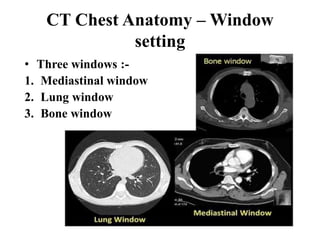
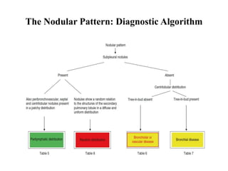
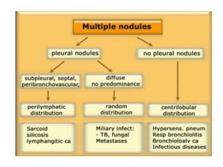
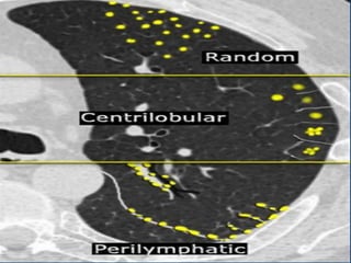
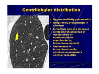
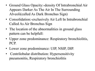
SlideShare a Scribd company logo
1 of 130
LUNG IMAGING CT SCAN
MODERATOR:-
Dr. AMAAN LATIF
(Assistant Professor)
M.D RESPIRATORY MEDICINE
Dr. MURARI LAL CHEST HOSPITAL
G.S.V.M MEDICAL COLLEGE, KANPUR
PRESENTED BY:-
Dr. Kranti Kumar
SEMINAR
TOMOGRAPHY
TOMO:-SLICE
GRAPHIC:-TO WRITE
DEFINITION:-IMAGING OF AN OBJECT BY
ANALYSING ITS SLICES
H/O TOMOGRAPHY
• 1924:-MATHEMATIC THEORY OF TOMOGRAPHIC
IMAGES RECONSTRUCTION(JOHANN RADON)
• 1930:-CONVENTIONAL
TOMOGRAPHY(A.VALLEBONE)
• 1963:-THEORETICAL BASIS OF CT (A MACLOED
CORMACK)
• 1971:-FIRST COMMERCIAL CT(SIR GODFREY
HOUNSFEILD)
CT Chest:-
1. CT Chest Type:-
2. CT Chest Anatomy
3. CT Chest Abnormalities
CT Chest Type:-
1. Standard or Conventional CT:- •Slice thickness: 3-10 mm
•scans a large volume,
very quickly
•Covers the full lung
•+/- contrast
2. High-Resolution Computed tomography (HRCT)
•Narrow x-ray beam
collimation: 1-1.3mm vs.
conventional 3-10mm
•Cross sections are further
apart: 10 mm
•High definition images of
lung parenchyma: vessels,
airspaces, airway and
interstitium
•No contrast
3. Low Dose CT
• Uses:
• Screening of lung cancer
•F/U: Infection, post lung
transplant, metastasis
•Premise: lower dose
radiation will not reduce
the diagnostic
functionality of the scan
(eg. 250 mAs 50 mAs)
•Detail is decreased
4. CT Angiography
Indications:-
• Pulmonary embolism
• Aortic aneurysm
• Aortic dissection
Risk :-
• HSR
• Contrast induced
nephropathy (CIN)
•Contrast injected into peripheral vein
•Injection timing/rate controlled automatically
•Dye is where you want it during scan
•Replaced conventional catheter angiogram
Indications
Std CT Chest:
• Chest x-ray (CXR)
abnormality
• Lung cancer staging
• F/U metastases
• Pleural and
mediastinal
abnormalities
• Empyema
HRCT
• ILD:
• Bronchiectasis
• Emphysema
• Normal or
equivocal CXR:
suspected lung
diseases and pattern
clarification
• Normal CXR with
abnormal PFT
• Evaluation of
hemoptysis
CECT
• Evaluation of the
mediastinum
(lymph nodes,
infection)
• Infection of the
chest wall
• Pleural thickening,
nodules, empyema
and evaluation of
metastatic or
primary malignancy
of the pleura.
• Pulmonary Lobar
Collapse
• Lung abscess,
CHEST CT :
GENERAL PRINCIPLES
• Scan levels : Lung apices to posterior costophrenic
angles.
• Patients position : Supine.
• Lung volume : Full inspiration ,single breath hold.
• Gantry rotation time : Approximately 0.5s in most
instances.
• Scan duration : Approximately 2.5s for the thorax
using MDCT and fast scanning.
• Detector width : The thinnest detector are typically
used for image acquisition
CT Chest Anatomy – Hounsfield
unit
CT Chest Anatomy - Views
CT Chest Anatomy – Window
setting
• Three windows :-
1. Mediastinal window
2. Lung window
3. Bone window
CT Chest Anatomy – Mediastinum
a) Supra-aortic mediastinum
Supra-aortic mediastinum
b) Aortic arch
Azygos & aorto-pulmonary window
c) Main pulmonary artery :-
Right pulmonary artery
d) Heart and paracardiac mediastinum
Heart
CT Chest Anatomy – Lung segments
a) At level of trachea (Upper Lobe)
• At level of trachea (Upper Lobe)
b) At level of main stem bronchi (Carina)
At level of main stem bronchi (Carina)
c) At the level of the heart.
• At the level of the heart.
SECONDARY LOBULE:-
Basic Anatomic Unit
Of Pulmonary
Structure And
Function
THE SECONDARY PULMONARY LOBULES
HAS THREE PRINCIPAL COMPONENTS :
1. The Interlobular Septa.
2. The Centrilobular Region.
3. The Lobular Lung Parenchyma.
INTERPRETATION OF CT SCAN
The three pillars on which the diagnosis of lung disease
on a chest CT is based:
1. The Appearance Pattern,
2. The Location And Distribution Pattern Of The
Abnormalities
3. Patient Data
APPEARANCE PATTERN OF DISEASE
• Abnormalities associated with an increase in
lung density, i.e. increased lung attenuation
• Abnormalities associated with a decrease in
lung density, i.e. decreased lung attenuation
• Abnormalities presenting as nodular opacities
• Abnormalities presenting as linear opacities
WHAT IS ATTENUATION ?
NODULAR PATTERN
•A nodule with a diameter less than 1 cm (small
nodule),larger than 1 cm (large nodule).
•The term “micronodule” smaller than 3 mm in
diameter.
•The term “miliary pattern” indicates the presence of
multiple small (1–3 mm) micronodules with sharp
contours distributed in a major part of the lungs.
The Nodular Pattern: Diagnostic Algorithm
Tree-in-bud
• In centrilobular nodules the recognition of 'tree-
in-bud' is of value for narrowing the differential
diagnosis.
• Tree-in-bud describes the appearance of an
irregular and often nodular branching structure,
most easily identified in the lung periphery.
• It represents dilated and impacted (mucus or pus-
filled) cen- trilobular bronchioles.
• Tree-in-bud almost always indicates the presence
of:
• Endobronchial spread of infection (TB, MAC, any
bacterial bronchopneumonia)
• Airway disease associated with infection (cystic
fibrosis, bronchiectasis)
• less often, an airway disease associated primarily
with mucus retention (allergic bronchopulmonary
aspergillosis, asthma).
INCREASED ATTENUATION
PATTERN
Increase in density of the lung parenchyma
• Increased Lung Attenuation Is Called As Ground
Glass Opacity - If There Is A Hazy Increase In Lung
Opacity Without Obscuration Of Underlying Vessels
And
• Consolidation If The Increase In Lung Opacity
Obscures The Vessels
• In Both Ground Glass And Consolidation The
Increase In Lung Density Is The Replacement Of Air
In The Alveoli By Fluid, cells or Fibrosis.
• Ground Glass Opacity:-density Of Intrabronchial Air
Appears Darker As The Air In The Surrounding
Alveoli(called As Dark Bronchus Sign)
• Consolidation:-exclusively Air Left In Intrabronchial
Called As Air Bronchus Sign
• The location of the abnormalities in ground glass
pattern can be helpfull:
• Upper zone predominance: Respiratory bronchiolitis,
PCP.
• Lower zone predominance: UIP, NSIP, DIP.
• Centrilobular distribution: Hypersensitivity
pneumonitis, Respiratory bronchiolitis
Fibrosis
Consolidation
DECREASED
ATTENUATION PATTERN
Generally four causes of decreased lung
attenuation can be found:
• Hypoperfusion
• Air trapping
• Cystic and cyst-like lesions
• Pulmonary emphysema
• Cysts are low-density thin-walled areas that
are well defined and circumscribed and that
have a cellular wall (usually less than 3 mm
thick)
• Most frequent causes of cystic lung changes is
advanced fibrosis giving rise to honeycombing
or honeycomb cysts
RETICULAR/LINEAR PATTERN:-
• TOO MANY LINES EITHER AS ARESULT OF
THICKENING OF LUNG INTERLOBULAR
SEPTA OR INTRALOBULAR SEPTA.
• THICKENING OF LUNG INTERSTITIUM BY
FLUID ,FIBROUS TISSUE OR INFILTRATION BY
CELLS RESULTS IN PATTERN OF RETICULAR
OPACITY DUE TO THICKENING OF SEPTA.
Linear opacities can develop:
• When the interstitium is thickened
• When lymphatics are involved
• When peripheral acinar alveoli are
filled or collapsed
• When blood vessels and airways are
involved
• When lung atelectasis or fibrosis occurs
RETICULAR
LINEAR
PARENCHYMAL SIGN
• Air Crescent Sign
• Monod Sign
• Halo Sign
• Atoll Sign
• Cheerio Sign
• Comet Tail Sign
• Mosaic Sign
• Head Cheese Sign
AIRWAY SIGN
• TREE IN BUD
• AIR BRONCHOGRAM SIGN
• TRAM TRACK SIGN
• BRONCHUS SIGN
• SINGNET RING SIGN
VASCULAR SIGN
• FEEDING VESSEL SIGN
• CT ANGIOGRAM SIGN
PLEURAL-BASED SIGNS
ABPA
• Allergic bronchopulmonary aspergillosis (ABPA) is
an idiopathic inflammatory disease of the lung,
characterized by an allergic inflammatory response
to colonization of the airways by Aspergillus
fumigatus or other fungi.
HRCT Finding :-
• Bronchiectasis is classically cylindric and proximal
(central),occurring within the proximal two-thirds of the
lung.
• Widespread proximal cylindrical bronchiectasis (mostly of
upper lobes)
• Bronchial wall thickening
• Mucous plugging
• Atelectasis
• Peripheral airspace consolidation
• Ground-glass attenuation
• Mosaic perfusion
• Air trapping
Central bronchiectasis and mucoid impaction
(finger-inglove sign), characteristic of ABPA.
Bronchiectasis
• Bronchiectasis is an entity characterized
pathologically by airway inflammation, and
permanent bronchial dilatation, and clinically by
cough, sputum production, and exacerbations with
recurrent respiratory tract infections.
CT signs of bronchiectasis
• Signet ring appearance on CT: normally, the vessel is larger
than the corresponding bronchus in bronchiectasis, the
bronchus is larger than the corresponding vessel.
• Bronchial dilation, the cardinal sign of bronchiectasis, is
characterised on HRCT by a bronchoarterial ratio (BAR) of .1,
lack of bronchial tapering, and visibility of airways within 1
cm of the pleural surface or abutting the mediastinal pleural
surface.
• "Tramlines" or "honeycombing represents dilated,
thickened bronchial walls.
Signet-ring sign.
Tram-track sign.
Characteristic radiologic findings of bronchiectasis: airway
dilation and bronchial wall thickening.
Types of Bronchiectasis
 Cylindrical (tram
track sign)
• This description applies to dilated
airways seen in a horizontal
orientation.
• Varicose
• Implies non uniform bronchial
dilatation. Cystic or saccular A
cluster of thin walled cystic
spaces
• Sacculer or Cystics
• A cluster of thin walled cystic spaces
Pleural Effusion
• Pleural effusion, a collection of fluid in the pleural
space, is rarely a primary disease process but is
usually secondary to other diseases.
• The pleural space normally contains only about 10-20
ml of serous fluid.
• Pleural fluid normally seeps continually into the
pleural space from the capillaries lining the parietal
pleura and is reabsorbed by the visceral pleural
capillaries and lymphatic system.
CT Finding :-
• Contrast-enhanced computed tomography: split
pleural sign .
• Split pleural sign refers to thickening & increased
contrast enhancement of the visceral & parietal pleura
separated by empyema or an exudative effusion.
Pneumothorax
• abnormal presence of
air in the pleural
cavity, separating
the visceral from the
parietal pleura, is
known as
pneumothorax.
COPD
• Chronic obstructive Pulmonary Disease (COPD) is a
Heterogeneous Lung condition characterized by
chronic respiratory symptoms (dyspnea, cough,
sputum production and/or exacerbation) due to
abnormalities of airways (bronchitis, bronchiolitis)
and/or alveoli (emphysema) that cause persistent,
often progressive, airflow obstruction.
CT Finding
•Emphysema ( Hypodense area) Enlarged Alveolar duct and Alveoli.
•Bronchitis Shows bronchial wall thickning.
Tuberculosis
• HRCT findings in TB :-
• Consolidation anywhere, lymphadenopathy and
pleural effusion. Usually regresses to calcified lung
nodule.
• Cavitation
• (Endobronchial spread) Tree in bud appearance)
• Miliary TB: 2-3 mm nodules with random
distribution.
Pulmonary Artery Hypertension
 Findings in CT:
• Extra cardiac vascular signs:
• Enlarged pulmonary trunk >29 mm diameter is often
used as a general predictive cut-off
• Enlarged pulmonary arteries
• Mural calcification in central pulmonary arteries
• Evidence of previous pulmonary emboli
• Cardiac signs:
• Right ventricular hypertrophy: defined as wall
thickness of more than 4 mm.
• Straightening or bowing (towards the left ventricle)
of the interventricular septum
• Right ventricular dilatation
• Decreased right ventricular ejection fraction
• Dilatation of the inferior vena cava and hepatic veins
• Pericardial effusion
• Parenchymal signs:
• Centrilobular ground-glass nodules (Cholesterol
granuloma).
• Neovascularity: tiny serpiginous intrapulmonary
vessels that often emerge from centrilobular
arterioles.
ILD (INTERSTITIAL LUNG
DISEASES)
• ILD refers to a heterogeneous collections of lung
disorder that are grouped together because they
share clinical radiographic and pathological
features , ILD also refers to as Diffuse
parenchymal lung Diseases.
• ILD involved the Parenchyma of Lung, Alveoli,
The Alveolar Epithelium, the capillary
endothelium, and the spaces between these
structure as well as perivascular and lymphatic
tissue.
Distribution of ILD
Pattern of ILD
Radiographic Signs in ILD
Anterior upper Lobe sign.
Concentration of fibrosis within the anterior aspect of upper lobe and
concomitant lower lobe involvement.
Exuberant honeycombing
Honeycomb cyst formation within the lung constituting greater than 70%
fibrotic person of Lung.
Straight edge sign
Isolation of fibrosis to the lung bases with sharp
demarcation in the craniocaudal plane without substantial
extension along the lateral margins
Three-density sign/
headcheese
CT imaging shows patchy ground-glass attenuation in the anterior left
upper lobe (blue circle). Other lobules are of preserved (normal)
attenuation (yellow circle), and there is lobular decreased attenuation in
the posterior left lung (red circle). No signs of fibrosis are present
Atoll sign/reverse halo
Central ground glass opacity surrounded by denser
consolidation in the shape of a crescent or ring.
Galaxy sign
CT images demonstrate perilymphatic irregular nodular thickening in an
upper/mid lung distribution with some areas of mass-like coalescing nodules,
more concentrated in the center than at the periphery the “galaxy sign.”
Sarcoidosis
• Sarcoidosis is a systemic disorder of unknown
origin.
• It is characterized by non-caseating granulomas in
multiple organs, that may resolve spontaneously
or progress to fibrosis.
• Pulmonary manifestations are present in 90% of
patients.
• Less than 5% of patients die from sarcoidosis
usually as a result of pulmonary fibrosis.
HRCT findings in Sarcoidosis
• Common findings:
-Small nodules in a per lymphatic distribution
(i.e. along subpleural surface and fissures, along
interlobular septa and the peribronchovascular
bundle).
-Upper and middle zone predominance.
-Lymphadenopathy in left hilus, right hilus and
paratracheal (1-2- 3 sign). Often with
calcifications.
• Uncommon findings:
-Conglomerate masses in a perihilar location.
-Larger nodules (> 1cm in diameter, in < 20%)
-Grouped nodules or coalescent nodules
surrounded by multiple satellite nodules (Galaxi
sign)
-Nodules so small and dense that they appear as
ground glass or even as consolidations (alveolar
sarcoidosis)
Typical presentation of sarcoidosis with mediastinal lymphadenopathy and
small nodules in a perilymphatic distribution along bronchovascular bundles
and along fissures (yellow arrows).
Always look for small nodules along the fissures, because this is a very specific
and typical sign of sarcoidosis.
Silicosis / Coal worker
pneumoconiosis
• Silicosis and Coal worker pneumoconiosis (CWP) are
pathologically distinct entities with differing
histology, resulting from the inhalation of different
inorganic dusts.
• The radiographic and HRCT appearances of these
diseases, however, may not be distinguishable from
each other and may be similar to sarcoidosis.
• Silicosis and CWP occur in a specific patient group
(construction workers, mining workers, workers
exposed to sandblasting, glass blowing and pottery).
HRCT findings in Silicosis/CWP
• Small well-defined nodules of 2 to 5mm in diameter
in both lungs.
• Upper lobe predominance
• Nodules may be calcified
• Centrilobular and subpleural distribution
• Sometimes random distribution
• Irregular conglomerate masses, known as progressive
massive fibrosis
• Masses may cavitate due to ischemic necrosis.
• Often hilar and mediastinal lymphnodes.
A case of silicosis showing nodules of varying sizes with a random and
subpleural distribution. One nodule contains calcification (arrow).
Note the absence of a lymphatic distribution pattern (peribronchovascular and
along fissures), which would be suggestive of sarcoidosis.
Hypersensitivity Pneumonitis
• Hypersensitivity pneumonitis (HP) is also known as
extrinsic allergic alveolitis (EAA).
HP is an allergic lung disease caused by the inhalation
of a variety of antigens (farmer's lung, bird fancier's
lung, 'hot tub' lung, humidifier lung).
• The radiographic and pathologic abnormalities in
patients can be classified into acute, subacute, and
chronic stages.
• Mostly HRCT is performed in the subacute stage of
HP, weeks to months following the first exposure to the
antigen or in the chronic phase.
Three-density pattern
• Ground glass opacification
• Mosaic attenuation with zonesof diminished
attenuation/hypoperfusion.
• Areas of normal-appearing parenchyma.
CT imaging shows patchy ground-glass attenuation in the anterior left
upper lobe (blue circle). Other lobules are of preserved (normal)
attenuation (yellow circle), and there is lobular decreased attenuation in
the posterior left lung (red circle). No signs of fibrosis are present
Usual interstitial pneumonia (UIP)
Computed tomography scan illustrates the “classic”
features of idiopathic pulmonary fibrosis (IPF), the usual interstitial
pneumonia (UIP) pattern. Bilateral, peripheral, and subpleural reticular
infiltrates are evident. The presence of advanced fibrosis is indicated by
honeycomb changes (arrowheads) and traction bronchiectasis (arrow).
Nonspecific interstitial pneumonia
(NSIP).
• Nonspecific interstitial pneumonia (NSIP). High-resolution CT
images through the upper (A) and lower (B) thorax
demonstrate peripheral and lower lobe–predominant ground-
glass opacities with mild reticular markings and minimal
traction bronchiectasis (arrows).
Central Tumor
Peripheral Tumor
Solitary Pulmonary Nodules
Corona radiata or the "sunburst appearance" - Presence of
spiculation associated with a nodule or a mass- Feature of
malignant lesion.
Pulmonary Metastasis
Lung metastases from non seminomatous germ cell tumour of testis. Seen as
multiple opacities in both the lung fields. ( Canon Ball appearance)
THANKYOU

More Related Content

Similar to LUNG IMAGING CT SCAN with classification.pptx

hrct.pptx high resolution ct patterns
hrct.pptx high resolution ct patterns hrct.pptx high resolution ct patterns
hrct.pptx high resolution ct patterns ranjitharadhakrishna3
 
Presentation1.pptx. interpretation of x ray chest.
Presentation1.pptx. interpretation of x ray chest.Presentation1.pptx. interpretation of x ray chest.
Presentation1.pptx. interpretation of x ray chest.Abdellah Nazeer
 
HRCT Low attenuation pattern
HRCT Low attenuation pattern HRCT Low attenuation pattern
HRCT Low attenuation pattern Sakher Alkhaderi
 
Rad Seminar CHEST IMAGING By Dr Siraj.pptx
Rad Seminar CHEST IMAGING By Dr Siraj.pptxRad Seminar CHEST IMAGING By Dr Siraj.pptx
Rad Seminar CHEST IMAGING By Dr Siraj.pptxImanuIliyas
 
Clinical Applications of Chest Sonography
Clinical Applications of Chest SonographyClinical Applications of Chest Sonography
Clinical Applications of Chest SonographyGamal Agmy
 
HRCT chest in Diffuse lung disease by Dr. Subash Pathak
HRCT chest in Diffuse lung disease by Dr. Subash PathakHRCT chest in Diffuse lung disease by Dr. Subash Pathak
HRCT chest in Diffuse lung disease by Dr. Subash PathakMilan Silwal
 
Hrct chest technique and interpretation
Hrct chest  technique and interpretationHrct chest  technique and interpretation
Hrct chest technique and interpretationthambimbbs
 
Tracheal pathologies
Tracheal pathologiesTracheal pathologies
Tracheal pathologiesJino Justin
 
Bronchiectasis and Role of Surgical Management.pptx
Bronchiectasis and Role of Surgical Management.pptxBronchiectasis and Role of Surgical Management.pptx
Bronchiectasis and Role of Surgical Management.pptxRohanReddy66
 
Mark rosemary. diagnosis of traumatic injury of the trachea
Mark rosemary. diagnosis of  traumatic injury of the tracheaMark rosemary. diagnosis of  traumatic injury of the trachea
Mark rosemary. diagnosis of traumatic injury of the tracheaRose Mary
 
MIM (Lung abscess).pptx
MIM (Lung abscess).pptxMIM (Lung abscess).pptx
MIM (Lung abscess).pptxNAMREEN5
 
Pediatric chest infection imaging considerations
Pediatric chest infection imaging considerationsPediatric chest infection imaging considerations
Pediatric chest infection imaging considerationsAhmed Bahnassy
 
Differential diagnosis of pulmonary cyst: 1. Bullous Emphysema. 2. Cystic Bro...
Differential diagnosis of pulmonary cyst: 1. Bullous Emphysema. 2. Cystic Bro...Differential diagnosis of pulmonary cyst: 1. Bullous Emphysema. 2. Cystic Bro...
Differential diagnosis of pulmonary cyst: 1. Bullous Emphysema. 2. Cystic Bro...Dr.Bijay Yadav
 
Airways disease abduljelil final
Airways disease abduljelil finalAirways disease abduljelil final
Airways disease abduljelil finalabduljelil nejmu
 
Diagnostic work of thoracic surgery patients
Diagnostic work of thoracic surgery patients Diagnostic work of thoracic surgery patients
Diagnostic work of thoracic surgery patients Dr Mengistu Kassa
 
Interstitial lung disease and Occupational lung disease HRCT
Interstitial lung disease and Occupational lung disease HRCTInterstitial lung disease and Occupational lung disease HRCT
Interstitial lung disease and Occupational lung disease HRCTSahroz Khan
 
Imaging of Airway Diseases
Imaging of Airway DiseasesImaging of Airway Diseases
Imaging of Airway DiseasesAbhineet Dey
 

Similar to LUNG IMAGING CT SCAN with classification.pptx (20)

hrct.pptx high resolution ct patterns
hrct.pptx high resolution ct patterns hrct.pptx high resolution ct patterns
hrct.pptx high resolution ct patterns
 
Presentation1.pptx. interpretation of x ray chest.
Presentation1.pptx. interpretation of x ray chest.Presentation1.pptx. interpretation of x ray chest.
Presentation1.pptx. interpretation of x ray chest.
 
HRCT Reticular pattern
HRCT Reticular pattern HRCT Reticular pattern
HRCT Reticular pattern
 
HRCT Low attenuation pattern
HRCT Low attenuation pattern HRCT Low attenuation pattern
HRCT Low attenuation pattern
 
Rad Seminar CHEST IMAGING By Dr Siraj.pptx
Rad Seminar CHEST IMAGING By Dr Siraj.pptxRad Seminar CHEST IMAGING By Dr Siraj.pptx
Rad Seminar CHEST IMAGING By Dr Siraj.pptx
 
Clinical Applications of Chest Sonography
Clinical Applications of Chest SonographyClinical Applications of Chest Sonography
Clinical Applications of Chest Sonography
 
HRCT chest in Diffuse lung disease by Dr. Subash Pathak
HRCT chest in Diffuse lung disease by Dr. Subash PathakHRCT chest in Diffuse lung disease by Dr. Subash Pathak
HRCT chest in Diffuse lung disease by Dr. Subash Pathak
 
Hrct chest technique and interpretation
Hrct chest  technique and interpretationHrct chest  technique and interpretation
Hrct chest technique and interpretation
 
Pneumothorax by Dr. Sookun Rajeev Kumar
Pneumothorax by Dr. Sookun Rajeev KumarPneumothorax by Dr. Sookun Rajeev Kumar
Pneumothorax by Dr. Sookun Rajeev Kumar
 
Tracheal pathologies
Tracheal pathologiesTracheal pathologies
Tracheal pathologies
 
Bronchiectasis and Role of Surgical Management.pptx
Bronchiectasis and Role of Surgical Management.pptxBronchiectasis and Role of Surgical Management.pptx
Bronchiectasis and Role of Surgical Management.pptx
 
Mark rosemary. diagnosis of traumatic injury of the trachea
Mark rosemary. diagnosis of  traumatic injury of the tracheaMark rosemary. diagnosis of  traumatic injury of the trachea
Mark rosemary. diagnosis of traumatic injury of the trachea
 
MIM (Lung abscess).pptx
MIM (Lung abscess).pptxMIM (Lung abscess).pptx
MIM (Lung abscess).pptx
 
Pediatric chest infection imaging considerations
Pediatric chest infection imaging considerationsPediatric chest infection imaging considerations
Pediatric chest infection imaging considerations
 
Differential diagnosis of pulmonary cyst: 1. Bullous Emphysema. 2. Cystic Bro...
Differential diagnosis of pulmonary cyst: 1. Bullous Emphysema. 2. Cystic Bro...Differential diagnosis of pulmonary cyst: 1. Bullous Emphysema. 2. Cystic Bro...
Differential diagnosis of pulmonary cyst: 1. Bullous Emphysema. 2. Cystic Bro...
 
Airways disease abduljelil final
Airways disease abduljelil finalAirways disease abduljelil final
Airways disease abduljelil final
 
Diagnostic work of thoracic surgery patients
Diagnostic work of thoracic surgery patients Diagnostic work of thoracic surgery patients
Diagnostic work of thoracic surgery patients
 
Abnormal x ray
Abnormal x rayAbnormal x ray
Abnormal x ray
 
Interstitial lung disease and Occupational lung disease HRCT
Interstitial lung disease and Occupational lung disease HRCTInterstitial lung disease and Occupational lung disease HRCT
Interstitial lung disease and Occupational lung disease HRCT
 
Imaging of Airway Diseases
Imaging of Airway DiseasesImaging of Airway Diseases
Imaging of Airway Diseases
 

Recently uploaded

Call Girls Horamavu WhatsApp Number 7001035870 Meeting With Bangalore Escorts
Call Girls Horamavu WhatsApp Number 7001035870 Meeting With Bangalore EscortsCall Girls Horamavu WhatsApp Number 7001035870 Meeting With Bangalore Escorts
Call Girls Horamavu WhatsApp Number 7001035870 Meeting With Bangalore Escortsvidya singh
 
Call Girls Aurangabad Just Call 9907093804 Top Class Call Girl Service Available
Call Girls Aurangabad Just Call 9907093804 Top Class Call Girl Service AvailableCall Girls Aurangabad Just Call 9907093804 Top Class Call Girl Service Available
Call Girls Aurangabad Just Call 9907093804 Top Class Call Girl Service AvailableDipal Arora
 
Kesar Bagh Call Girl Price 9548273370 , Lucknow Call Girls Service
Kesar Bagh Call Girl Price 9548273370 , Lucknow Call Girls ServiceKesar Bagh Call Girl Price 9548273370 , Lucknow Call Girls Service
Kesar Bagh Call Girl Price 9548273370 , Lucknow Call Girls Servicemakika9823
 
Call Girls Cuttack Just Call 9907093804 Top Class Call Girl Service Available
Call Girls Cuttack Just Call 9907093804 Top Class Call Girl Service AvailableCall Girls Cuttack Just Call 9907093804 Top Class Call Girl Service Available
Call Girls Cuttack Just Call 9907093804 Top Class Call Girl Service AvailableDipal Arora
 
Night 7k to 12k Navi Mumbai Call Girl Photo 👉 BOOK NOW 9833363713 👈 ♀️ night ...
Night 7k to 12k Navi Mumbai Call Girl Photo 👉 BOOK NOW 9833363713 👈 ♀️ night ...Night 7k to 12k Navi Mumbai Call Girl Photo 👉 BOOK NOW 9833363713 👈 ♀️ night ...
Night 7k to 12k Navi Mumbai Call Girl Photo 👉 BOOK NOW 9833363713 👈 ♀️ night ...aartirawatdelhi
 
Call Girls Service Surat Samaira ❤️🍑 8250192130 👄 Independent Escort Service ...
Call Girls Service Surat Samaira ❤️🍑 8250192130 👄 Independent Escort Service ...Call Girls Service Surat Samaira ❤️🍑 8250192130 👄 Independent Escort Service ...
Call Girls Service Surat Samaira ❤️🍑 8250192130 👄 Independent Escort Service ...CALL GIRLS
 
Bangalore Call Girls Hebbal Kempapura Number 7001035870 Meetin With Bangalor...
Bangalore Call Girls Hebbal Kempapura Number 7001035870  Meetin With Bangalor...Bangalore Call Girls Hebbal Kempapura Number 7001035870  Meetin With Bangalor...
Bangalore Call Girls Hebbal Kempapura Number 7001035870 Meetin With Bangalor...narwatsonia7
 
Artifacts in Nuclear Medicine with Identifying and resolving artifacts.
Artifacts in Nuclear Medicine with Identifying and resolving artifacts.Artifacts in Nuclear Medicine with Identifying and resolving artifacts.
Artifacts in Nuclear Medicine with Identifying and resolving artifacts.MiadAlsulami
 
VIP Mumbai Call Girls Hiranandani Gardens Just Call 9920874524 with A/C Room ...
VIP Mumbai Call Girls Hiranandani Gardens Just Call 9920874524 with A/C Room ...VIP Mumbai Call Girls Hiranandani Gardens Just Call 9920874524 with A/C Room ...
VIP Mumbai Call Girls Hiranandani Gardens Just Call 9920874524 with A/C Room ...Garima Khatri
 
Call Girls Darjeeling Just Call 9907093804 Top Class Call Girl Service Available
Call Girls Darjeeling Just Call 9907093804 Top Class Call Girl Service AvailableCall Girls Darjeeling Just Call 9907093804 Top Class Call Girl Service Available
Call Girls Darjeeling Just Call 9907093804 Top Class Call Girl Service AvailableDipal Arora
 
Night 7k to 12k Chennai City Center Call Girls 👉👉 7427069034⭐⭐ 100% Genuine E...
Night 7k to 12k Chennai City Center Call Girls 👉👉 7427069034⭐⭐ 100% Genuine E...Night 7k to 12k Chennai City Center Call Girls 👉👉 7427069034⭐⭐ 100% Genuine E...
Night 7k to 12k Chennai City Center Call Girls 👉👉 7427069034⭐⭐ 100% Genuine E...hotbabesbook
 
Call Girls Nagpur Just Call 9907093804 Top Class Call Girl Service Available
Call Girls Nagpur Just Call 9907093804 Top Class Call Girl Service AvailableCall Girls Nagpur Just Call 9907093804 Top Class Call Girl Service Available
Call Girls Nagpur Just Call 9907093804 Top Class Call Girl Service AvailableDipal Arora
 
Vip Call Girls Anna Salai Chennai 👉 8250192130 ❣️💯 Top Class Girls Available
Vip Call Girls Anna Salai Chennai 👉 8250192130 ❣️💯 Top Class Girls AvailableVip Call Girls Anna Salai Chennai 👉 8250192130 ❣️💯 Top Class Girls Available
Vip Call Girls Anna Salai Chennai 👉 8250192130 ❣️💯 Top Class Girls AvailableNehru place Escorts
 
Call Girls Colaba Mumbai ❤️ 9920874524 👈 Cash on Delivery
Call Girls Colaba Mumbai ❤️ 9920874524 👈 Cash on DeliveryCall Girls Colaba Mumbai ❤️ 9920874524 👈 Cash on Delivery
Call Girls Colaba Mumbai ❤️ 9920874524 👈 Cash on Deliverynehamumbai
 
High Profile Call Girls Coimbatore Saanvi☎️ 8250192130 Independent Escort Se...
High Profile Call Girls Coimbatore Saanvi☎️  8250192130 Independent Escort Se...High Profile Call Girls Coimbatore Saanvi☎️  8250192130 Independent Escort Se...
High Profile Call Girls Coimbatore Saanvi☎️ 8250192130 Independent Escort Se...narwatsonia7
 
Book Paid Powai Call Girls Mumbai 𖠋 9930245274 𖠋Low Budget Full Independent H...
Book Paid Powai Call Girls Mumbai 𖠋 9930245274 𖠋Low Budget Full Independent H...Book Paid Powai Call Girls Mumbai 𖠋 9930245274 𖠋Low Budget Full Independent H...
Book Paid Powai Call Girls Mumbai 𖠋 9930245274 𖠋Low Budget Full Independent H...Call Girls in Nagpur High Profile
 
Call Girl Coimbatore Prisha☎️ 8250192130 Independent Escort Service Coimbatore
Call Girl Coimbatore Prisha☎️  8250192130 Independent Escort Service CoimbatoreCall Girl Coimbatore Prisha☎️  8250192130 Independent Escort Service Coimbatore
Call Girl Coimbatore Prisha☎️ 8250192130 Independent Escort Service Coimbatorenarwatsonia7
 
VIP Service Call Girls Sindhi Colony 📳 7877925207 For 18+ VIP Call Girl At Th...
VIP Service Call Girls Sindhi Colony 📳 7877925207 For 18+ VIP Call Girl At Th...VIP Service Call Girls Sindhi Colony 📳 7877925207 For 18+ VIP Call Girl At Th...
VIP Service Call Girls Sindhi Colony 📳 7877925207 For 18+ VIP Call Girl At Th...jageshsingh5554
 

Recently uploaded (20)

Call Girls Horamavu WhatsApp Number 7001035870 Meeting With Bangalore Escorts
Call Girls Horamavu WhatsApp Number 7001035870 Meeting With Bangalore EscortsCall Girls Horamavu WhatsApp Number 7001035870 Meeting With Bangalore Escorts
Call Girls Horamavu WhatsApp Number 7001035870 Meeting With Bangalore Escorts
 
Call Girls Aurangabad Just Call 9907093804 Top Class Call Girl Service Available
Call Girls Aurangabad Just Call 9907093804 Top Class Call Girl Service AvailableCall Girls Aurangabad Just Call 9907093804 Top Class Call Girl Service Available
Call Girls Aurangabad Just Call 9907093804 Top Class Call Girl Service Available
 
Escort Service Call Girls In Sarita Vihar,, 99530°56974 Delhi NCR
Escort Service Call Girls In Sarita Vihar,, 99530°56974 Delhi NCREscort Service Call Girls In Sarita Vihar,, 99530°56974 Delhi NCR
Escort Service Call Girls In Sarita Vihar,, 99530°56974 Delhi NCR
 
Kesar Bagh Call Girl Price 9548273370 , Lucknow Call Girls Service
Kesar Bagh Call Girl Price 9548273370 , Lucknow Call Girls ServiceKesar Bagh Call Girl Price 9548273370 , Lucknow Call Girls Service
Kesar Bagh Call Girl Price 9548273370 , Lucknow Call Girls Service
 
Call Girls Cuttack Just Call 9907093804 Top Class Call Girl Service Available
Call Girls Cuttack Just Call 9907093804 Top Class Call Girl Service AvailableCall Girls Cuttack Just Call 9907093804 Top Class Call Girl Service Available
Call Girls Cuttack Just Call 9907093804 Top Class Call Girl Service Available
 
Night 7k to 12k Navi Mumbai Call Girl Photo 👉 BOOK NOW 9833363713 👈 ♀️ night ...
Night 7k to 12k Navi Mumbai Call Girl Photo 👉 BOOK NOW 9833363713 👈 ♀️ night ...Night 7k to 12k Navi Mumbai Call Girl Photo 👉 BOOK NOW 9833363713 👈 ♀️ night ...
Night 7k to 12k Navi Mumbai Call Girl Photo 👉 BOOK NOW 9833363713 👈 ♀️ night ...
 
Call Girls Service Surat Samaira ❤️🍑 8250192130 👄 Independent Escort Service ...
Call Girls Service Surat Samaira ❤️🍑 8250192130 👄 Independent Escort Service ...Call Girls Service Surat Samaira ❤️🍑 8250192130 👄 Independent Escort Service ...
Call Girls Service Surat Samaira ❤️🍑 8250192130 👄 Independent Escort Service ...
 
Bangalore Call Girls Hebbal Kempapura Number 7001035870 Meetin With Bangalor...
Bangalore Call Girls Hebbal Kempapura Number 7001035870  Meetin With Bangalor...Bangalore Call Girls Hebbal Kempapura Number 7001035870  Meetin With Bangalor...
Bangalore Call Girls Hebbal Kempapura Number 7001035870 Meetin With Bangalor...
 
Artifacts in Nuclear Medicine with Identifying and resolving artifacts.
Artifacts in Nuclear Medicine with Identifying and resolving artifacts.Artifacts in Nuclear Medicine with Identifying and resolving artifacts.
Artifacts in Nuclear Medicine with Identifying and resolving artifacts.
 
VIP Mumbai Call Girls Hiranandani Gardens Just Call 9920874524 with A/C Room ...
VIP Mumbai Call Girls Hiranandani Gardens Just Call 9920874524 with A/C Room ...VIP Mumbai Call Girls Hiranandani Gardens Just Call 9920874524 with A/C Room ...
VIP Mumbai Call Girls Hiranandani Gardens Just Call 9920874524 with A/C Room ...
 
Call Girls Darjeeling Just Call 9907093804 Top Class Call Girl Service Available
Call Girls Darjeeling Just Call 9907093804 Top Class Call Girl Service AvailableCall Girls Darjeeling Just Call 9907093804 Top Class Call Girl Service Available
Call Girls Darjeeling Just Call 9907093804 Top Class Call Girl Service Available
 
Night 7k to 12k Chennai City Center Call Girls 👉👉 7427069034⭐⭐ 100% Genuine E...
Night 7k to 12k Chennai City Center Call Girls 👉👉 7427069034⭐⭐ 100% Genuine E...Night 7k to 12k Chennai City Center Call Girls 👉👉 7427069034⭐⭐ 100% Genuine E...
Night 7k to 12k Chennai City Center Call Girls 👉👉 7427069034⭐⭐ 100% Genuine E...
 
Call Girls Nagpur Just Call 9907093804 Top Class Call Girl Service Available
Call Girls Nagpur Just Call 9907093804 Top Class Call Girl Service AvailableCall Girls Nagpur Just Call 9907093804 Top Class Call Girl Service Available
Call Girls Nagpur Just Call 9907093804 Top Class Call Girl Service Available
 
Vip Call Girls Anna Salai Chennai 👉 8250192130 ❣️💯 Top Class Girls Available
Vip Call Girls Anna Salai Chennai 👉 8250192130 ❣️💯 Top Class Girls AvailableVip Call Girls Anna Salai Chennai 👉 8250192130 ❣️💯 Top Class Girls Available
Vip Call Girls Anna Salai Chennai 👉 8250192130 ❣️💯 Top Class Girls Available
 
Call Girls Colaba Mumbai ❤️ 9920874524 👈 Cash on Delivery
Call Girls Colaba Mumbai ❤️ 9920874524 👈 Cash on DeliveryCall Girls Colaba Mumbai ❤️ 9920874524 👈 Cash on Delivery
Call Girls Colaba Mumbai ❤️ 9920874524 👈 Cash on Delivery
 
Russian Call Girls in Delhi Tanvi ➡️ 9711199012 💋📞 Independent Escort Service...
Russian Call Girls in Delhi Tanvi ➡️ 9711199012 💋📞 Independent Escort Service...Russian Call Girls in Delhi Tanvi ➡️ 9711199012 💋📞 Independent Escort Service...
Russian Call Girls in Delhi Tanvi ➡️ 9711199012 💋📞 Independent Escort Service...
 
High Profile Call Girls Coimbatore Saanvi☎️ 8250192130 Independent Escort Se...
High Profile Call Girls Coimbatore Saanvi☎️  8250192130 Independent Escort Se...High Profile Call Girls Coimbatore Saanvi☎️  8250192130 Independent Escort Se...
High Profile Call Girls Coimbatore Saanvi☎️ 8250192130 Independent Escort Se...
 
Book Paid Powai Call Girls Mumbai 𖠋 9930245274 𖠋Low Budget Full Independent H...
Book Paid Powai Call Girls Mumbai 𖠋 9930245274 𖠋Low Budget Full Independent H...Book Paid Powai Call Girls Mumbai 𖠋 9930245274 𖠋Low Budget Full Independent H...
Book Paid Powai Call Girls Mumbai 𖠋 9930245274 𖠋Low Budget Full Independent H...
 
Call Girl Coimbatore Prisha☎️ 8250192130 Independent Escort Service Coimbatore
Call Girl Coimbatore Prisha☎️  8250192130 Independent Escort Service CoimbatoreCall Girl Coimbatore Prisha☎️  8250192130 Independent Escort Service Coimbatore
Call Girl Coimbatore Prisha☎️ 8250192130 Independent Escort Service Coimbatore
 
VIP Service Call Girls Sindhi Colony 📳 7877925207 For 18+ VIP Call Girl At Th...
VIP Service Call Girls Sindhi Colony 📳 7877925207 For 18+ VIP Call Girl At Th...VIP Service Call Girls Sindhi Colony 📳 7877925207 For 18+ VIP Call Girl At Th...
VIP Service Call Girls Sindhi Colony 📳 7877925207 For 18+ VIP Call Girl At Th...
 

LUNG IMAGING CT SCAN with classification.pptx

  • 1. LUNG IMAGING CT SCAN MODERATOR:- Dr. AMAAN LATIF (Assistant Professor) M.D RESPIRATORY MEDICINE Dr. MURARI LAL CHEST HOSPITAL G.S.V.M MEDICAL COLLEGE, KANPUR PRESENTED BY:- Dr. Kranti Kumar SEMINAR
  • 3. H/O TOMOGRAPHY • 1924:-MATHEMATIC THEORY OF TOMOGRAPHIC IMAGES RECONSTRUCTION(JOHANN RADON) • 1930:-CONVENTIONAL TOMOGRAPHY(A.VALLEBONE) • 1963:-THEORETICAL BASIS OF CT (A MACLOED CORMACK) • 1971:-FIRST COMMERCIAL CT(SIR GODFREY HOUNSFEILD)
  • 4. CT Chest:- 1. CT Chest Type:- 2. CT Chest Anatomy 3. CT Chest Abnormalities
  • 5. CT Chest Type:- 1. Standard or Conventional CT:- •Slice thickness: 3-10 mm •scans a large volume, very quickly •Covers the full lung •+/- contrast
  • 6. 2. High-Resolution Computed tomography (HRCT) •Narrow x-ray beam collimation: 1-1.3mm vs. conventional 3-10mm •Cross sections are further apart: 10 mm •High definition images of lung parenchyma: vessels, airspaces, airway and interstitium •No contrast
  • 7. 3. Low Dose CT • Uses: • Screening of lung cancer •F/U: Infection, post lung transplant, metastasis •Premise: lower dose radiation will not reduce the diagnostic functionality of the scan (eg. 250 mAs 50 mAs) •Detail is decreased
  • 8. 4. CT Angiography Indications:- • Pulmonary embolism • Aortic aneurysm • Aortic dissection Risk :- • HSR • Contrast induced nephropathy (CIN) •Contrast injected into peripheral vein •Injection timing/rate controlled automatically •Dye is where you want it during scan •Replaced conventional catheter angiogram
  • 9. Indications Std CT Chest: • Chest x-ray (CXR) abnormality • Lung cancer staging • F/U metastases • Pleural and mediastinal abnormalities • Empyema HRCT • ILD: • Bronchiectasis • Emphysema • Normal or equivocal CXR: suspected lung diseases and pattern clarification • Normal CXR with abnormal PFT • Evaluation of hemoptysis CECT • Evaluation of the mediastinum (lymph nodes, infection) • Infection of the chest wall • Pleural thickening, nodules, empyema and evaluation of metastatic or primary malignancy of the pleura. • Pulmonary Lobar Collapse • Lung abscess,
  • 10. CHEST CT : GENERAL PRINCIPLES • Scan levels : Lung apices to posterior costophrenic angles. • Patients position : Supine. • Lung volume : Full inspiration ,single breath hold. • Gantry rotation time : Approximately 0.5s in most instances. • Scan duration : Approximately 2.5s for the thorax using MDCT and fast scanning. • Detector width : The thinnest detector are typically used for image acquisition
  • 11. CT Chest Anatomy – Hounsfield unit
  • 12. CT Chest Anatomy - Views
  • 13.
  • 14.
  • 15. CT Chest Anatomy – Window setting • Three windows :- 1. Mediastinal window 2. Lung window 3. Bone window
  • 16. CT Chest Anatomy – Mediastinum a) Supra-aortic mediastinum
  • 21. c) Main pulmonary artery :-
  • 23. d) Heart and paracardiac mediastinum
  • 24. Heart
  • 25. CT Chest Anatomy – Lung segments a) At level of trachea (Upper Lobe)
  • 26. • At level of trachea (Upper Lobe)
  • 27. b) At level of main stem bronchi (Carina)
  • 28. At level of main stem bronchi (Carina)
  • 29. c) At the level of the heart.
  • 30. • At the level of the heart.
  • 31. SECONDARY LOBULE:- Basic Anatomic Unit Of Pulmonary Structure And Function
  • 32. THE SECONDARY PULMONARY LOBULES HAS THREE PRINCIPAL COMPONENTS : 1. The Interlobular Septa. 2. The Centrilobular Region. 3. The Lobular Lung Parenchyma.
  • 33.
  • 34. INTERPRETATION OF CT SCAN The three pillars on which the diagnosis of lung disease on a chest CT is based: 1. The Appearance Pattern, 2. The Location And Distribution Pattern Of The Abnormalities 3. Patient Data
  • 35.
  • 36.
  • 37.
  • 38.
  • 39.
  • 40. APPEARANCE PATTERN OF DISEASE • Abnormalities associated with an increase in lung density, i.e. increased lung attenuation • Abnormalities associated with a decrease in lung density, i.e. decreased lung attenuation • Abnormalities presenting as nodular opacities • Abnormalities presenting as linear opacities
  • 42. NODULAR PATTERN •A nodule with a diameter less than 1 cm (small nodule),larger than 1 cm (large nodule). •The term “micronodule” smaller than 3 mm in diameter. •The term “miliary pattern” indicates the presence of multiple small (1–3 mm) micronodules with sharp contours distributed in a major part of the lungs.
  • 43. The Nodular Pattern: Diagnostic Algorithm
  • 44.
  • 45.
  • 46.
  • 47.
  • 48.
  • 49. Tree-in-bud • In centrilobular nodules the recognition of 'tree- in-bud' is of value for narrowing the differential diagnosis. • Tree-in-bud describes the appearance of an irregular and often nodular branching structure, most easily identified in the lung periphery. • It represents dilated and impacted (mucus or pus- filled) cen- trilobular bronchioles.
  • 50. • Tree-in-bud almost always indicates the presence of: • Endobronchial spread of infection (TB, MAC, any bacterial bronchopneumonia) • Airway disease associated with infection (cystic fibrosis, bronchiectasis) • less often, an airway disease associated primarily with mucus retention (allergic bronchopulmonary aspergillosis, asthma).
  • 51.
  • 52. INCREASED ATTENUATION PATTERN Increase in density of the lung parenchyma
  • 53.
  • 54. • Increased Lung Attenuation Is Called As Ground Glass Opacity - If There Is A Hazy Increase In Lung Opacity Without Obscuration Of Underlying Vessels And • Consolidation If The Increase In Lung Opacity Obscures The Vessels • In Both Ground Glass And Consolidation The Increase In Lung Density Is The Replacement Of Air In The Alveoli By Fluid, cells or Fibrosis.
  • 55. • Ground Glass Opacity:-density Of Intrabronchial Air Appears Darker As The Air In The Surrounding Alveoli(called As Dark Bronchus Sign) • Consolidation:-exclusively Air Left In Intrabronchial Called As Air Bronchus Sign • The location of the abnormalities in ground glass pattern can be helpfull: • Upper zone predominance: Respiratory bronchiolitis, PCP. • Lower zone predominance: UIP, NSIP, DIP. • Centrilobular distribution: Hypersensitivity pneumonitis, Respiratory bronchiolitis
  • 56.
  • 57.
  • 60. DECREASED ATTENUATION PATTERN Generally four causes of decreased lung attenuation can be found: • Hypoperfusion • Air trapping • Cystic and cyst-like lesions • Pulmonary emphysema
  • 61.
  • 62.
  • 63. • Cysts are low-density thin-walled areas that are well defined and circumscribed and that have a cellular wall (usually less than 3 mm thick) • Most frequent causes of cystic lung changes is advanced fibrosis giving rise to honeycombing or honeycomb cysts
  • 64.
  • 65. RETICULAR/LINEAR PATTERN:- • TOO MANY LINES EITHER AS ARESULT OF THICKENING OF LUNG INTERLOBULAR SEPTA OR INTRALOBULAR SEPTA. • THICKENING OF LUNG INTERSTITIUM BY FLUID ,FIBROUS TISSUE OR INFILTRATION BY CELLS RESULTS IN PATTERN OF RETICULAR OPACITY DUE TO THICKENING OF SEPTA.
  • 66. Linear opacities can develop: • When the interstitium is thickened • When lymphatics are involved • When peripheral acinar alveoli are filled or collapsed • When blood vessels and airways are involved • When lung atelectasis or fibrosis occurs
  • 68. PARENCHYMAL SIGN • Air Crescent Sign • Monod Sign • Halo Sign • Atoll Sign • Cheerio Sign • Comet Tail Sign • Mosaic Sign • Head Cheese Sign
  • 69.
  • 70.
  • 71.
  • 72.
  • 73. AIRWAY SIGN • TREE IN BUD • AIR BRONCHOGRAM SIGN • TRAM TRACK SIGN • BRONCHUS SIGN • SINGNET RING SIGN
  • 74.
  • 75.
  • 76.
  • 77. VASCULAR SIGN • FEEDING VESSEL SIGN • CT ANGIOGRAM SIGN
  • 78.
  • 80. ABPA • Allergic bronchopulmonary aspergillosis (ABPA) is an idiopathic inflammatory disease of the lung, characterized by an allergic inflammatory response to colonization of the airways by Aspergillus fumigatus or other fungi.
  • 81. HRCT Finding :- • Bronchiectasis is classically cylindric and proximal (central),occurring within the proximal two-thirds of the lung. • Widespread proximal cylindrical bronchiectasis (mostly of upper lobes) • Bronchial wall thickening • Mucous plugging • Atelectasis • Peripheral airspace consolidation • Ground-glass attenuation • Mosaic perfusion • Air trapping
  • 82.
  • 83. Central bronchiectasis and mucoid impaction (finger-inglove sign), characteristic of ABPA.
  • 84. Bronchiectasis • Bronchiectasis is an entity characterized pathologically by airway inflammation, and permanent bronchial dilatation, and clinically by cough, sputum production, and exacerbations with recurrent respiratory tract infections.
  • 85. CT signs of bronchiectasis • Signet ring appearance on CT: normally, the vessel is larger than the corresponding bronchus in bronchiectasis, the bronchus is larger than the corresponding vessel. • Bronchial dilation, the cardinal sign of bronchiectasis, is characterised on HRCT by a bronchoarterial ratio (BAR) of .1, lack of bronchial tapering, and visibility of airways within 1 cm of the pleural surface or abutting the mediastinal pleural surface. • "Tramlines" or "honeycombing represents dilated, thickened bronchial walls.
  • 86. Signet-ring sign. Tram-track sign. Characteristic radiologic findings of bronchiectasis: airway dilation and bronchial wall thickening.
  • 87. Types of Bronchiectasis  Cylindrical (tram track sign) • This description applies to dilated airways seen in a horizontal orientation.
  • 88. • Varicose • Implies non uniform bronchial dilatation. Cystic or saccular A cluster of thin walled cystic spaces
  • 89. • Sacculer or Cystics • A cluster of thin walled cystic spaces
  • 90. Pleural Effusion • Pleural effusion, a collection of fluid in the pleural space, is rarely a primary disease process but is usually secondary to other diseases. • The pleural space normally contains only about 10-20 ml of serous fluid. • Pleural fluid normally seeps continually into the pleural space from the capillaries lining the parietal pleura and is reabsorbed by the visceral pleural capillaries and lymphatic system.
  • 91. CT Finding :- • Contrast-enhanced computed tomography: split pleural sign . • Split pleural sign refers to thickening & increased contrast enhancement of the visceral & parietal pleura separated by empyema or an exudative effusion.
  • 92. Pneumothorax • abnormal presence of air in the pleural cavity, separating the visceral from the parietal pleura, is known as pneumothorax.
  • 93. COPD • Chronic obstructive Pulmonary Disease (COPD) is a Heterogeneous Lung condition characterized by chronic respiratory symptoms (dyspnea, cough, sputum production and/or exacerbation) due to abnormalities of airways (bronchitis, bronchiolitis) and/or alveoli (emphysema) that cause persistent, often progressive, airflow obstruction.
  • 94. CT Finding •Emphysema ( Hypodense area) Enlarged Alveolar duct and Alveoli. •Bronchitis Shows bronchial wall thickning.
  • 95. Tuberculosis • HRCT findings in TB :- • Consolidation anywhere, lymphadenopathy and pleural effusion. Usually regresses to calcified lung nodule. • Cavitation • (Endobronchial spread) Tree in bud appearance) • Miliary TB: 2-3 mm nodules with random distribution.
  • 96.
  • 97.
  • 98. Pulmonary Artery Hypertension  Findings in CT: • Extra cardiac vascular signs: • Enlarged pulmonary trunk >29 mm diameter is often used as a general predictive cut-off • Enlarged pulmonary arteries • Mural calcification in central pulmonary arteries • Evidence of previous pulmonary emboli
  • 99. • Cardiac signs: • Right ventricular hypertrophy: defined as wall thickness of more than 4 mm. • Straightening or bowing (towards the left ventricle) of the interventricular septum • Right ventricular dilatation • Decreased right ventricular ejection fraction • Dilatation of the inferior vena cava and hepatic veins • Pericardial effusion
  • 100. • Parenchymal signs: • Centrilobular ground-glass nodules (Cholesterol granuloma). • Neovascularity: tiny serpiginous intrapulmonary vessels that often emerge from centrilobular arterioles.
  • 101.
  • 102.
  • 103. ILD (INTERSTITIAL LUNG DISEASES) • ILD refers to a heterogeneous collections of lung disorder that are grouped together because they share clinical radiographic and pathological features , ILD also refers to as Diffuse parenchymal lung Diseases. • ILD involved the Parenchyma of Lung, Alveoli, The Alveolar Epithelium, the capillary endothelium, and the spaces between these structure as well as perivascular and lymphatic tissue.
  • 107. Anterior upper Lobe sign. Concentration of fibrosis within the anterior aspect of upper lobe and concomitant lower lobe involvement.
  • 108. Exuberant honeycombing Honeycomb cyst formation within the lung constituting greater than 70% fibrotic person of Lung.
  • 109. Straight edge sign Isolation of fibrosis to the lung bases with sharp demarcation in the craniocaudal plane without substantial extension along the lateral margins
  • 110. Three-density sign/ headcheese CT imaging shows patchy ground-glass attenuation in the anterior left upper lobe (blue circle). Other lobules are of preserved (normal) attenuation (yellow circle), and there is lobular decreased attenuation in the posterior left lung (red circle). No signs of fibrosis are present
  • 111. Atoll sign/reverse halo Central ground glass opacity surrounded by denser consolidation in the shape of a crescent or ring.
  • 112. Galaxy sign CT images demonstrate perilymphatic irregular nodular thickening in an upper/mid lung distribution with some areas of mass-like coalescing nodules, more concentrated in the center than at the periphery the “galaxy sign.”
  • 113. Sarcoidosis • Sarcoidosis is a systemic disorder of unknown origin. • It is characterized by non-caseating granulomas in multiple organs, that may resolve spontaneously or progress to fibrosis. • Pulmonary manifestations are present in 90% of patients. • Less than 5% of patients die from sarcoidosis usually as a result of pulmonary fibrosis.
  • 114. HRCT findings in Sarcoidosis • Common findings: -Small nodules in a per lymphatic distribution (i.e. along subpleural surface and fissures, along interlobular septa and the peribronchovascular bundle). -Upper and middle zone predominance. -Lymphadenopathy in left hilus, right hilus and paratracheal (1-2- 3 sign). Often with calcifications.
  • 115. • Uncommon findings: -Conglomerate masses in a perihilar location. -Larger nodules (> 1cm in diameter, in < 20%) -Grouped nodules or coalescent nodules surrounded by multiple satellite nodules (Galaxi sign) -Nodules so small and dense that they appear as ground glass or even as consolidations (alveolar sarcoidosis)
  • 116. Typical presentation of sarcoidosis with mediastinal lymphadenopathy and small nodules in a perilymphatic distribution along bronchovascular bundles and along fissures (yellow arrows). Always look for small nodules along the fissures, because this is a very specific and typical sign of sarcoidosis.
  • 117. Silicosis / Coal worker pneumoconiosis • Silicosis and Coal worker pneumoconiosis (CWP) are pathologically distinct entities with differing histology, resulting from the inhalation of different inorganic dusts. • The radiographic and HRCT appearances of these diseases, however, may not be distinguishable from each other and may be similar to sarcoidosis. • Silicosis and CWP occur in a specific patient group (construction workers, mining workers, workers exposed to sandblasting, glass blowing and pottery).
  • 118. HRCT findings in Silicosis/CWP • Small well-defined nodules of 2 to 5mm in diameter in both lungs. • Upper lobe predominance • Nodules may be calcified • Centrilobular and subpleural distribution • Sometimes random distribution • Irregular conglomerate masses, known as progressive massive fibrosis • Masses may cavitate due to ischemic necrosis. • Often hilar and mediastinal lymphnodes.
  • 119. A case of silicosis showing nodules of varying sizes with a random and subpleural distribution. One nodule contains calcification (arrow). Note the absence of a lymphatic distribution pattern (peribronchovascular and along fissures), which would be suggestive of sarcoidosis.
  • 120. Hypersensitivity Pneumonitis • Hypersensitivity pneumonitis (HP) is also known as extrinsic allergic alveolitis (EAA). HP is an allergic lung disease caused by the inhalation of a variety of antigens (farmer's lung, bird fancier's lung, 'hot tub' lung, humidifier lung). • The radiographic and pathologic abnormalities in patients can be classified into acute, subacute, and chronic stages. • Mostly HRCT is performed in the subacute stage of HP, weeks to months following the first exposure to the antigen or in the chronic phase.
  • 121. Three-density pattern • Ground glass opacification • Mosaic attenuation with zonesof diminished attenuation/hypoperfusion. • Areas of normal-appearing parenchyma.
  • 122. CT imaging shows patchy ground-glass attenuation in the anterior left upper lobe (blue circle). Other lobules are of preserved (normal) attenuation (yellow circle), and there is lobular decreased attenuation in the posterior left lung (red circle). No signs of fibrosis are present
  • 123. Usual interstitial pneumonia (UIP) Computed tomography scan illustrates the “classic” features of idiopathic pulmonary fibrosis (IPF), the usual interstitial pneumonia (UIP) pattern. Bilateral, peripheral, and subpleural reticular infiltrates are evident. The presence of advanced fibrosis is indicated by honeycomb changes (arrowheads) and traction bronchiectasis (arrow).
  • 124. Nonspecific interstitial pneumonia (NSIP). • Nonspecific interstitial pneumonia (NSIP). High-resolution CT images through the upper (A) and lower (B) thorax demonstrate peripheral and lower lobe–predominant ground- glass opacities with mild reticular markings and minimal traction bronchiectasis (arrows).
  • 127. Solitary Pulmonary Nodules Corona radiata or the "sunburst appearance" - Presence of spiculation associated with a nodule or a mass- Feature of malignant lesion.
  • 128.
  • 129. Pulmonary Metastasis Lung metastases from non seminomatous germ cell tumour of testis. Seen as multiple opacities in both the lung fields. ( Canon Ball appearance)