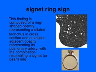
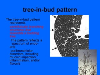
Terms for Thoracic
Imaging ..Unifying the
     language.
        Part I
        Dr/Ahmed Bahnassy
    Consultant Radiologist –RMH.
         MBCHB-MD-FRCR.
Members of the Fleischner Society
compiled a glossary of terms for thoracic
imaging that replaces previous glossaries
published in 1984 and 1996 for thoracic
radiography and computed tomography
(CT), respectively.
bronchiectasis
Bronchiectasis is
  irreversible
localized or diffuse
  bronchial dilatation,
usually resulting from
  chronic infection,
proximal airway
  obstruction, or
congenital bronchial
  abnormality
signet ring sign
This finding is
composed of a ring-
shaped opacity
representing a dilated
bronchus in cross
section and a smaller
adjacent opacity
representing its
pulmonary artery, with
the combination
resembling a signet (or
pearl) ring
bronchocele
A bronchocele is
   bronchial
dilatation due to
   retained secretions
(mucoid impaction)
   usually caused by
proximal obstruction,
   either congenital
(eg, bronchial atresia)
   or acquired (eg,
obstructing cancer)
bronchocentric
This descriptor is applied to
disease that is conspicuously
   centered on macroscopic
   bronchovascular bundles


Examples of diseases with
a bronchocentric distribution
   include :

sarcoidosis , Kaposi
   sarcoma ,
and organizing pneumonia .
bulla
Airspace measuring
more than 1 cm—usually
 several centimeters—
in diameter, sharply
 demarcated by a thin wall
 that is no greater
than 1 mm in thickness. A
 bulla is usually
 accompanied by
 emphysematous
 changes in the adjacent
 lung.
cavity
A cavity is
  a gas-filled space,
  seen as a lucency
  or low-attenuation
  area, within
  pulmonary
  consolidation, a
  mass, or a nodule
centrilobular emphysema
Centrilobular
emphysema
 is characterized by
:destroyed
centrilobular
alveolar walls and
enlargement
of respiratory
bronchioles and
associated
  alveoli
cyst
A cyst is any
  round
  circumscribed
  space that is
  surrounded by an
  epithelial or
  fibrous wall of
  variable thickness
mycetoma
A mycetoma is a discrete
mass of intertwined
 hyphae, usually of
an Aspergillus species,
 matted together
by mucus, fibrin, and
 cellular debris colonizing
 a cavity, usually from
 prior fibrocavitary
 disease (eg,
 tuberculosis or
 sarcoidosis)
tree-in-bud pattern
The tree-in-bud pattern
  represents
  centrilobular branching
  structures that
  resemble a budding
  tree.
   The pattern reflects a
    spectrum of endo-
  and
   peribronchiolar
  disorders, including
  mucoid impaction,
  inflammation, and/or
  fibrosis
Thoracic imaging terms (I)

Thoracic imaging terms (I)

  • 1.
    Terms for Thoracic Imaging..Unifying the language. Part I Dr/Ahmed Bahnassy Consultant Radiologist –RMH. MBCHB-MD-FRCR.
  • 2.
    Members of theFleischner Society compiled a glossary of terms for thoracic imaging that replaces previous glossaries published in 1984 and 1996 for thoracic radiography and computed tomography (CT), respectively.
  • 3.
    bronchiectasis Bronchiectasis is irreversible localized or diffuse bronchial dilatation, usually resulting from chronic infection, proximal airway obstruction, or congenital bronchial abnormality
  • 4.
    signet ring sign Thisfinding is composed of a ring- shaped opacity representing a dilated bronchus in cross section and a smaller adjacent opacity representing its pulmonary artery, with the combination resembling a signet (or pearl) ring
  • 5.
    bronchocele A bronchocele is bronchial dilatation due to retained secretions (mucoid impaction) usually caused by proximal obstruction, either congenital (eg, bronchial atresia) or acquired (eg, obstructing cancer)
  • 6.
    bronchocentric This descriptor isapplied to disease that is conspicuously centered on macroscopic bronchovascular bundles Examples of diseases with a bronchocentric distribution include : sarcoidosis , Kaposi sarcoma , and organizing pneumonia .
  • 7.
    bulla Airspace measuring more than1 cm—usually several centimeters— in diameter, sharply demarcated by a thin wall that is no greater than 1 mm in thickness. A bulla is usually accompanied by emphysematous changes in the adjacent lung.
  • 8.
    cavity A cavity is a gas-filled space, seen as a lucency or low-attenuation area, within pulmonary consolidation, a mass, or a nodule
  • 9.
    centrilobular emphysema Centrilobular emphysema ischaracterized by :destroyed centrilobular alveolar walls and enlargement of respiratory bronchioles and associated alveoli
  • 10.
    cyst A cyst isany round circumscribed space that is surrounded by an epithelial or fibrous wall of variable thickness
  • 11.
    mycetoma A mycetoma isa discrete mass of intertwined hyphae, usually of an Aspergillus species, matted together by mucus, fibrin, and cellular debris colonizing a cavity, usually from prior fibrocavitary disease (eg, tuberculosis or sarcoidosis)
  • 12.
    tree-in-bud pattern The tree-in-budpattern represents centrilobular branching structures that resemble a budding tree. The pattern reflects a spectrum of endo- and peribronchiolar disorders, including mucoid impaction, inflammation, and/or fibrosis