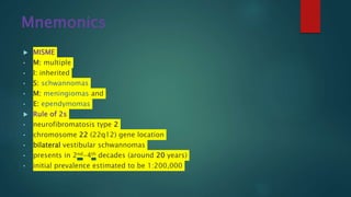
Radiology Spotters collection by Dr Pradeep. Nice collection Radiology spotters mixed collection ppt made by or collected by Dr. Pradeep, this is a collection of confusing spotter and very important spotter commonly asked in exams, our references is radiopaedia, learning radiology and Aunt Minnie.. Thanks
Radiology Spotters mixed Bag Collection for post graduates student .PPTDr pradeep Kumar
Radiology Spotters collection by Dr Pradeep. nice collection of radiology spotter made by or collected by Dr. Pradeep, this is a collection of confusing spotter and very important spotter commonly asked in exams, our references is radiopaedia, learning radiology and Aunt Minnie.. Thanks.
Its important to recognise the myelination pattern in neonates and infants. This presentation talks about the myelination pattern and imaging of white matter diseases in children.
Imaging assessment of malignant focal and diffuse liver lesions from Ultrasound to Mri with overview of interventional modalities and diagnostic snippets,
Skeletal dysplasia musculoskeletal radiology is very concise and it cover the all-important topic of skeletal dysplasia with their characteristic feature and radiological findings with a proper radiographic image. Starting from classification and approach. It includes nosology classification. Thanks.
Abnormal abdominal CT is best powerpoint presentation for radiologist, radiology resident and gastroenterologist, this include pancreatitis, all abdominal trauma grading with systemic manner. Thanks
Role of hrct in interstitial lung diseases pk uploadDr pradeep Kumar
Role of hrct in interstitial lung diseases pk , This is best powerpoint slides presentation including Latest American thoracic society and fleishners society guidelines . this includes radiographic images a well HRCT chest findings of various ILD. This will help alot for md pg radiology resident and radiologist. Thanks
Solitary pulmonary nodules radiology ppt is very good power point presentation from various source radiology assistant and latest guidelines. this power-point also includes many sign with multiple xray, ct and mri images. this will help alot. Thanks.
Jaw lesion radiology ppt ppt . This powerpoint presentation includes important anatomy, radiographs and important pathology of jaw lesion with its imaging feature as well as its Xray ct mri image. This will help alot. this will help for radiology resident as well as ent resident and event dentist.
Skull base tumors & perineural spread radiology pptDr pradeep Kumar
Skull base tumors & perineural spread radiology ppt This powerpoint presentation includes important anatomy and important pathology of skull base lesion with its imaging feature as well as its ct mri image. This will help alot. this will help for radiology resident as well as ent .
Salivary gland imaging radiology ppt . This powerpoint presentation includes important anatomy and important pathology of salivary gland with its imaging feature as well as its ct mri image. This will help alot. this will help for radiology resident as well as ent .
MRI anatomy of ankle radiology ppt pk is nice presentation that covers cross sectional anatomy as well as relevant anatomy from standard radiology book like CT MRI whole body by Hagga . cross section of mri is taken from mrimaster.com. This will help for radiology resident as well radiographers.
Congenital neck mass radiology pk final is very good power point presentation for radiologist, radiology resident, student and even ent surgeon or resident doctor.. Every disease of neck lesion is properly describe with multi usg, ct and MRI images. this will help a lot. thanks.
Imaging of paranasal sinuses (including anatomy and varaints)pk1 pdf pptDr pradeep Kumar
This is very good powerpoint presentation of imaging anatomy and variants of paranasal sinuses and imaging pathology as well as multiple pathological imaging findings and images.it will helps for radiologist and radiology resident and even ent resident. our references is CT and mri whole body by Haaga and various internet sources. THANKS.
Important radiological classification of fracture and AVNDr pradeep Kumar
This is Important radio-logical classification of fracture and AVN, I made this from various references like radiopaedia and radiology website , It will help for radiology resident, radiologist and even orthopedics resident. Thanks.
This slide includes various CT protocol , liver ct triple phase protocol , with important findings, this power-point presentation help a lot for radiologist, radiology resident, radiographers, technician. Thanks.
this power-point presentation includes knee and ankle MRI anatomy with cross sectional axial saggital and coronal views images. this also includes some pathology. this slide will help a lot for radiologist, radiographers, technician radiology resident, thanks.
This presentation include biliary anatomy ,indication, contraindication post op care of percutaneus transhepatic biliary drainage with important technique. and advantage and disadvantage of different technique. This is important for radiologist, radiographers, intervention radiologist radiology resident. Thanks
This power-point presentation is very important for radiology resident radiologist and radiographers and technician. this includes principles, technique , biological effects of radiation and how to protect, whats should normal radiation dose with latest update. This slide also includes ALARA PRINCIPLE thanks.
this power-point slide presentation includes lots of information like how MRI coil works. what is shimming, magnet, fringe, and design of mri coil and also magnet. this will help a lot for radiologist and technician radiographers.. thanks.
Barium meal ppt presentation is very important for radiology resident , radiologist and radiographers. this slide contents lots of barium image and technique, position, indication and modification and lots of information. this presentation help alot thanks .
Sellar, Suprasellar and Pineal tumor final pk .pptDr pradeep Kumar
this is very good presentation slide for radiologist and radiology resident. our references is authentic and most are from osborn brain imaging 2nd edition. This deal with sellar, suprasellar and pineal tumor . This help alot. thanks
Imaging of intracranial infections including COVID 19 pk2 ppt, pdfDr pradeep Kumar
This is nice presentation covers most of imporant intrancranial ( Brain) infection with many ct mri images . This presentation also includes cns (brain) manifestation of COVID-19 latest hot topic. This is very helpful for radiologist or radiology resident. Thanks.
This presentation includes stroke and infarct latest defination an pathophysiology and CT MRI imaging features and management . This presntation help alot. Thanks
Unit 8 - Information and Communication Technology (Paper I).pdfThiyagu K
This slides describes the basic concepts of ICT, basics of Email, Emerging Technology and Digital Initiatives in Education. This presentations aligns with the UGC Paper I syllabus.
Instructions for Submissions thorugh G- Classroom.pptxJheel Barad
This presentation provides a briefing on how to upload submissions and documents in Google Classroom. It was prepared as part of an orientation for new Sainik School in-service teacher trainees. As a training officer, my goal is to ensure that you are comfortable and proficient with this essential tool for managing assignments and fostering student engagement.
How to Make a Field invisible in Odoo 17Celine George
It is possible to hide or invisible some fields in odoo. Commonly using “invisible” attribute in the field definition to invisible the fields. This slide will show how to make a field invisible in odoo 17.
How to Split Bills in the Odoo 17 POS ModuleCeline George
Bills have a main role in point of sale procedure. It will help to track sales, handling payments and giving receipts to customers. Bill splitting also has an important role in POS. For example, If some friends come together for dinner and if they want to divide the bill then it is possible by POS bill splitting. This slide will show how to split bills in odoo 17 POS.
Read| The latest issue of The Challenger is here! We are thrilled to announce that our school paper has qualified for the NATIONAL SCHOOLS PRESS CONFERENCE (NSPC) 2024. Thank you for your unwavering support and trust. Dive into the stories that made us stand out!
This is a presentation by Dada Robert in a Your Skill Boost masterclass organised by the Excellence Foundation for South Sudan (EFSS) on Saturday, the 25th and Sunday, the 26th of May 2024.
He discussed the concept of quality improvement, emphasizing its applicability to various aspects of life, including personal, project, and program improvements. He defined quality as doing the right thing at the right time in the right way to achieve the best possible results and discussed the concept of the "gap" between what we know and what we do, and how this gap represents the areas we need to improve. He explained the scientific approach to quality improvement, which involves systematic performance analysis, testing and learning, and implementing change ideas. He also highlighted the importance of client focus and a team approach to quality improvement.
Operation “Blue Star” is the only event in the history of Independent India where the state went into war with its own people. Even after about 40 years it is not clear if it was culmination of states anger over people of the region, a political game of power or start of dictatorial chapter in the democratic setup.
The people of Punjab felt alienated from main stream due to denial of their just demands during a long democratic struggle since independence. As it happen all over the word, it led to militant struggle with great loss of lives of military, police and civilian personnel. Killing of Indira Gandhi and massacre of innocent Sikhs in Delhi and other India cities was also associated with this movement.
The Roman Empire A Historical Colossus.pdfkaushalkr1407
The Roman Empire, a vast and enduring power, stands as one of history's most remarkable civilizations, leaving an indelible imprint on the world. It emerged from the Roman Republic, transitioning into an imperial powerhouse under the leadership of Augustus Caesar in 27 BCE. This transformation marked the beginning of an era defined by unprecedented territorial expansion, architectural marvels, and profound cultural influence.
The empire's roots lie in the city of Rome, founded, according to legend, by Romulus in 753 BCE. Over centuries, Rome evolved from a small settlement to a formidable republic, characterized by a complex political system with elected officials and checks on power. However, internal strife, class conflicts, and military ambitions paved the way for the end of the Republic. Julius Caesar’s dictatorship and subsequent assassination in 44 BCE created a power vacuum, leading to a civil war. Octavian, later Augustus, emerged victorious, heralding the Roman Empire’s birth.
Under Augustus, the empire experienced the Pax Romana, a 200-year period of relative peace and stability. Augustus reformed the military, established efficient administrative systems, and initiated grand construction projects. The empire's borders expanded, encompassing territories from Britain to Egypt and from Spain to the Euphrates. Roman legions, renowned for their discipline and engineering prowess, secured and maintained these vast territories, building roads, fortifications, and cities that facilitated control and integration.
The Roman Empire’s society was hierarchical, with a rigid class system. At the top were the patricians, wealthy elites who held significant political power. Below them were the plebeians, free citizens with limited political influence, and the vast numbers of slaves who formed the backbone of the economy. The family unit was central, governed by the paterfamilias, the male head who held absolute authority.
Culturally, the Romans were eclectic, absorbing and adapting elements from the civilizations they encountered, particularly the Greeks. Roman art, literature, and philosophy reflected this synthesis, creating a rich cultural tapestry. Latin, the Roman language, became the lingua franca of the Western world, influencing numerous modern languages.
Roman architecture and engineering achievements were monumental. They perfected the arch, vault, and dome, constructing enduring structures like the Colosseum, Pantheon, and aqueducts. These engineering marvels not only showcased Roman ingenuity but also served practical purposes, from public entertainment to water supply.
11. Bovine arch is the most common variant of the aortic arch and occurs when
the brachiocephalic (innominate) artery shares a common origin with the
left common carotid artery
29. NF1 or von Recklinghausen
disease,
• CAFE SPOT
Mnemonic
• C: café-au-lait spots (greater than six seen during one year)
• A: axillary or inguinal freckling
• F: fibromas (neurofibroma (two or more) or plexiform
neurofibroma (one))
• E: eye hamartomas (Lisch nodules)
• S: skeletal abnormalities, e.g. sphenoid wing dysplasia, leg bowing
• P: positive family history
• OT: optic tumour (optic nerve glioma)
64. Diastematomyelia
Diastematomyelia, also known as a split cord malformation, refers to a
type of spinal dysraphism (spina bifida occulta) when there is a longitudinal
split in the spinal cord.
72. Signs of Paget’s disease
There are many Paget disease-related signs,
listed here and described in the modality-
specific sections below:
•banana fracture
•blade of grass sign
•cotton wool appearance of bone
•ivory vertebra sign
•jigsaw pattern bone or mosaic pattern bone
•Lincoln sign
•Looser zones
•Mickey Mouse sign
•osteoporosis circumscripta
•picture frame vertebra
•Tam o' Shanter sign
78. EROSIVE
Erosive osteoarthritis has a predilection for the hands. The
dominant features are those of osteoarthritis, particularly in
terms of distribution:
• distal interphalangeal (DIP) joints
• proximal interphalangeal (PIP) joints
• first carpometacarpal (CMC) joint
Additional characteristic features include:
• diffuse cartilage loss, with joint space narrowing
• subchondral erosions (at least two central erosions affecting
separate interphalangeal joints); typical central location of the
erosions produces the classic "gull-wing" appearance
• joint ankylosis
• absence of 2
• marginal erosions
• fusiform soft-tissue swelling
• osteopenia
79.
80. RA
MRI is particularly sensitive to the early and subtle features of RA.
Commonly used sequences include T1-weighted contrast-enhanced spin-echo with fat saturation and T2-weighted spin-echo or
gradient-echo sequences 2.
Features of RA best demonstrated with MRI include 2:
• synovial hyperaemia: an indication of acute inflammation
• synovial hyperplasia (rice bodies)
• pannus formation
• decreased thickness of cartilage
• subchondral cysts and erosions:
• MRI is much more sensitive than radiography
• it is thought that subchondral cysts in RA eventually progress to erosions (i.e. constitute "pre-erosions")
• contrast enhancement may distinguish erosions or pre-erosions from degenerative subchondral cysts
• juxta-articular bone marrow oedema
• joint effusions
85. NEUROPATHIC
Charcot joint, also known as a neuropathic joint or Charcot (neuro/osteo)arthropathy, refers to a
progressive degenerative/destructive joint disorder in patients with abnormal pain sensation and
proprioception.
88. Osteochondroma/exostosis
They are usually sporadic, but can be part of:
• hereditary multiple exostoses (HME): also known as diaphyseal aclasis
• Trevor disease: also known as dysplasia epiphysealis hemimelica
125. Croup
Croup, also known as acute laryngotracheobronchitis, is due to viral infection of
the upper airway by parainfluenza virus or respiratory syncytial virus (RSV)
Digital subtraction angiogram of the internal carotid artery, lateral view, demonstrates a large tortuous vessel (arrow) passing from the internal carotid artery (open arrow) to the basilar artery (arrowhead), and supplying the posterior circulationLarge persistent trigeminal artery passing from the left ICA to the terminal basilar artery via the cavernous sinus. Note the grossly hypoplastic vertebro-basilar arterial system as a result of this congenital anomaly with most posterior circulation blood coming from the left ICA via the trigeminal artery
Persistent primitive trigeminal artery (PPTA) is one of the persistent carotid-vertebrobasilar anastomoses.
There is direct communication between the basilar artery and left internal carotid artery, in keeping with a persistent left trigeminal artery
Persistent smooth posterior oesophageal indentation, suggestive of vascular impression
First vessel from the arch is the left commmon carotid artery. Bilateral calcified pleural plaques.Thrombosed ligamentum arteriosum. Kommerell's diverticulum/dilatation of the origin of the left subclavian artery causes compression of the left brachiocephalic vein resulting in contrast entering the heart via alternative routes
This anomaly is caused by persistence of the right and left embryonic fourth aortic arches, which results in formation of a vascular ring from the splitting of the ascending aorta into two limbs that pass to either side of the trachea and oesophagus 5 (both of which get encircled), which then join as a single descending aorta
Aberrant right subclavian arteries (ARSA), also known as arteria lusoria, are one of the commonest of the aortic arch anomalies.
Instead of being the first branch (with the right common carotid as the brachiocephalic artery), it arises on its own as the fourth branch, distal to the left subclavian artery. It then hooks back to reach the right side with its relationship to the oesophagus variable:
80% posterior to the oesophagus
15% between oesophagus and trachea
5% anterior to the trachea
aneurysmal dilatation (aberrant subclavian arterial aneurysms) of the proximal portion of an aberrant right subclavian artery can occur, a pouch-like aneurysmal dilatation is called a diverticulum of Kommerell
Aberrant left pulmonary artery, also known as pulmonary sling, represents an anatomical variant characterised by the left pulmonary artery arising from the right pulmonary artery and passing above the right main bronchus and in between the trachea and oesophagus to reach the left lung. It may lead to compression and focal stenosis of the trachea
Fluoroscopy
In most instances, the barium oesophagogram characteristically shows a mass between the trachea and the oesophagus just above the level of the carina, usually seen as an anterior indentation over the oesophagus
Post contrast T1 axial imaging demonstrates bilateral enhancing masses in the cerebellopontine angle, larger on the left. Both can be seen extending into the internal acoustic meatii and are consistent with acoustic schwannomas. The fact that they are bilateral almost certainly indicates that the patient has NF2.
There are multiple enhancing nodular lesions spread throughout all the spinal canal and going through some vertebral foramina
Mixed solid-cystic mass arising from the fourth ventricle and causing mass effect on the brainstem. There was also resultant obstructive hydrocephalus, not shown here. The mass appears to ‘ooze’ out of the foramen of Magendie. There was no associated spinal lesion. Classical ‘plastic’ ependymoma
Pancake kidney (also known as discoid kidney, disc kidney, lump kidney, fused pelvic kidney or cake kidney) is a rare renal fusion anomaly of the kidneys of the crossed fused variety.
Characterised by bilateral symmetrical confluent areas of periventricular deep white matter signal change, in particular around the atria and frontal horns with sparing of subcortical U fibres leading to a "butterfly pattern". Progression can lead to cortical and subcortical atrophy
T2:affected areas are high signal and may show a "tigroid pattern" on axial plane or "leopard pattern" on sagittal plane representing sparing along the venules
subcortical U-fibres are usually spared
five different MRI patterns of adrenoleukodystrophy based on the involved anatomic locations and MR patterns of progression:
deep white matter in the parieto-occipital lobes and splenium of the corpus callosum (66% of cases, chiefly in children); may include lesions of the visual and auditory pathways
frontal lobe or genu of the corpus callosum (15.5%, mostly in adolescents)
frontopontine or corticospinal projection fibres (12%, mostly in adults)
cerebellar white matter (1%, mostly in adolescents)
combined parieto-occipital and frontal white matter (2.5%, mostly children)
There tends to be cortical and subcortical U-fibre sparing
MRI confirms the megalencephalic appearance and provides more detail of the white matter disease, which is typically diffuse, bilateral, and involving the subcortical U-fibres
Diffuse T2 hyperintensities and restricted DWI signal of the pons with extension into the middle cerebellar peduncles. Also similar increased T2 signal of the symmetric basal ganglia, including the caudate nuclei, putamina, as well as the ventrolateral thalami, without the restriction of diffusion.
2. On MRI images seen T2, FLAIR hyperintensity in medulla oblangata, pons, both thalami and putamen, and the caudate nuclei.
Additionally an cavum septum pellucidum and cavum vergae.
There is symmetric marked hyperintense signal intensity on T2/FLAIR imaging and restricted diffusion in the dentate nuclei, inferior cerebellar peduncles, lateral fourth ventricle floor in the expected locations of the vestibular tract and in the expected tract of the vestibular cochlear nerve. Also present, is diffusion restriction in the posterior columns of the proximal cervical cord, anteriorly in the inferior medulla, and punctate symmetric foci of restricted diffusion in the inferior colliculi of the quadrigeminal plate and medial geniculate bodies. There is mild high signal on FLAIR and diffusion weighted imaging involving the bilateral putamen
2nd image is dwi image
1. Selected FLAIR MRI images of the brain demonstrates hyperintensities in the periaqueductal region and the medial thalami. Note that this is on a background of global cerebral atrophy with CSF spaces more prominent than would normally be expected for a patient of 45 years old.
2.Hyperintense signals in the bilateral medial thalami and periventricular regions of third ventricle on FLAIR images, consistent with Wernicke encephalopathy
3.Abnormal signal bilateral and symmetrical involving the medial and posterior aspect of both thalami displaying bright signal in DWI, T2 and FLAIR.
Focal inflammatory disorder of the spinal cord resulting in motor, sensory and autonomic dysfunction.
Imaging findings:
More than 2/3 of the cross sectional area is involved.
Focal enlargement.
T2WI hyperintensity
Enhancement + / -.
Two forms of TM:
Acute partial transverse myelitis - APTMLesions extending less than two Segments.These patients are at risk of developing MS.
Acute complete transverse myelitis - ACTMLesions extending more than two Segments
Arterial infarction
Spinal cord ischemia is typically seen as a complication of aortic aneurysm surgery or stenting.The images are of a patient who developed a paraparesis after stenting of an aortic aneurysm.Notice the high signal ventrally in the chord, which is typical for arterial infarction.On transverse images a typical snake-eye appearance can be seen.
Notice the high signal in the lower thoracic cord and the surrounding dilated vessels on the T2WI.On the enhanced T1WI there is subtle enhancement.
T2 hyperintense expansion of the thoracic cord is demonstrated. The main mass effect is centred between T5 and T8 but abnormal high T2 signal extends between T4 and T11. Postcontrast images demonstrate patchy irregular peripheral enhancement of this lesion, and central low intensity. No definite evidence of blood products/haemorrhage although some T2 hypointensity is seen.No further intra medullary lesion is identified.
Lower thoracic cord is a vividly enhancing nodule with associated prominent serpiginous flow voids both within the mass and over the surface of the cord, and among the cauda equina. It is an isolated abnormality, associated with marked expansion of the entire cord by a large syrinx.
Selected MRI images demonstrate multiple enhancing intramedullary lesions, the largest at C4/5, with an associated syrinx.
1.Paget involvement of the skull, with widening of the diploic space, typical "cotton wool" appearance and over-riding enlarged frontal bone (Tam o' Shanter sign).
2. Marked thickening of the calvarium. The diploic space is widened and there are ill-defined sclerotic and lucent areas throughout. The cortex is thickened and irregular.
3. Osteosarcoma in Paget disease
1.chalk stick fracture-Single sagittal image of the lumbar spine demonstrates a fracture extending from the anterior disc margin, through the inferior body of L1 and through the posterior elements. Note extensive syndesmophytic fusion of the discs spaces and posterior elements consistent with ankylosing spondylitis.
2.Typical features of ankylosing spondylitis with syndesmophytes, bamboo spine and bony fusion of posterior elements (dagger sign).
3.Throughout the spine, the vertebral bodies are fused by marginal syndesmophytes giving the appearance of Bamboo spine.
1. Severe joint space narrowing at the first carpometacarpal joint and the interphalangeal joints. At the first carpometacarpal joint there is associated subchondral sclerosis and cyst formation. There are also subchondral central erosions at the heads of the proximal phalanges with marginal osteophytes involving the distal phalangeal bases, demonstrating gull wing deformity through the second and fifth digits. Severe joint space narrowing, subchondral sclerosis, and erosive changes of the scaphoid-trapezium and scaphoid-trapezoid joints. Narrowing of the radiocarpal joint. Diffuse osteopenia. Findings are keeping with erosive osteoarthritic changes
2. Extensive erosive changes and alignment abnormalities are observed along the articular surfaces of the 2nd through 5th PIP and DIP joints bilaterally, as well as at the 1st IP, MCP and carpometacarpal joints bilaterally. These erosive changes assume a gull wing appearance. The alignment abnormalities involving the 1st digits resemble a hitchhiker's thumb. There are no significant erosive changes involving the radiocarpal or intercapral joints, and the 2nd-5th MCP joints are also spared.
Almost total loss of radiocarpal, carpometacarpal and intercarpal joint spaces associated with multiple radial and carpal bone small subchondral cysts and bony fusion of trapezium, trapezoid and scaphoid bones.
Diagnosis: Advanced rheumatoid arthriti
Erosive changes of intercarpal, carpometacarpal anX-ray (both hands) findings:
Loss of intercarpal, carpometacarpal and distal radio-ulnar joint spaces
ulnar deviation of MCP joints
extension deformity of DIPs (Boutonierre deformity) and hitchhiker thumb deformit
d metacarpophalangeal of the 2nd finger of right hand. Left elbow and left knee are affected as well.
Punched-out juxta-articular erosions of the first and fifth metatarsal heads with overhanging edges are typical for gout. Overlying soft tissue swelling and increased density represent tophi.
There is destruction and deformity of the tarsal bones characterised by sclerosis, joint space narrowing, cortical irregularities, and loose bodies. Lisfranc injury involving the second to fifth tarsometatarsal joints with lateral dislocation. Cortical thickening and sclerosis involving the proximal second to fifth metatarsal bones. Local soft tissue oedema.
Destruction, disorganisation and fragmentation of the subtalar and inter-tarsal joints, associated with soft tissue tumefaction and multiple bone fragments.
1. A sharply defined lucent lesion is centred on the epiphysis of the distal femur, and appears to transgress the growth plate (which remains open). It has a narrow zone of transition and no convincing matrix calcification. Anteriorly it appears to abut the articular surface, possibly breaching it. A joint effusion is present. No periosteal reaction is present
2.Chondroblastoma in right lateral femoral condyle with low T1W and high T2W/STIR internal signal and surrounding bone marrow oedema
2. A large posteromedial bony projection is demonstrated involving the distal one quarter of the femoral shaft. It is continuous with the medullary cavity and has an irregular medial margin without large soft tissue component or fracture. Severe narrowing of the medial and patellofemoral joint compartments and marginal osteophyte formations indicate degenerative disease of the knee.
There are numerous similar-sized intra-articular loose bodies of variable signal intensity, some of them are showing intermediate to high signal intensity that is of cartilage. Others have low signal intensity at the periphery, which represents ossification.
The joint capsule is distended with a little fluid and these numerous loose bodies
Synovial chondromatosis (osteochondromatosis or synovial chondrometaplasia) also known as Reichel syndrome, is a disorder characterised by loose cartilaginous bodies which may, or may not be calcified or ossified.
It is classified under two main types:
primary synovial chondromatosis: predominantly monoarticular disorder of unknown aetiology
secondary synovial chondromatosis: resulting in intra-articular loose bodies from causes such as trauma, osteoarthrosis and neuropathic arthropathy
"Cloud-like" calcifications in the region of the triceps insertion, olecranon, and olecranon bursa (3.3 x 7.7 x 5.7 cm). There is no connection to the underlying humerus or ulna.
Bilateral extensive soft tissue calcification posterior to the elbow joint around the insertion of triceps. US confirmed that it was extra-articular. Limitation of extension at the elbow.
Hypertrophic phenomena and gross heterogeneous muscle calcifications that extend from the pelvis to the middle third of the thighs. A firearm projectile is located in the soft tissues in left inguinal region. Notice that the bone cortical isn't affected
Myositis ossificans is a benign process characterised by heterotopic ossification usually within large muscles. The diagnosis is usually based on the patient's history (of trauma),
he distal half of the femur is occupied and expanded with a heterogeneous mass with areas of bone formation. Posterior the periosteum is elevated (Codman's triangle).
There is a lobulated juxtacortical mass with dense calcification and smooth borders arising from the lateral cortex of the right femoral metaphysis measuring approximately 5.8 x 3.3 x 9.3 cm. There is no aggressive periosteal reaction. Superiorly, a thin radiolucent line separates the tumour from the cortex.
Ill-defined calcified/ossified mass arising from the lateral aspect of distal femu
Synovial sarcomas are relatively common intermediate-to-high grade malignant soft tissue tumours, often with an initial indolent course, affecting young patients, and most commonly involving the soft tissue surrounding the knees
Lobulated mass-like density in the left lung adjacent to the heart border. A prominent vessel is noted extending to the mass from the pulmonary hilum. Appearance is suggestive of a pulmonary arteriovenous malformation. After adjusting the image contrast, a similar lesion is seen in the right lung base behind the diaphragm.
Large wide necked diverticulum arising several centrimetres proximal to the GOJ.
The diverticulum contains a progressively filling air fluid-level following barium consumption.
Normal calibre oesophagus - no stricture.
No hiatus hernia.
Characteristic tapering of the upper trachea (steeple sign) on an AP radiograph which was suspicious for croup in this clinical context.
Lateral x-ray of the neck demonstrates distension of hypopharynx and subglottic oedema consistent with croup.
Epiglottitis. Lateral radiograph of the neck demonstrates and enlarged epiglottis (red arrow) and thickening of the aryepiglottic folds (yellow arrow). There is also reversal of the normal lordotic curve in the cervical spine and slight dilatation of the hypopharynx
Annotations showing the normal width of the pre-vertebral soft tissue at the various cervical levels.
The annotated pathological images reveals the magnitude of the soft tissue widening.
Huge fluid-gas filled rim enhancing abscess in the retropharyngeal space.
Its extends over nearly the whole length of the cervical spine.