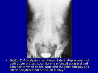
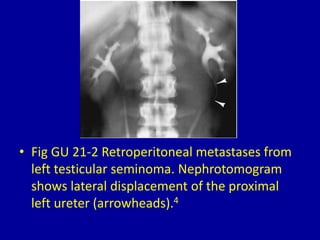
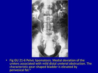
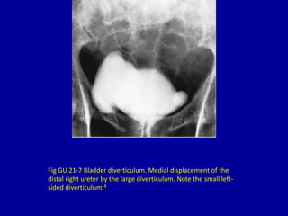
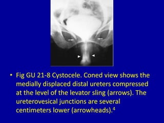
2. CLINICAL IMAGAGING
AN ATLAS OF DIFFERENTIAL DAIGNOSIS
EISENBERG
DR. Muhammad Bin Zulfiqar
PGR-FCPS III SIMS/SHL
3. • Fig GU 21-1 Hodgkin's lymphoma. Lateral displacement of
both upper ureters, secondary to enlarged paracaval and
para-aortic lymph nodes. Note also the splenomegaly and
inferior displacement of the left kidney.4
4. • Fig GU 21-2 Retroperitoneal metastases from
left testicular seminoma. Nephrotomogram
shows lateral displacement of the proximal
left ureter (arrowheads).4
5. • Fig GU 21-3 Aortic and common iliac aneurysms.
Aortography faintly opacifies massively dilated
vessels (arrowheads) because of contrast
dilution. The deviated ureters typically follow the
lateral borders of the aortic and common iliac
artery aneurysms. The medial borders of the
common iliac aneurysms are indicated (arrows).4
6. • Fig GU 21-4 Ureterosciatic hernia. Excretory
urogram demonstrates a “horizontal curlicue”
or horizontal looping of the left ureter in the
region of the greater sciatic foramen
(arrows).22
7. • Fig GU 21-5 Retroperitoneal fibrosis. Bilateral
retrograde pyelogram shows dilatation of the collecting
systems and ureters to the level of the L4-L5 disk space
with medial deviation of the ureters.4
8. • Fig GU 21-6 Pelvic lipomatosis. Medial deviation of the
ureters associated with mild distal ureteral obstruction. The
characteristic pear-shaped bladder is elevated by
perivesical fat.4
9. Fig GU 21-7 Bladder diverticulum. Medial displacement of the
distal right ureter by the large diverticulum. Note the small left-
sided diverticulum.4
10. • Fig GU 21-8 Cystocele. Coned view shows the
medially displaced distal ureters compressed
at the level of the levator sling (arrows). The
ureterovesical junctions are several
centimeters lower (arrowheads).4