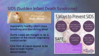
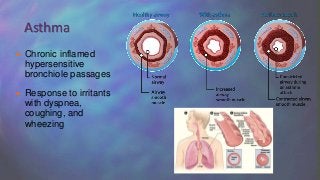
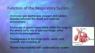
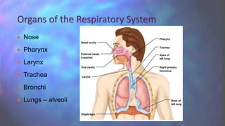
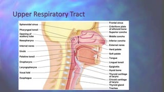
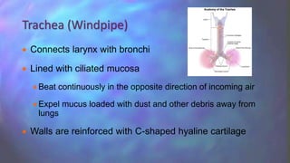
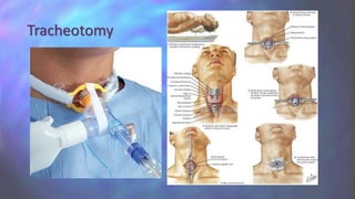
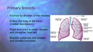
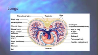
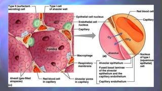
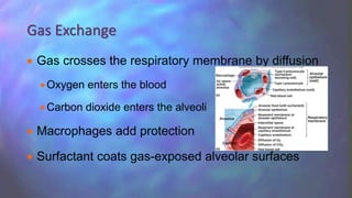
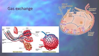
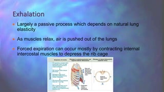
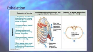
The respiratory system oversees gas exchange between the blood and external environment through the lungs. Air passes through the nasal cavity, pharynx, larynx, trachea, bronchi and into the lungs where gas exchange occurs in the alveoli. The respiratory system works with the cardiovascular system to oxygenate blood and remove carbon dioxide. Key organs include the nose, pharynx, larynx, trachea, lungs and diaphragm.


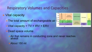
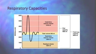
![ Normal breathing moves about 500 ml of air with each breath (tidal
volume [TV])
Many factors that affect respiratory capacity
A person’s size
Sex
Age
Physical condition
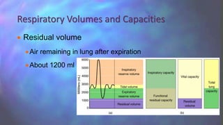
Residual volume of air – after exhalation, about 1200 ml of air
remains in the lungs
Respiratory Volumes and Capacities](https://image.slidesharecdn.com/therespiratorytoupload-160329025636/85/The-Respiratory-System-41-320.jpg)
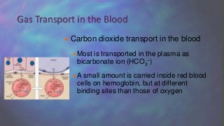
![ Oxygen transport in the blood
Inside red blood cells attached to
hemoglobin (oxyhemoglobin
[HbO2])
A small amount is carried dissolved
in the plasma
Gas Transport in the Blood](https://image.slidesharecdn.com/therespiratorytoupload-160329025636/85/The-Respiratory-System-49-320.jpg)