Downloaded 814 times

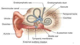
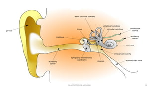
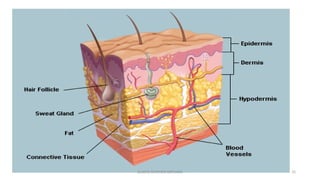
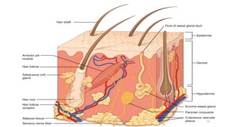
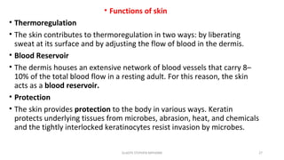
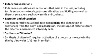

The document outlines the anatomy and physiology of the sense organs, focusing on the eye, ear, and skin, and their roles in sensory perception and homeostasis. It details the structures and functions of these organs, including vision, hearing, and equilibrium, along with common disorders associated with each. The integumentary system is also discussed, highlighting its functions in protection, sensation, and temperature regulation.


