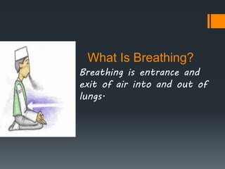
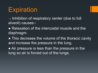
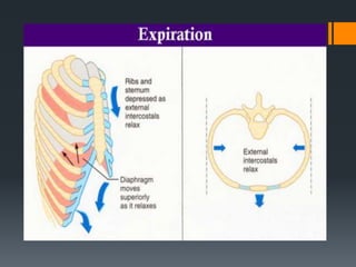
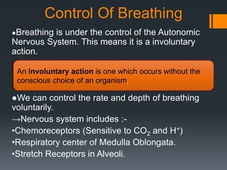
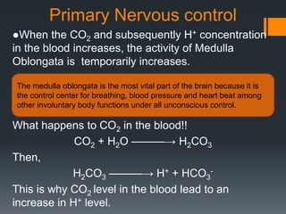
This document discusses control of breathing. It begins by defining breathing and respiration, noting that breathing is the process of inhaling and exhaling air while respiration involves cellular oxidation reactions. It then describes the mechanics of breathing, including inspiration driven by diaphragm and intercostal muscle contraction, and expiration triggered by relaxation of these muscles. The document outlines three levels of nervous control over breathing: primary control by the medulla oblongata in response to rising blood CO2 and H+ levels, secondary control via chemoreceptors responding to blood O2, and alveolar control through stretch receptors signaling full alveoli to the medulla.
lecture 5: it's good for as to take a breif about how does atmospheric air will pass to our lungs then to blood, for transportation and utilization of oxygen and excretion of carbon dioxide. Many issue are related when gas exchange is performed.
The apparatus used to measure
Volume of air exchanged during breathing
Respiratory rate
The record is called a spirogram
Upward deflection inhalation
Downward deflection exhalation
Gas exchange between the alveoli and the pulmonary capillary blood occurs by diffusion, as will be discussed in the next chapter. Diffusion of oxygen and carbon dioxide occurs passively, according to their concentration differences across the alveolar-capillary barrier. These concentration differences must be maintained by ventilation of the alveoli and perfusion of the pulmonary capillaries.
Alveolar ventilation brings oxygen into the lung and removes carbon dioxide from it. Similarly, the mixed venous blood brings carbon dioxide into the lung and takes up alveolar oxygen. The alveolar Image not available. and Image not available. are thus determined by the relationship between alveolar ventilation and pulmonary capillary perfusion. Alterations in the ratio of ventilation to perfusion, called the Image not available., will result in changes in the alveolar Image not available. and Image not available., as well as in gas delivery to or removal from the lung.
Alveolar ventilation is normally about 4 to 6 L/min and pulmonary blood flow (which is equal to cardiac output) has a similar range, and so the Image not available. for the whole lung is in the range of 0.8 to 1.2. Image not available. However, ventilation and perfusion must be matched on the alveolar-capillary level, and the Image not available. for the whole lung is really of interest only as an approximation of the situation in all the alveolar-capillary units of the lung. For instance, suppose that all 5 L/min of the cardiac output went to the left lung and all 5 L/min of alveolar ventilation went to the right lung. The whole lung Image not available. would be 1.0, but there would be no gas exchange because there could be no gas diffusion between the ventilated alveoli and the perfused pulmonary capillaries.
Oxygen is delivered to the alveolus by alveolar ventilation, is removed from the alveolus as it diffuses into the pulmonary capillary blood, and is carried away by blood flow. Similarly, carbon dioxide is delivered to the alveolus in the mixed venous blood and diffuses into the alveolus in the pulmonary capillary. The carbon dioxide is removed from the alveolus by alveolar ventilation. As will be discussed in Chapter 6, at resting cardiac outputs the diffusion of both oxygen and carbon dioxide is normally limited by pulmonary perfusion. Thus, the alveolar partial pressures of both oxygen and carbon dioxide are determined by the Image not available. If the Image not available. in an alveolar-capillary unit increases, the delivery of oxygen relative to its removal will increase, as will the removal ...
Ventilation perfusion ratio (The guyton and hall physiology)Maryam Fida
Ventilation perfusion ratio is :
“The ratio of alveolar ventilation and the amount of blood that perfuse the alveoli”.
FORMULA
It is expressed as VA/Q.
VA is alveolar ventilation
Q is the blood flow (perfusion)
Normal value of ventilation perfusion ratio is about
0.8
VA is 4.2 L /min
Q is 5.5 L/min (Same as Cardiac output)
So VA/Q = 4.2/5.5 = 0.8
If VA becomes zero ratio becomes zero
If Q becomes zero ratio becomes infinite.
If ratio becomes zero or infinite then there is no gaseous exchange. So this ratio indicates the efficiency of gaseous exchange in lungs.
In standing or sitting position this ratio is not uniform in all parts of the lungs.
In standing position, in upper parts of lungs there is almost no blood flow so normally in upper parts of lungs the ratio is higher may be near 3.
In lower part of lungs, there is more blood flow so the ratio is decreased may be 0.6.
In certain diseases the VA/Q ratio is higher which means perfusion is inadequate i.e. in some parts of lungs the alveoli are non functional or partially functional. This is seen in cases of pulmonary thrombosis or embolism.
When there is higher VA/Q ratio, PO2 and PCO2 in the alveolar air resembles the values in the inspired air.
When exchange is not occurring because of lack of perfusion, inspired air goes to alveoli, as there is no exchange occurring so the same values of PCO2 and PO2 as in inspired air.
it contains all the physiology of lung volume and capacity.
in this we study:-
introduction
lung volume
lung capacities
measurements of lung volume and capacities.
measurement of FRC and RV.
vital capacity.
FEV
RMV
MBC
PEFR
restrictive and obstructive respiratory disease.
Ventilation and Perfusion in different zones of lungs.Gyaltsen Gurung
This powerpoint presentation will make you explore about the Perfusion and Ventilation in different zones of lungs with its co-relation with pulmonary tuberculosis.
Inspiratory Muscle Training or Respiratory Muscle Training or Ventilatory Muscle Training. IMT is the physiotherapy technique, with the help of different breathing exercises.
Like heartbeat, breathing must occur in a continuous, cyclic pattern to sustain life processes.
Inspiratory muscles must rhythmically contract and relax to alternately fill the lungs with air and empty them.
The rhythmic pattern of breathing is established by cyclic neural activity to the respiratory muscles
describes the structure of hb, its variants in detail. Oxygen dissociation curve is explained with graph. Hemoglobinopathy is explained with diagram. myoglobin is also explained.
Hyperventilation
Respiration
Muscles of respiration
control of respiration
central and peripheral chemoreceptors
effect of exercise on respiration
Effect of respiration on heart rate
Regulation of respiration (the guyton and hall physiology)Maryam Fida
Normal respiration is spontaneous and unconscious.
There are 4 groups of neurons on each side in the Pons and medulla oblongata which are involved in regulation of respiration. These include
1. Medullary centers
Dorsal respiratory group of neurons
Ventral respiratory group of neurons
2. Pontine centers
Pneumotaxic centre
Apneustic centre.
It contains “I”neurons which are inspiratory neurons.
It’s located in dorsal portion of medulla oblongata.
It also includes the nucleus of tractus solitarius which is the sensory termination of afferent fibers in 9th ( GLOSSOPHARYNGEAL NERVE) and 10th (VAGUS NERVE) cranial nerves.
They receive impulses from peripheral chemoreceptors, carotid and aortic baroreceptors and also other receptors in the lungs.
In this group inspiratory ramp signals are produced spontaneously.
If we cut the medulla oblongata from other parts of brain and also the afferent nerves which enter the medulla, still inspiratory ramp signals are produced which indicate it’s the inherent property of medulla.
Initially the signal is weak and then it progressively increases and then fades away.
Each ramp signal’s duration is 2 sec and then for 3 seconds there is no ramp signal.
So each cycle lasts for 5 seconds and there are 12 cycles /minute which is the respiratory rate.
Significance of the signal in the form of ramp is that it causes progressive expansion of the lungs. After production, these ramp signals are transmitted to the contra lateral motor neurons supplying the inspiratory muscles.
Rate and duration of inspiratory ramp signals is controlled by impulses from the Pneumotaxic centre and impulses from the lungs via vagi.
Microspheres are spherical & free flowing particles ranging in average particle size from 1 to 50 microns which consist of proteins or synthetic polymers. Some of the problems of overcome by producing control drug delivery system which enhances the therapeutic efficacy of a given drug. One such approach is using microspheres as carriers for drugs. The target site drug deliver with Specificity & maintain the concentration at site of interest without untoward effects. It will find the central place in novel drug delivery. Drugs can be targeted to specific sites in the body using microspheres. Degree of targeting can be achieved by localization of the drug to a specific area in body (for example in lungs), to a particular group of cells and even to the intracellular structures. The rate of drug release from the microspheres dictates their therapeutic action.
Methods For Assesment Of Bioavailability Anindya Jana
Bioavailability means the rate and extent to which the active ingredient or active moiety is absorbed from a drug product and becomes available at the site of action. For drug products that are not intended to be absorbed into the bloodstream, bioavailability may be assessed by measurements intended to reflect the rate and extent to which the active ingredient or active moiety becomes available at the site of action.
Bioavailability studies are important in the Primary stages of development of a suitable dosage form for a new drug entity, determination of influence of excipients, patient related factors & possible interaction with other drugs on the efficiency of absorption, development of new formulations of the existing drugs, control of quality of a drug product during the early stages of marketing in order to determine the influence of processing factors, storage & stability on drug absorption
Pharmaceutical aerosols have been playing a crucial role in the health and wellbeing of millions of people throughout the world for many years. These products include pressurized metered dose inhalers (MDIs), dry powder inhalers (DPIs), nebulizers, sublingual’s, skin sprays (coolants, anaesthetics, etc.) and dental sprays. The technology’s continual advancement, the ease of use, and the more desirable pulmonary-rather-than-needle delivery for systemic drugs has increased the attraction for the pharmaceutical aerosol in recent years.
Many of the tests required for the evaluation of MDIs are similar to those used for other dosage forms. These include description, identification, and assay of the active ingredient; microbial limits; moisture content; net weight, degradation products and impurities (if any); extractable; and any other tests deemed appropriate for the active ingredient.
Application Of Polymer In Controlled Release FormulationAnindya Jana
Polymers are becoming increasingly important in the field of drug delivery. The pharmaceutical applications of polymers range from their use as binders in tablets to viscosity and flow controlling agents in liquids, suspensions and emulsions. Polymers can be used as film coatings to disguise the unpleasant taste of a drug, to enhance drug stability and to modify drug release characteristics.
As a consequence, increasing attention has been focused on methods of giving drugs continually for a prolonged time periods and in a controlled fashion.
This technology now spans many fields and includes pharmaceutical, food and agricultural applications, pesticides, cosmetics, and household products.
Radioimmunoassay allows for the measurement of wide range of materials of clinical and biological importance. This technique has a significant impact on medical diagnosis due to the ease with which the tests can be carried out, while assuring precision, specificity and sensitivity.
The radioimmunoassay technique, as the name implies, achieves sensitivity through the use of radionuclides and specificity that is uniquely associated with immunochemical reactions. It can detect substance from a range of Nano gram(ng) to Pico gram(pg).
WHO Guideline & Stability Protocols for Liquid Dosage FormsAnindya Jana
These guidelines seek to exemplify the core stability data package required for registration of active pharmaceutical ingredient (APIs) & finished pharmaceutical protocols (FPPs), replacing the previous WHO guidelines in this area. However, alternative approaches can be used when they are scientifically justified. Further guidelines can be found in International Conference on Harmonisation (ICH) guidelines and in the who guidelines on the active pharmaceutical ingredient master file procedure.
Stability studies ensuring the maintenance of product quality, safety and efficacy throughout the shelf life are considered as pre-requisite for the acceptance and approval of any pharmaceutical product. Stability testing is a routine procedure performed on drug substances and products and is employed at various stages of the product development.
Lung Cancer: Artificial Intelligence, Synergetics, Complex System Analysis, S...Oleg Kshivets
RESULTS: Overall life span (LS) was 2252.1±1742.5 days and cumulative 5-year survival (5YS) reached 73.2%, 10 years – 64.8%, 20 years – 42.5%. 513 LCP lived more than 5 years (LS=3124.6±1525.6 days), 148 LCP – more than 10 years (LS=5054.4±1504.1 days).199 LCP died because of LC (LS=562.7±374.5 days). 5YS of LCP after bi/lobectomies was significantly superior in comparison with LCP after pneumonectomies (78.1% vs.63.7%, P=0.00001 by log-rank test). AT significantly improved 5YS (66.3% vs. 34.8%) (P=0.00000 by log-rank test) only for LCP with N1-2. Cox modeling displayed that 5YS of LCP significantly depended on: phase transition (PT) early-invasive LC in terms of synergetics, PT N0—N12, cell ratio factors (ratio between cancer cells- CC and blood cells subpopulations), G1-3, histology, glucose, AT, blood cell circuit, prothrombin index, heparin tolerance, recalcification time (P=0.000-0.038). Neural networks, genetic algorithm selection and bootstrap simulation revealed relationships between 5YS and PT early-invasive LC (rank=1), PT N0—N12 (rank=2), thrombocytes/CC (3), erythrocytes/CC (4), eosinophils/CC (5), healthy cells/CC (6), lymphocytes/CC (7), segmented neutrophils/CC (8), stick neutrophils/CC (9), monocytes/CC (10); leucocytes/CC (11). Correct prediction of 5YS was 100% by neural networks computing (area under ROC curve=1.0; error=0.0).
CONCLUSIONS: 5YS of LCP after radical procedures significantly depended on: 1) PT early-invasive cancer; 2) PT N0--N12; 3) cell ratio factors; 4) blood cell circuit; 5) biochemical factors; 6) hemostasis system; 7) AT; 8) LC characteristics; 9) LC cell dynamics; 10) surgery type: lobectomy/pneumonectomy; 11) anthropometric data. Optimal diagnosis and treatment strategies for LC are: 1) screening and early detection of LC; 2) availability of experienced thoracic surgeons because of complexity of radical procedures; 3) aggressive en block surgery and adequate lymph node dissection for completeness; 4) precise prediction; 5) adjuvant chemoimmunoradiotherapy for LCP with unfavorable prognosis.
Couples presenting to the infertility clinic- Do they really have infertility...Sujoy Dasgupta
Dr Sujoy Dasgupta presented the study on "Couples presenting to the infertility clinic- Do they really have infertility? – The unexplored stories of non-consummation" in the 13th Congress of the Asia Pacific Initiative on Reproduction (ASPIRE 2024) at Manila on 24 May, 2024.
Flu Vaccine Alert in Bangalore Karnatakaaddon Scans
As flu season approaches, health officials in Bangalore, Karnataka, are urging residents to get their flu vaccinations. The seasonal flu, while common, can lead to severe health complications, particularly for vulnerable populations such as young children, the elderly, and those with underlying health conditions.
Dr. Vidisha Kumari, a leading epidemiologist in Bangalore, emphasizes the importance of getting vaccinated. "The flu vaccine is our best defense against the influenza virus. It not only protects individuals but also helps prevent the spread of the virus in our communities," he says.
This year, the flu season is expected to coincide with a potential increase in other respiratory illnesses. The Karnataka Health Department has launched an awareness campaign highlighting the significance of flu vaccinations. They have set up multiple vaccination centers across Bangalore, making it convenient for residents to receive their shots.
To encourage widespread vaccination, the government is also collaborating with local schools, workplaces, and community centers to facilitate vaccination drives. Special attention is being given to ensuring that the vaccine is accessible to all, including marginalized communities who may have limited access to healthcare.
Residents are reminded that the flu vaccine is safe and effective. Common side effects are mild and may include soreness at the injection site, mild fever, or muscle aches. These side effects are generally short-lived and far less severe than the flu itself.
Healthcare providers are also stressing the importance of continuing COVID-19 precautions. Wearing masks, practicing good hand hygiene, and maintaining social distancing are still crucial, especially in crowded places.
Protect yourself and your loved ones by getting vaccinated. Together, we can help keep Bangalore healthy and safe this flu season. For more information on vaccination centers and schedules, residents can visit the Karnataka Health Department’s official website or follow their social media pages.
Stay informed, stay safe, and get your flu shot today!
Explore natural remedies for syphilis treatment in Singapore. Discover alternative therapies, herbal remedies, and lifestyle changes that may complement conventional treatments. Learn about holistic approaches to managing syphilis symptoms and supporting overall health.
Tom Selleck Health: A Comprehensive Look at the Iconic Actor’s Wellness Journeygreendigital
Tom Selleck, an enduring figure in Hollywood. has captivated audiences for decades with his rugged charm, iconic moustache. and memorable roles in television and film. From his breakout role as Thomas Magnum in Magnum P.I. to his current portrayal of Frank Reagan in Blue Bloods. Selleck's career has spanned over 50 years. But beyond his professional achievements. fans have often been curious about Tom Selleck Health. especially as he has aged in the public eye.
Follow us on: Pinterest
Introduction
Many have been interested in Tom Selleck health. not only because of his enduring presence on screen but also because of the challenges. and lifestyle choices he has faced and made over the years. This article delves into the various aspects of Tom Selleck health. exploring his fitness regimen, diet, mental health. and the challenges he has encountered as he ages. We'll look at how he maintains his well-being. the health issues he has faced, and his approach to ageing .
Early Life and Career
Childhood and Athletic Beginnings
Tom Selleck was born on January 29, 1945, in Detroit, Michigan, and grew up in Sherman Oaks, California. From an early age, he was involved in sports, particularly basketball. which played a significant role in his physical development. His athletic pursuits continued into college. where he attended the University of Southern California (USC) on a basketball scholarship. This early involvement in sports laid a strong foundation for his physical health and disciplined lifestyle.
Transition to Acting
Selleck's transition from an athlete to an actor came with its physical demands. His first significant role in "Magnum P.I." required him to perform various stunts and maintain a fit appearance. This role, which he played from 1980 to 1988. necessitated a rigorous fitness routine to meet the show's demands. setting the stage for his long-term commitment to health and wellness.
Fitness Regimen
Workout Routine
Tom Selleck health and fitness regimen has evolved. adapting to his changing roles and age. During his "Magnum, P.I." days. Selleck's workouts were intense and focused on building and maintaining muscle mass. His routine included weightlifting, cardiovascular exercises. and specific training for the stunts he performed on the show.
Selleck adjusted his fitness routine as he aged to suit his body's needs. Today, his workouts focus on maintaining flexibility, strength, and cardiovascular health. He incorporates low-impact exercises such as swimming, walking, and light weightlifting. This balanced approach helps him stay fit without putting undue strain on his joints and muscles.
Importance of Flexibility and Mobility
In recent years, Selleck has emphasized the importance of flexibility and mobility in his fitness regimen. Understanding the natural decline in muscle mass and joint flexibility with age. he includes stretching and yoga in his routine. These practices help prevent injuries, improve posture, and maintain mobilit
Title: Sense of Smell
Presenter: Dr. Faiza, Assistant Professor of Physiology
Qualifications:
MBBS (Best Graduate, AIMC Lahore)
FCPS Physiology
ICMT, CHPE, DHPE (STMU)
MPH (GC University, Faisalabad)
MBA (Virtual University of Pakistan)
Learning Objectives:
Describe the primary categories of smells and the concept of odor blindness.
Explain the structure and location of the olfactory membrane and mucosa, including the types and roles of cells involved in olfaction.
Describe the pathway and mechanisms of olfactory signal transmission from the olfactory receptors to the brain.
Illustrate the biochemical cascade triggered by odorant binding to olfactory receptors, including the role of G-proteins and second messengers in generating an action potential.
Identify different types of olfactory disorders such as anosmia, hyposmia, hyperosmia, and dysosmia, including their potential causes.
Key Topics:
Olfactory Genes:
3% of the human genome accounts for olfactory genes.
400 genes for odorant receptors.
Olfactory Membrane:
Located in the superior part of the nasal cavity.
Medially: Folds downward along the superior septum.
Laterally: Folds over the superior turbinate and upper surface of the middle turbinate.
Total surface area: 5-10 square centimeters.
Olfactory Mucosa:
Olfactory Cells: Bipolar nerve cells derived from the CNS (100 million), with 4-25 olfactory cilia per cell.
Sustentacular Cells: Produce mucus and maintain ionic and molecular environment.
Basal Cells: Replace worn-out olfactory cells with an average lifespan of 1-2 months.
Bowman’s Gland: Secretes mucus.
Stimulation of Olfactory Cells:
Odorant dissolves in mucus and attaches to receptors on olfactory cilia.
Involves a cascade effect through G-proteins and second messengers, leading to depolarization and action potential generation in the olfactory nerve.
Quality of a Good Odorant:
Small (3-20 Carbon atoms), volatile, water-soluble, and lipid-soluble.
Facilitated by odorant-binding proteins in mucus.
Membrane Potential and Action Potential:
Resting membrane potential: -55mV.
Action potential frequency in the olfactory nerve increases with odorant strength.
Adaptation Towards the Sense of Smell:
Rapid adaptation within the first second, with further slow adaptation.
Psychological adaptation greater than receptor adaptation, involving feedback inhibition from the central nervous system.
Primary Sensations of Smell:
Camphoraceous, Musky, Floral, Pepperminty, Ethereal, Pungent, Putrid.
Odor Detection Threshold:
Examples: Hydrogen sulfide (0.0005 ppm), Methyl-mercaptan (0.002 ppm).
Some toxic substances are odorless at lethal concentrations.
Characteristics of Smell:
Odor blindness for single substances due to lack of appropriate receptor protein.
Behavioral and emotional influences of smell.
Transmission of Olfactory Signals:
From olfactory cells to glomeruli in the olfactory bulb, involving lateral inhibition.
Primitive, less old, and new olfactory systems with different path
Prix Galien International 2024 Forum ProgramLevi Shapiro
June 20, 2024, Prix Galien International and Jerusalem Ethics Forum in ROME. Detailed agenda including panels:
- ADVANCES IN CARDIOLOGY: A NEW PARADIGM IS COMING
- WOMEN’S HEALTH: FERTILITY PRESERVATION
- WHAT’S NEW IN THE TREATMENT OF INFECTIOUS,
ONCOLOGICAL AND INFLAMMATORY SKIN DISEASES?
- ARTIFICIAL INTELLIGENCE AND ETHICS
- GENE THERAPY
- BEYOND BORDERS: GLOBAL INITIATIVES FOR DEMOCRATIZING LIFE SCIENCE TECHNOLOGIES AND PROMOTING ACCESS TO HEALTHCARE
- ETHICAL CHALLENGES IN LIFE SCIENCES
- Prix Galien International Awards Ceremony
Title: Sense of Taste
Presenter: Dr. Faiza, Assistant Professor of Physiology
Qualifications:
MBBS (Best Graduate, AIMC Lahore)
FCPS Physiology
ICMT, CHPE, DHPE (STMU)
MPH (GC University, Faisalabad)
MBA (Virtual University of Pakistan)
Learning Objectives:
Describe the structure and function of taste buds.
Describe the relationship between the taste threshold and taste index of common substances.
Explain the chemical basis and signal transduction of taste perception for each type of primary taste sensation.
Recognize different abnormalities of taste perception and their causes.
Key Topics:
Significance of Taste Sensation:
Differentiation between pleasant and harmful food
Influence on behavior
Selection of food based on metabolic needs
Receptors of Taste:
Taste buds on the tongue
Influence of sense of smell, texture of food, and pain stimulation (e.g., by pepper)
Primary and Secondary Taste Sensations:
Primary taste sensations: Sweet, Sour, Salty, Bitter, Umami
Chemical basis and signal transduction mechanisms for each taste
Taste Threshold and Index:
Taste threshold values for Sweet (sucrose), Salty (NaCl), Sour (HCl), and Bitter (Quinine)
Taste index relationship: Inversely proportional to taste threshold
Taste Blindness:
Inability to taste certain substances, particularly thiourea compounds
Example: Phenylthiocarbamide
Structure and Function of Taste Buds:
Composition: Epithelial cells, Sustentacular/Supporting cells, Taste cells, Basal cells
Features: Taste pores, Taste hairs/microvilli, and Taste nerve fibers
Location of Taste Buds:
Found in papillae of the tongue (Fungiform, Circumvallate, Foliate)
Also present on the palate, tonsillar pillars, epiglottis, and proximal esophagus
Mechanism of Taste Stimulation:
Interaction of taste substances with receptors on microvilli
Signal transduction pathways for Umami, Sweet, Bitter, Sour, and Salty tastes
Taste Sensitivity and Adaptation:
Decrease in sensitivity with age
Rapid adaptation of taste sensation
Role of Saliva in Taste:
Dissolution of tastants to reach receptors
Washing away the stimulus
Taste Preferences and Aversions:
Mechanisms behind taste preference and aversion
Influence of receptors and neural pathways
Impact of Sensory Nerve Damage:
Degeneration of taste buds if the sensory nerve fiber is cut
Abnormalities of Taste Detection:
Conditions: Ageusia, Hypogeusia, Dysgeusia (parageusia)
Causes: Nerve damage, neurological disorders, infections, poor oral hygiene, adverse drug effects, deficiencies, aging, tobacco use, altered neurotransmitter levels
Neurotransmitters and Taste Threshold:
Effects of serotonin (5-HT) and norepinephrine (NE) on taste sensitivity
Supertasters:
25% of the population with heightened sensitivity to taste, especially bitterness
Increased number of fungiform papillae
Pulmonary Thromboembolism - etilogy, types, medical- Surgical and nursing man...VarunMahajani
Disruption of blood supply to lung alveoli due to blockage of one or more pulmonary blood vessels is called as Pulmonary thromboembolism. In this presentation we will discuss its causes, types and its management in depth.
Anti ulcer drugs and their Advance pharmacology ||
Anti-ulcer drugs are medications used to prevent and treat ulcers in the stomach and upper part of the small intestine (duodenal ulcers). These ulcers are often caused by an imbalance between stomach acid and the mucosal lining, which protects the stomach lining.
||Scope: Overview of various classes of anti-ulcer drugs, their mechanisms of action, indications, side effects, and clinical considerations.
micro teaching on communication m.sc nursing.pdfAnurag Sharma
Microteaching is a unique model of practice teaching. It is a viable instrument for the. desired change in the teaching behavior or the behavior potential which, in specified types of real. classroom situations, tends to facilitate the achievement of specified types of objectives.
Ozempic: Preoperative Management of Patients on GLP-1 Receptor Agonists Saeid Safari
Preoperative Management of Patients on GLP-1 Receptor Agonists like Ozempic and Semiglutide
ASA GUIDELINE
NYSORA Guideline
2 Case Reports of Gastric Ultrasound
2. What Is Breathing?
Breathing is entrance and
exit of air into and out of
lungs.
3. Mechanics Of Breathing
Breathing, called ventilation consists of two phases :-
1st – Inspiration
2nd - Expiration
Inspiration
→ To increase temporarily the activity of
respiratory center causes:
● Contraction of the Diaphragm (downwards) and
intercostal muscle (up and outwards)
● This increase the volume of thoracic cavity and
decrease the pressure in the lungs.
● Air pressure is greater than the pressure in the
lungs and alveoli fill with air.
4. Expiration
→ Inhibition of respiratory center (due to full
alveoli) causes:-
● Relaxation of the intercostal muscle and the
diaphragm.
● This decrease the volume of the thoracic cavity
and increase the pressure in the lung.
● Air pressure is less than the pressure in the
lung so air is forced out of the lungs.
5.
6. What is the difference between
breathing and respiration?
Generally, breathing and respiration are often
considered the same; however there is a big
difference in the meaning of the two.
Breathing is when you consistently breathing air in
and out. It is the process of taking in oxygen and
expelling carbon dioxide.
Respiration is a process in which the C-H bonds
(carbon-hydrogen bonds) in food are broken down
by oxidation reduction reactions and the energy is
transformed in to ATP
7. Control Of Breathing
●Breathing is under the control of the Autonomic
Nervous System. This means it is a involuntary
action.
An involuntary action is one which occurs without the
conscious choice of an organism.
●We can control the rate and depth of breathing
voluntarily.
→Nervous system includes :-
•Chemoreceptors (Sensitive to CO2 and H+)
•Respiratory center of Medulla Oblongata.
•Stretch Receptors in Alveoli.
8. Primary Nervous control
●When the CO2 and subsequently H+ concentration
in the blood increases, the activity of Medulla
Oblongata is temporarily increases.
The medulla oblongata is the most vital part of the brain because it is
the control center for breathing, blood pressure and heart beat among
other involuntary body functions under all unconscious control.
What happens to CO2 in the blood!!
CO2 + H2O ———→ H2CO3
Then,
H2CO3 ———→ H+ + HCO3
-
This is why CO2 level in the blood lead to an
increase in H+ level.
9. Secondary Nervous Control
●Chemoreceptors located in the carotid arteries and the
aorta respond O2 level in the blood.
A chemoreceptor, also known as chemo sensor, is
a sensory receptor that produce a chemical signal into
an action potential.
●When O2 level in the blood are low, a nerve impulse is
stimulate the respiratory center of medulla oblongata.
10. Alveolar Control
● The alveoli contain stretch receptors.
Stretch Receptors:
They are the mechanoreceptors responsive to various
organs and muscles, and are neurologically linked to
the medulla in the brain via afferent nerve fibers.
● When the alveoli are full, the stretch receptors sent a
signal to the respiratory center.
●This signal inhibits the respiratory center (negative
feedback).