
Recommended
More Related Content
What's hot
What's hot (20)
Similar to Respiratory system presentation
Similar to Respiratory system presentation (20)
Recently uploaded
Recently uploaded (20)
Respiratory system presentation
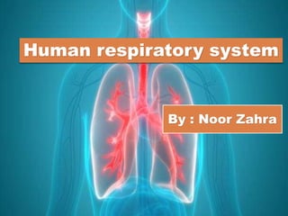
- 1. Human respiratory system By : Noor Zahra
- 2. Respiratory System Definition. “Human Respiratory System is the organ system that involves inhaling of oxygen and exhaling of carbon dioxide to meet the energy requirements.”
- 3. The Design Of The Respiratory System. The human gas-exchanging organ, the lung is located in the thorax, where its delicate tissues are protected by the bony and muscular thoracic cage. The lung provides the tissues of the human body with a continuous flow of oxygen and clears the blood of the gaseous waste product, carbon dioxide. Atmospheric air is pumped in and out regularly through a system of pipes, called conducting airways, which join the gas-exchange region with the outside of the body.
- 4. Division of airways. The airways can be divided into upper and lower airway systems. The transition between the two systems is located where the pathways of the respiratory and digestive systems cross, just at the top of the larynx.
- 6. Upper and lower Airways systems The upper airway system comprises the nose and the paranasal cavities (or sinuses), the pharynx (or throat), and partly also the oral cavity, since it may be used for breathing. The lower airway system consists of the larynx, the trachea, the stem bronchi, and all the airways ramifying intensively within the lungs, such as the intrapulmonary bronchi, the bronchioles, and the alveolar ducts.
- 7. For respiration, the collaboration of other organ systems is clearly essential. The diaphragm, as the main respiratory muscle, and the intercostal muscles of the chest wall play an essential role by generating, under the control of the central nervous system, the pumping action on the lung The muscles expand and contract the internal space of the thorax, the bony framework of which is formed by the ribs and the thoracic vertebrae.
- 8. The Mechanics of Breathing. The contribution of the lung and chest wall (ribs and muscles) to respiration is described below in The mechanics of breathing. The blood, as a carrier for the gases, and the circulatory system (i.e., the heart and the blood vessels) are mandatory elements of a working respiratory system ( blood; cardiovascular system).
- 9. Respiratory System parts. The lungs are the main part of the respiratory system. This system is divided into the upper respiratory tract and the lower respiratory tract. The upper respiratory tract includes the: Mouth and nose. Nasal cavity. Throat (pharynx). Voice box (larynx).
- 10. Respiratory System parts. The lower respiratory tract is made up of the: lungs trachea (windpipe) bronchi bronchioles alveoli Other parts of the respiratory system help your lungs to expand and contract as you breathe. These include the ribs around the lungs and the dome-shaped diaphragm muscle.
- 11. Respiratory System Parts and functions. Nose The nose is the external protuberance of an internal space, the nasal cavity. It is subdivided into a left and right canal by a thin medial cartilaginous and bony wall, the nasal septum. Each canal opens to the face by a nostril and into the pharynx by the choana. The floor of the nasal cavity is formed by the palate, which also forms the roof of the oral cavity. The complex shape of the nasal cavity is due to projections of bony ridges, the superior, middle, and inferior turbinate bones (or conchae), from the lateral wall. The passageways thus formed below each ridge are called the superior, middle, and inferior nasal meatuses.
- 12. On each side, the intranasal space communicates with a series of neighbouring air-filled cavities within the skull (the paranasal sinuses) and also, via the nasolacrimal duct, with the lacrimal apparatus in the corner of the eye. The duct drains the lacrimal fluid into the nasal cavity. The nasal cavity with its adjacent spaces is lined by a respiratory mucosa. Typically, the mucosa of the nose contains mucus-secreting glands and venous plexuses; its top cell layer, the epithelium, consists principally of two cell types, ciliated and secreting cells. They clean, moisten, and warm the inspired air, preparing it for intimate contact with the delicate tissues of the gas-exchange area.
- 13. Pharynx pharynx can be divided into three floors. The upper floor, the nasopharynx, is primarily a passageway for air and secretions from the nose to the oral pharynx. It is also connected to the tympanic cavity of the middle ear through the auditory tubes that open on both lateral walls. . When it is enlarged (as in tonsil hypertrophy or adenoid vegetation), it may interfere with nasal respiration and alter the resonance pattern of the voice
- 14. The middle floor of the pharynx connects anteriorly to the mouth and is therefore called the oral pharynx or oropharynx. It is delimited from the nasopharynx by the soft palate, which roofs the posterior part of the oral cavity. The lower floor of the pharynx is called the hypopharynx. Air from the nasal cavity flows into the larynx, and food from the oral cavity is routed to the esophagus directly behind the larynx. The epiglottis, a cartilaginous, leaf-shaped flap, functions as a lid to the larynx and, during the act of swallowing, controls the traffic of air and food.
- 15. Larynx The larynx is an organ of complex structure that serves a dual function: as an air canal to the lungs and a controller of its access, and as the organ of phonation. Two cartilaginous chords lay the framework for the larynx. They are situated at the point of joining the pharynx and trachea. It is also termed as Adam’s apple or the voice box. It is the portion which rises and falls duringSound is produced by forcing air through a sagittal slit formed by the vocal cords, the glottis. This causes not only the vocal cords but also the column of air above them to vibrate swallowing of food particles.
- 17. Trachea The trachea and the stem bronchi Below the larynx lies the trachea, a tube about 10 to 12 cm (3.9 to 4.7 inches) long and 2 cm (0.8 inch) wide. Its wall is stiffened by 16 to 20 characteristic horseshoe- shaped, incomplete cartilage rings that open toward the back and are embedded in a dense connective tissue.
- 18. Trachea The dorsal wall contains a strong layer of transverse smooth muscle fibres that spans the gap of the cartilage. The interior of the trachea is lined by the typical respiratory epithelium. The mucosal layer contains mucous glands. At its lower end, the trachea divides in an inverted Y into the two stem (or main) bronchi, one each for the left and right lung
- 19. Bronchi The trachea splits into two tubes termed as bronchi, which enter each lung individually. The bronchi divide into secondary, tertiary, and to bronchioles, which is again further divided into small air-sacs called the alveoli. The alveoli are minute sacs of air with thin walls and single-celled manner. It enables the exchange of oxygen and carbon dioxide molecules into or away from the bloodstream.
- 20. Lungs This spongy, pinkish organ looks like two upside-down cones in your chest. The right lung is made up of three lobes. The left lung has only two lobes to make room for your heart. Its superior region called the Apex projects superiorly to a point that is slightly superior and posterior to the clavicle. Both lungs are bordered by thoracic wall anteriorly, laterally, and posteriorly, and supported by the rib cage.
- 21. lungs Toward the midline, the lungs are seorated from each other by the Mediastinum. The relatively broad, rounded surface in contact with thoracic wall is called Coastal surface of the lung. Left Lung Divided into two lobes by oblique fissure Smaller than the right lung Cardiac notch accommodates the heart Right Lung Divided into three lobes by oblique and horizontal fissure. Located more superiorly in the body due to liver on right side.
- 22. Lung
- 23. Pleura The outer surface of each lung and adjacent internal thoracic wall are lined by a serous membrane Called Pleura. The outer surface of each lung is tightly covered by the Visceral pleura. While the internal thoracic walls, the lateral surface of the mediastinum, and superior surface of the diaphragm are lined up by the Parietal pleura. The parietal and visceral layers are continuous at the Hilus of each Lung.
- 24. Pleural cavity The potential space between the serous membrane is a Pleural Cavity. The pleural membrane produces thin, serous pleural fluid that circulates In the pleural cavity and acts as lubricant, ensuring minimal friction during breathing. Pleural effusion pleurisies with too much fluid.
- 25. The air that we inhale has the following composition Nitrogen – 78% Oxygen – 21% Carbon dioxide – 0.03 – 0.04% Traces of Hydrogen and Noble gases Air contains more oxygen than carbon dioxide. The air is inhaled with the help of nostrils, and in the nasal cavity, the air is cleansed by the fine hair follicles present within them. The cavity also has a collective group of blood vessels that keep the air warm. This air then passes to the pharynx, then to the larynx and into the trachea. Once the air reaches bronchus, it moves into the bronchioles, and then into the alveoli. From the alveoli, the formation of respiratory surfaces occurs in humans
- 26. Respiratory System Functions The human respiratory system functions are mentioned below: Inhalation and Exhalation The respiratory system helps in breathing, known as pulmonary ventilation. The air inhaled through the nose moves through the pharynx, larynx, and trachea into the lungs. The air is exhaled back through the same pathway. Changes in the volume and pressure in the lungs aid in pulmonary ventilation. Exchange of Gases between Lungs and Bloodstream Inside the lungs, the oxygen is exchanged for carbon dioxide waste through millions of microscopic sacs called alveoli. The inhaled oxygen diffuses into the pulmonary capillaries, binds to haemoglobin and is pumped through the bloodstream. The carbon dioxide from the blood diffuses into the alveoli and is expelled through exhalation.
- 27. Exchange of Gases between Bloodstream and Body Tissues The blood carries the oxygen from the lungs around the body and releases the oxygen when it reaches the capillaries. The oxygen is diffused through the capillary walls into the body tissues. The carbon dioxide also diffuses into the blood and is carried back to the lungs for release. The vibration of the Vocal Cords While speaking, the muscles in the larynx move the arytenoid cartilage. These cartilages push the vocal cords together. During exhalation, when the air passes through the vocal cords, it makes them vibrate and creates sound. Olfaction or Smelling During inhalation, when the air enters the nasal cavities, some chemicals present in the air bind to it and activate the receptors of the nervous system on the cilia. The signals are sent to the olfactory bulbs via the brain.
- 28. Breathing (or ventilation) is the process of moving air into and out of the lungs to facilitate gas exchange with the internal environment, mostly to bring in oxygen and flush out carbon dioxide.
- 29. Breathing mechanism Process of Inspiration Inspiration is the phase of ventilation in which air enters the lungs. It is initiated by contraction of the inspiratory muscles: Diaphragm – flattens, extending the superior/inferior dimension of the thoracic cavity. External intercostal muscles – elevates the ribs and sternum, extending the anterior/posterior dimension of the thoracic cavity. The action of the inspiratory muscles results in an increase in the volume of the thoracic cavity. As the lungs are held against the inner thoracic wall by the pleural seal, they also undergo an increase in volume.
- 30. Process of Passive Expiration Expiration is the phase of ventilation in which air is expelled from the lungs. It is initiated by relaxation of the inspiratory muscles: Diaphragm – relaxes to return to its resting position, reducing the superior/inferior dimension of the thoracic cavity. External intercostal muscles – relax to depress the ribs and sternum, reducing the anterior/posterior dimension of the thoracic cavity. The relaxation of the inspiratory muscles results in a decrease in the volume of the thoracic cavity. The elastic recoil of the previously expanded lung tissue allows them to return to their original size.
- 31. Forced Breathing Forced breathing is an active mode of breathing which utilises additional muscles to rapidly expand and contract the thoracic cavity volume. It most commonly occurs during exercise. Active Inspiration; involves in the contraction of the accessory muscles of breathing (in addition to those of quiet inspiration, the diaphragm and external intercostals). All of these muscles act to increase the volume of the thoracic cavity: Scalenes – elevates the upper ribs. Sternocleidomastoid – elevates the sternum. Pectoralis major and minor – pulls ribs outwards. Serratus anterior – elevates the ribs (when the scapulae are fixed). Latissimus dorsi – elevates the lower ribs.
- 32. Active Expiration Active expiration utilises the contraction of several thoracic and abdominal muscles. These muscles act to decrease the volume of the thoracic cavity: Anterolateral abdominal wall – increases the intra- abdominal pressure, pushing the diaphragm further upwards into the thoracic cavity. Internal intercostal – depresses the ribs. Innermost intercostal – depresses the ribs Variation in Breathing mechanism By Exercise By Sleep
- 33. Chemoreceptor A chemoreceptor, also known as chemosensor, is a specialized sensory receptor cell which transduces a chemical substance (endogenous or induced) to generate a biological signal. The main chemoreceptors involved in respiratory feedback are: Central chemoreceptors: These are located on the ventrolateral surface of medulla oblongata and detect changes in the pH of spinal fluid. They can be desensitized over time from chronic hypoxia (oxygen deficiency) and increased carbon dioxide
- 34. There are two kinds of Respiratory Chemoreceptor Arterial chemoreceptors: which monitor and respond to changes in the partial pressure of oxygen and carbon dioxide in the arterial blood. Central chemoreceptors in the brain, which respond to changes in the partial pressure of carbon dioxide in their immediate environment How do Chemoreceptors regulate respiration? The respiratory centers contain chemoreceptors that detect pH levels in the blood and send signals to the respiratory centers of the brain to adjust the ventilation rate to change acidity by increasing or decreasing the removal of carbon dioxide (since carbon dioxide is linked to higher levels of hydrogen ions in blood .