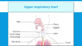
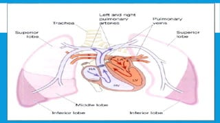
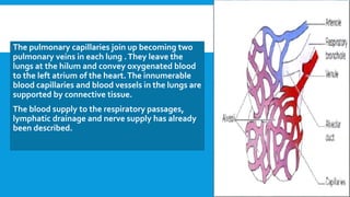
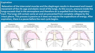
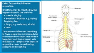
RESPIRATORY SYSTEM
Dr. Luma Alzamel
Upper respiratory tract
The nose
Respiratory function of the nose
 The nose is the first of the respiratory
passages through which the inspired air
passes.The function of the nose is to begin
the process by which the air is warmed,
moistened humidity and 'filtered’.
Olfactory function of the nose
 The nose is the organ of the sense of smell
by olfactory nerve.
Pharynx
 Functions
1. Passageway for air and food.The pharynx is an organ involved in both the
respiratory and the digestive systems: air passes through the nasal and oral parts,
and food through the oral and laryngeal parts.
2. Warming and humidifying.
3. Taste. By olfactory nerve.
4. Hearing.The auditory tube, extending from the nasal part and allows air to enter
the middle ear.
5. Protection.The lymphatic tissue of the pharyngeal and laryngeal tonsils produces
antibodies in response to antigens.
6. Speech.
Larynx
 Function
1. Production of sound. Sound has the
properties of pitch, volume and resonance.
2. Speech.This occurs during expiration when
the sounds produced by the vocal cords are
manipulated by the tongue, cheeks and lips.
3. Protection of the lower respiratory tract.
4. Passageway for air.This is between the
pharynx and trachea.
5. Humidifying, filtering and warming.These
continue as inspired air travels through the
larynx.
Trachea
 Functions
1. Support and patency.The arrangement
of cartilage and elastic tissue prevents
kinking and obstruction of the airway as
the head and neck move.
2. Cough reflex. Nerve endings in the
larynx, trachea and bronchi are sensitive
to irritation.
3. Warming, humidifying and filtering of
air.These continue as in the nose,
although air is normally saturated and
at body temperature when it reaches
the trachea.
Lower respiratory track
Air passages
 Functions of air passages not involved in gaseous exchange. Control of air
entry.The diameter of the respiratory passages may be altered by contraction
or relaxation of the involuntary muscles in their walls, thus regulating the
volume of air entering the lungs.These changes are controlled by the
autonomic nerve supply: parasympathetic stimulation causes constriction
and sympathetic stimulation causes dilatation.
 The following functions continue as in the upper airways:
• warming and humidifying
• support and patency
• removal of particulate matter
• cough reflex
Functions of respiratory bronchioles and alveoli
1. External respiration.
2. Defense against microbes.
ciliated epithelium, goblet cells
and mucus are present.
3. Warming and humidifying.These
continue as in the upper airways.
Lungs
Interior of the lungs
The lungs are composed of the bronchi and smaller air passages, alveoli,
connective tissue, blood vessels, lymph vessels and nerves.The left lung is
divided into two lobes and the right, into three. Each lobe is made up of many
lobules.
Pulmonary blood supply
The pulmonary artery divides into two, one branch conveying deoxygenated
blood to each lung. Within the lungs each pulmonary artery divides into many
branches which eventually end in a dense capillary network around the walls of
the alveoli.The walls of the alveoli and those of the capillaries each consist of
only one layer of flattened epithelial cells.The exchange of gases between air in
the alveoli and blood in the capillaries takes place across these two very fine
membranes.
The pulmonary capillaries join up becoming two
pulmonary veins in each lung .They leave the
lungs at the hilum and convey oxygenated blood
to the left atrium of the heart.The innumerable
blood capillaries and blood vessels in the lungs are
supported by connective tissue.
The blood supply to the respiratory passages,
lymphatic drainage and nerve supply has already
been described.
CYCLE OF RESPIRATION
This occurs 12 to 15 times per minute and consists of three phases:
• inspiration
• expiration
• pause.
Inspiration
When the capacity of the thoracic cavity is increased by simultaneous contraction of
the intercostal muscles and the diaphragm, the parietal pleura moves with the walls of
the thorax and the diaphragm.This reduces the pressure in the pleural cavity to a level
considerably lower than atmospheric pressure.The visceral pleura follows the parietal
pleura pulling the lung with it.The process of inspiration is active, as it requires
expenditure of energy for muscle contraction.The negative pressure created in the
thoracic cavity aids venous return to the heart and is known as the respiratory pump.
Expiration
Relaxation of the intercostal muscles and the diaphragm results in downward and inward
movement of the rib cage and elastic recoil of the lungs. As this occurs, pressure inside the
lungs exceeds that in the atmosphere and therefore air is expelled from the respiratory
tract.The lungs still contain some air and are prevented from complete collapse by the
intact pleura.This process is passive as it does not require the expenditure of energy. After
expiration, there is a pause before the next cycle begins.
DIFFUSION OF GASES
Exchange of gases occurs when a difference in partial pressure exists across semipermeable
membranes. Gases move by diffusion from the higher concentration to the lower until
equilibrium is established. Atmospheric nitrogen is not used by the body, so its partial pressure
remains unchanged and is the same in inspired and expired air, alveolar air and in the blood.
External respiration
This is exchange of gases by diffusion between the alveoli and the blood. Each alveolar wall is
one cell thick and is surrounded by a network of tiny capillaries (the walls of which are also only
one cell thick).The total area for gas exchange in the lungs is 70 to 80 square meters.Venous
blood arriving at the lungs has travelled from all the active tissues of the body and contains high
levels of CO2 and low levels of O2. Carbon dioxide diffuses from venous blood down its
concentration gradient into the alveoli until equilibrium with alveolar air is reached. By the same
process, oxygen diffuses from the alveoli into the blood.The slow flow of blood through the
capillaries increases the time available for diffusion to occur. When blood leaves the alveolar
capillaries, the oxygen and carbon dioxide concentrations are in equilibrium with those of
alveolar air.
Internal respiration
This is exchange of gases by diffusion between blood in
the capillaries and the body cells. Gaseous exchange
does not occur across the walls of the arteries carrying
blood from the heart to the tissues, because their walls
are too thick. PO2 of blood arriving at the capillary bed
is therefore the same as blood leaving the lungs. Blood
arriving at the tissues has been cleansed of its CO2 and
saturated with O2 during its passage through the lungs,
and therefore has a higher PO2 and a lower PCO2 than
the tissues.
This creates concentration gradients between the blood
and the tissues, and gaseous exchange therefore
occurs. O2 diffuses from the bloodstream through the
capillary wall into the tissues. CO2 diffuses from the
cells into the extracellular fluid then into the
bloodstream towards the venous end of the capillary.
Oxygen
Oxygen is carried in the blood:
• in chemical combination with haemoglobin as oxyhaemoglobin (98.5% of blood O2)
• in solution in plasma water (1.5% of blood O2).
Oxyhaemoglobin is an unstable compound that under certain conditions readily dissociates
releasing oxygen. Factors that increase dissociation include raised carbon dioxide content of
tissue fluid and raised temperature. In active tissues there is increased production of carbon
dioxide and heat which leads to increased release of oxygen. In this way oxygen is available to
tissues in greatest need.When oxygen leaves the erythrocyte, the deoxygenated haemoglobin
turns purplish in color.
Carbon dioxide
Carbon dioxide is one of the waste products of metabolism. It is excreted by the lungs and is
transported by three mechanisms:
• most of it is in the form of bicarbonate ions (HCO3) in the plasma (70% of blood CO2)
• some is dissolved in the plasma (7% of blood CO2)
• some is carried in erythrocytes, loosely combined with haemoglobin as
carbaminohaemoglobin (23% of blood CO2).
CONTROL OF RESPIRATION
Control of respiration is normally involuntary.Voluntary control is exerted during
activities such as speaking and singing but is overridden if blood CO2 rises (hypercapnia).
The respiratory center
This is formed by groups of nerve cells that control the rate and depth of respiration (Fig.
10.25).They are situated in the brain stem, in the medulla oblongata and the pons.The
interrelationship between these groups of cells is complex. In the medulla there are
inspiratory neurons and expiratory neurons. Neurons in the pneumotaxic and apneustic
centers, situated in the pons, influence the inspiratory and expiratory neurons of the
medulla.
Motor impulses leaving the respiratory center pass in the phrenic and intercostal nerves
to the diaphragm and intercostal muscles, respectively.
Chemoreceptors
These are receptors that respond to changes in the partial pressures of oxygen and carbon
dioxide in the blood and cerebrospinal fluid.They are located centrally and peripherally.
Central chemoreceptors.These are on the surface of the medulla oblongata and are bathed
in cerebrospinal fluid. When the arterial PCO2 rises (hypercapnia), even slightly, the central
chemoreceptors respond by stimulating the respiratory center, increasing ventilation of
the lungs and reducing arterial PCO2.The sensitivity of the central chemoreceptors to
raised arterial PCO2 is the most important factor in maintaining homeostasis of blood
gases in health. A small reduction in PO2 (hypoxaemia) has the same, but less pronounced
effect, but a substantial reduction has a depressing effect.
Peripheral chemoreceptors. These are situated in the arch of the aorta and in the carotid
bodies.They are more sensitive to small rises in arterial PCO2 than to similarly low arterial
PO2 levels. Nerve impulses, generated in the peripheral chemoreceptors, are conveyed by
the glossopharyngeal and vagus nerves to the medulla and stimulate the respiratory
center.The rate and depth of breathing are then increased. An increase in blood acidity
(decreased pH or raised [H+]) stimulates the peripheral chemoreceptors, resulting in
increased ventilation, increased CO2 excretion and increased blood pH.
Other factors that influence
respiration
Breathing may be modified by the
higher centers in the brain by:
• speech, singing
• emotional displays, e.g. crying,
laughing, fear
• drugs, e.g. sedatives, alcohol
• sleep.
Temperature influences breathing.
In fever respiration is increased due
to increased metabolic rate while in
hypothermia it is depressed, as is
metabolism.Temporary changes in
respiration occur in swallowing,
sneezing and coughing.
GOOD Luck

Respiratory system.ppsx

  • 1.
  • 2.
  • 3.
    The nose Respiratory functionof the nose  The nose is the first of the respiratory passages through which the inspired air passes.The function of the nose is to begin the process by which the air is warmed, moistened humidity and 'filtered’. Olfactory function of the nose  The nose is the organ of the sense of smell by olfactory nerve.
  • 4.
    Pharynx  Functions 1. Passagewayfor air and food.The pharynx is an organ involved in both the respiratory and the digestive systems: air passes through the nasal and oral parts, and food through the oral and laryngeal parts. 2. Warming and humidifying. 3. Taste. By olfactory nerve. 4. Hearing.The auditory tube, extending from the nasal part and allows air to enter the middle ear. 5. Protection.The lymphatic tissue of the pharyngeal and laryngeal tonsils produces antibodies in response to antigens. 6. Speech.
  • 5.
    Larynx  Function 1. Productionof sound. Sound has the properties of pitch, volume and resonance. 2. Speech.This occurs during expiration when the sounds produced by the vocal cords are manipulated by the tongue, cheeks and lips. 3. Protection of the lower respiratory tract. 4. Passageway for air.This is between the pharynx and trachea. 5. Humidifying, filtering and warming.These continue as inspired air travels through the larynx.
  • 6.
    Trachea  Functions 1. Supportand patency.The arrangement of cartilage and elastic tissue prevents kinking and obstruction of the airway as the head and neck move. 2. Cough reflex. Nerve endings in the larynx, trachea and bronchi are sensitive to irritation. 3. Warming, humidifying and filtering of air.These continue as in the nose, although air is normally saturated and at body temperature when it reaches the trachea.
  • 7.
  • 8.
    Air passages  Functionsof air passages not involved in gaseous exchange. Control of air entry.The diameter of the respiratory passages may be altered by contraction or relaxation of the involuntary muscles in their walls, thus regulating the volume of air entering the lungs.These changes are controlled by the autonomic nerve supply: parasympathetic stimulation causes constriction and sympathetic stimulation causes dilatation.  The following functions continue as in the upper airways: • warming and humidifying • support and patency • removal of particulate matter • cough reflex
  • 9.
    Functions of respiratorybronchioles and alveoli 1. External respiration. 2. Defense against microbes. ciliated epithelium, goblet cells and mucus are present. 3. Warming and humidifying.These continue as in the upper airways.
  • 10.
    Lungs Interior of thelungs The lungs are composed of the bronchi and smaller air passages, alveoli, connective tissue, blood vessels, lymph vessels and nerves.The left lung is divided into two lobes and the right, into three. Each lobe is made up of many lobules. Pulmonary blood supply The pulmonary artery divides into two, one branch conveying deoxygenated blood to each lung. Within the lungs each pulmonary artery divides into many branches which eventually end in a dense capillary network around the walls of the alveoli.The walls of the alveoli and those of the capillaries each consist of only one layer of flattened epithelial cells.The exchange of gases between air in the alveoli and blood in the capillaries takes place across these two very fine membranes.
  • 12.
    The pulmonary capillariesjoin up becoming two pulmonary veins in each lung .They leave the lungs at the hilum and convey oxygenated blood to the left atrium of the heart.The innumerable blood capillaries and blood vessels in the lungs are supported by connective tissue. The blood supply to the respiratory passages, lymphatic drainage and nerve supply has already been described.
  • 13.
    CYCLE OF RESPIRATION Thisoccurs 12 to 15 times per minute and consists of three phases: • inspiration • expiration • pause. Inspiration When the capacity of the thoracic cavity is increased by simultaneous contraction of the intercostal muscles and the diaphragm, the parietal pleura moves with the walls of the thorax and the diaphragm.This reduces the pressure in the pleural cavity to a level considerably lower than atmospheric pressure.The visceral pleura follows the parietal pleura pulling the lung with it.The process of inspiration is active, as it requires expenditure of energy for muscle contraction.The negative pressure created in the thoracic cavity aids venous return to the heart and is known as the respiratory pump.
  • 14.
    Expiration Relaxation of theintercostal muscles and the diaphragm results in downward and inward movement of the rib cage and elastic recoil of the lungs. As this occurs, pressure inside the lungs exceeds that in the atmosphere and therefore air is expelled from the respiratory tract.The lungs still contain some air and are prevented from complete collapse by the intact pleura.This process is passive as it does not require the expenditure of energy. After expiration, there is a pause before the next cycle begins.
  • 15.
    DIFFUSION OF GASES Exchangeof gases occurs when a difference in partial pressure exists across semipermeable membranes. Gases move by diffusion from the higher concentration to the lower until equilibrium is established. Atmospheric nitrogen is not used by the body, so its partial pressure remains unchanged and is the same in inspired and expired air, alveolar air and in the blood. External respiration This is exchange of gases by diffusion between the alveoli and the blood. Each alveolar wall is one cell thick and is surrounded by a network of tiny capillaries (the walls of which are also only one cell thick).The total area for gas exchange in the lungs is 70 to 80 square meters.Venous blood arriving at the lungs has travelled from all the active tissues of the body and contains high levels of CO2 and low levels of O2. Carbon dioxide diffuses from venous blood down its concentration gradient into the alveoli until equilibrium with alveolar air is reached. By the same process, oxygen diffuses from the alveoli into the blood.The slow flow of blood through the capillaries increases the time available for diffusion to occur. When blood leaves the alveolar capillaries, the oxygen and carbon dioxide concentrations are in equilibrium with those of alveolar air.
  • 16.
    Internal respiration This isexchange of gases by diffusion between blood in the capillaries and the body cells. Gaseous exchange does not occur across the walls of the arteries carrying blood from the heart to the tissues, because their walls are too thick. PO2 of blood arriving at the capillary bed is therefore the same as blood leaving the lungs. Blood arriving at the tissues has been cleansed of its CO2 and saturated with O2 during its passage through the lungs, and therefore has a higher PO2 and a lower PCO2 than the tissues. This creates concentration gradients between the blood and the tissues, and gaseous exchange therefore occurs. O2 diffuses from the bloodstream through the capillary wall into the tissues. CO2 diffuses from the cells into the extracellular fluid then into the bloodstream towards the venous end of the capillary.
  • 17.
    Oxygen Oxygen is carriedin the blood: • in chemical combination with haemoglobin as oxyhaemoglobin (98.5% of blood O2) • in solution in plasma water (1.5% of blood O2). Oxyhaemoglobin is an unstable compound that under certain conditions readily dissociates releasing oxygen. Factors that increase dissociation include raised carbon dioxide content of tissue fluid and raised temperature. In active tissues there is increased production of carbon dioxide and heat which leads to increased release of oxygen. In this way oxygen is available to tissues in greatest need.When oxygen leaves the erythrocyte, the deoxygenated haemoglobin turns purplish in color. Carbon dioxide Carbon dioxide is one of the waste products of metabolism. It is excreted by the lungs and is transported by three mechanisms: • most of it is in the form of bicarbonate ions (HCO3) in the plasma (70% of blood CO2) • some is dissolved in the plasma (7% of blood CO2) • some is carried in erythrocytes, loosely combined with haemoglobin as carbaminohaemoglobin (23% of blood CO2).
  • 18.
    CONTROL OF RESPIRATION Controlof respiration is normally involuntary.Voluntary control is exerted during activities such as speaking and singing but is overridden if blood CO2 rises (hypercapnia). The respiratory center This is formed by groups of nerve cells that control the rate and depth of respiration (Fig. 10.25).They are situated in the brain stem, in the medulla oblongata and the pons.The interrelationship between these groups of cells is complex. In the medulla there are inspiratory neurons and expiratory neurons. Neurons in the pneumotaxic and apneustic centers, situated in the pons, influence the inspiratory and expiratory neurons of the medulla. Motor impulses leaving the respiratory center pass in the phrenic and intercostal nerves to the diaphragm and intercostal muscles, respectively.
  • 19.
    Chemoreceptors These are receptorsthat respond to changes in the partial pressures of oxygen and carbon dioxide in the blood and cerebrospinal fluid.They are located centrally and peripherally. Central chemoreceptors.These are on the surface of the medulla oblongata and are bathed in cerebrospinal fluid. When the arterial PCO2 rises (hypercapnia), even slightly, the central chemoreceptors respond by stimulating the respiratory center, increasing ventilation of the lungs and reducing arterial PCO2.The sensitivity of the central chemoreceptors to raised arterial PCO2 is the most important factor in maintaining homeostasis of blood gases in health. A small reduction in PO2 (hypoxaemia) has the same, but less pronounced effect, but a substantial reduction has a depressing effect. Peripheral chemoreceptors. These are situated in the arch of the aorta and in the carotid bodies.They are more sensitive to small rises in arterial PCO2 than to similarly low arterial PO2 levels. Nerve impulses, generated in the peripheral chemoreceptors, are conveyed by the glossopharyngeal and vagus nerves to the medulla and stimulate the respiratory center.The rate and depth of breathing are then increased. An increase in blood acidity (decreased pH or raised [H+]) stimulates the peripheral chemoreceptors, resulting in increased ventilation, increased CO2 excretion and increased blood pH.
  • 20.
    Other factors thatinfluence respiration Breathing may be modified by the higher centers in the brain by: • speech, singing • emotional displays, e.g. crying, laughing, fear • drugs, e.g. sedatives, alcohol • sleep. Temperature influences breathing. In fever respiration is increased due to increased metabolic rate while in hypothermia it is depressed, as is metabolism.Temporary changes in respiration occur in swallowing, sneezing and coughing.
  • 21.