Student’s Prayer (St.Thomas
Aquinas)
Creatorof all things, true source of light
and wisdom, origin of all being,
graciously let a ray of Your brilliance
penetrate the darkness of my
understanding.
Take from me the double darkness in
which I have been born, sin and
ignorance.
2.
Give me akeen understanding, a retentive
memory, and the ability to grasp things
correctly and fundamentally.
Grant me the talent of being exact in my
explanations and the ability to express myself
with thoroughness and charm.
Point out the beginning, direct the progress,
and aid in the completion. I ask through Christ
Jesus, our Lord, AMEN.
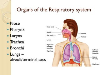
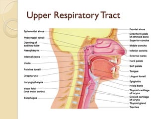
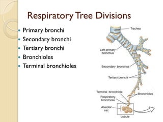
Organs of theRespiratory system
— Nose
— Pharynx
— Larynx
— Trachea
— Bronchi
— Lungs –
alveoli/terminal sacs
5.
Function of theRespiratory System
— Oversees gas exchanges between the
blood and external environment
— Exchange of gasses takes place within the
lungs in the alveoli
— Passageways to the lungs purify, warm, and
humidify the incoming air
6.
The Nose
— Theonly externally visible part of the
respiratory system
— Air enters the nose through the external
nares (nostrils)
— The interior of the nose consists of a
nasal cavity divided by a nasal septum
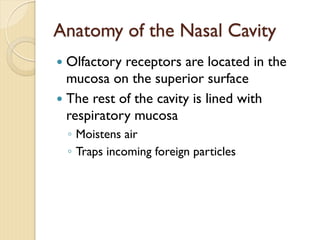
Anatomy of theNasal Cavity
— Olfactory receptors are located in the
mucosa on the superior surface
— The rest of the cavity is lined with
respiratory mucosa
◦ Moistens air
◦ Traps incoming foreign particles
9.
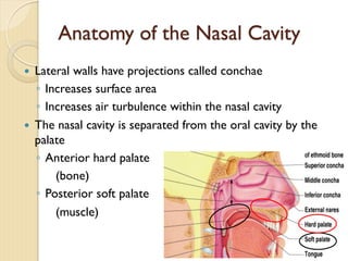
Anatomy of theNasal Cavity
— Lateral walls have projections called conchae
◦ Increases surface area
◦ Increases air turbulence within the nasal cavity
— The nasal cavity is separated from the oral cavity by the
palate
◦ Anterior hard palate
(bone)
◦ Posterior soft palate
(muscle)
10.
— The geneticdefect cleft palate (failure of
the bones forming the palate to fuse
medially) results in breathing difficulty as
well as problems with oral cavity
functions such as chewing and speaking.
11.
Paranasal Sinuses
— Cavitieswithin bones surrounding the
nasal cavity
◦ Frontal bone
◦ Sphenoid bone
◦ Ethmoid bone
◦ Maxillary bone
12.
Paranasal Sinuses
— Functionof the sinuses
◦ Lighten the skull
◦ Act as resonance chambers for speech
◦ Produce mucus that drains into the nasal
cavity
13.
— Cold virusesand various allergens can
cause rhinitis – inflammation of the nasal
mucosa.
◦ The excessive mucus produced results in
nasal congestion and postnasal drip.
— Sinusitis or sinus inflammation
14.
Pharynx (Throat)
— Muscularpassage from nasal cavity to larynx
— Three regions of the pharynx
◦ Nasopharynx – superior region behind nasal
cavity
◦ Oropharynx – middle region behind mouth
◦ Laryngopharynx – inferior region attached to
larynx
— The oropharynx and laryngopharynx are
common passageways for air and food
15.
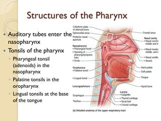
Structures of thePharynx
— Auditory tubes enter the
nasopharynx
— Tonsils of the pharynx
◦ Pharyngeal tonsil
(adenoids) in the
nasopharynx
◦ Palatine tonsils in the
oropharynx
◦ Lingual tonsils at the base
of the tongue
16.
— Tonsillitis
◦ Ifthe pharyngeal tonsil becomes inflamed and
swollen (as during a bacterial infection), it
obstructs the nasopharynx and forces the
person to breathe through the mouth.
17.
Larynx (Voice Box)
—Routes air and food into proper channels
— Plays a role in speech
— Made of eight rigid hyaline cartilages and a
spoon-shaped flap of elastic cartilage
(epiglottis)
18.
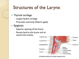
Structures of theLarynx
— Thyroid cartilage
◦ Largest hyaline cartilage
◦ Protrudes anteriorly (Adam’s apple)
— Epiglottis
◦ Superior opening of the larynx
◦ Routes food to the larynx and air
toward the trachea
19.
Structures of theLarynx
— Vocal cords (vocal folds)
◦ Vibrate with expelled air to create sound
(speech)
— Glottis – opening between vocal cords
20.
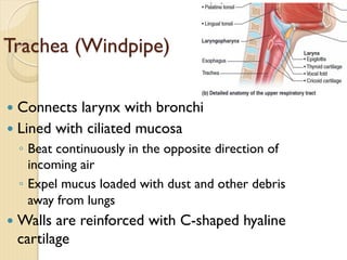
Trachea (Windpipe)
— Connectslarynx with bronchi
— Lined with ciliated mucosa
◦ Beat continuously in the opposite direction of
incoming air
◦ Expel mucus loaded with dust and other debris
away from lungs
— Walls are reinforced with C-shaped hyaline
cartilage
21.
Main (Primary) Bronchi
—Formed by division of the trachea
— Enters the lung at the hilus
(medial depression)
— Right bronchus is wider, shorter,
and straighter than left
— Bronchi subdivide into smaller
and smaller branches
22.
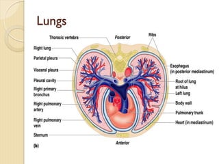
Lungs
— Occupy mostof the thoracic cavity
◦ Apex is near the clavicle (superior portion)
– Base rests on the diaphragm (inferior portion)
◦ Each lung is divided into lobes by fissures
– Left lung – two lobes
– Right lung – three lobes
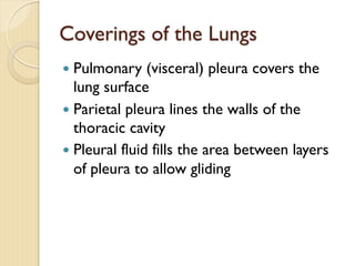
Coverings of theLungs
— Pulmonary (visceral) pleura covers the
lung surface
— Parietal pleura lines the walls of the
thoracic cavity
— Pleural fluid fills the area between layers
of pleura to allow gliding
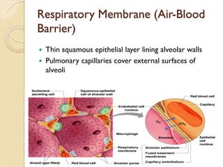
Gas Exchange
— Gascrosses the respiratory membrane by
diffusion
◦ Oxygen enters the blood
◦ Carbon dioxide enters the alveoli
— Macrophages add protection
— Surfactant coats gas-exposed alveolar
surfaces
30.
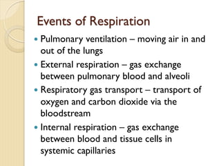
Events of Respiration
—Pulmonary ventilation – moving air in and
out of the lungs
— External respiration – gas exchange
between pulmonary blood and alveoli
— Respiratory gas transport – transport of
oxygen and carbon dioxide via the
bloodstream
— Internal respiration – gas exchange
between blood and tissue cells in
systemic capillaries
31.
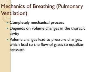
Mechanics of Breathing(Pulmonary
Ventilation)
— Completely mechanical process
— Depends on volume changes in the thoracic
cavity
— Volume changes lead to pressure changes,
which lead to the flow of gases to equalize
pressure
32.
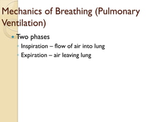
Mechanics of Breathing(Pulmonary
Ventilation)
— Two phases
◦ Inspiration – flow of air into lung
◦ Expiration – air leaving lung
33.
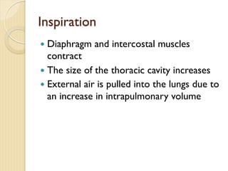
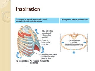
Inspiration
— Diaphragm andintercostal muscles
contract
— The size of the thoracic cavity increases
— External air is pulled into the lungs due to
an increase in intrapulmonary volume
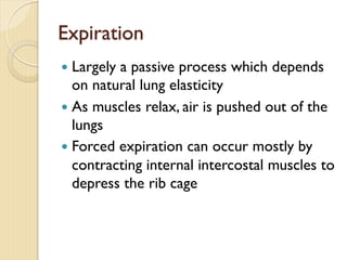
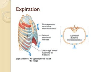
Expiration
— Largely apassive process which depends
on natural lung elasticity
— As muscles relax, air is pushed out of the
lungs
— Forced expiration can occur mostly by
contracting internal intercostal muscles to
depress the rib cage
Pressure Differences inthe Thoracic
Cavity
— Normal pressure within the pleural space is
always negative (intrapleural pressure)
— Differences in lung and pleural space
pressures keep lungs from collapsing
38.
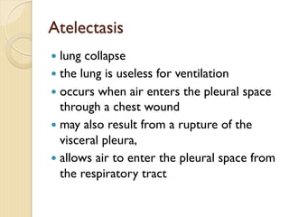
Atelectasis
— lung collapse
—the lung is useless for ventilation
— occurs when air enters the pleural space
through a chest wound
— may also result from a rupture of the
visceral pleura,
— allows air to enter the pleural space from
the respiratory tract
39.
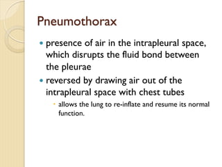
Pneumothorax
— presence ofair in the intrapleural space,
which disrupts the fluid bond between
the pleurae
— reversed by drawing air out of the
intrapleural space with chest tubes
– allows the lung to re-inflate and resume its normal
function.
RespiratoryVolumes and Capacities
•Normal breathing moves about 500 ml of air with
each breath (tidal volume [TV])
• Many factors that affect respiratory capacity
• A person’s size
• Sex
• Age
• Physical condition
• Residual volume of air – after exhalation, about
1200 ml of air remains in the lungs
42.
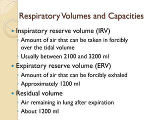
RespiratoryVolumes and Capacities
—Inspiratory reserve volume (IRV)
◦ Amount of air that can be taken in forcibly
over the tidal volume
◦ Usually between 2100 and 3200 ml
— Expiratory reserve volume (ERV)
◦ Amount of air that can be forcibly exhaled
◦ Approximately 1200 ml
— Residual volume
◦ Air remaining in lung after expiration
◦ About 1200 ml
43.
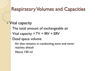
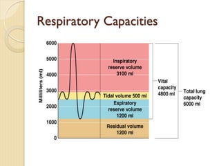
RespiratoryVolumes and Capacities
—Vital capacity
◦ The total amount of exchangeable air
◦ Vital capacity = TV + IRV + ERV
◦ Dead space volume
– Air that remains in conducting zone and never
reaches alveoli
– About 150 ml
44.
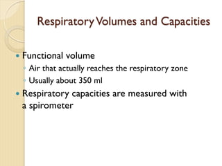
RespiratoryVolumes and Capacities
—Functional volume
◦ Air that actually reaches the respiratory zone
◦ Usually about 350 ml
— Respiratory capacities are measured with
a spirometer
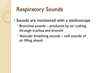
Respiratory Sounds
— Soundsare monitored with a stethoscope
◦ Bronchial sounds – produced by air rushing
through trachea and bronchi
◦ Vesicular breathing sounds – soft sounds of
air filling alveoli
47.
Abnormal Breath Sounds
—Diseased respiratory tissue, mucus, or pus
can produce abnormal sounds
◦ Crackle - a bubbling sound
◦ Wheezing - a whistling sound
◦ Rales - abnormal bronchial sounds produced
by the presence of mucus or exudate in the
lung passages or by thickening of the
bronchial walls
External Respiration
— Oxygenmovement into the blood
◦ The alveoli always has more oxygen than the
blood
◦ Oxygen moves by diffusion towards the area
of lower concentration
◦ Pulmonary capillary blood gains oxygen
50.
External Respiration
— Carbondioxide movement out of the
blood
◦ Blood returning from tissues has higher
concentrations of carbon dioxide than air in
the alveoli
◦ Pulmonary capillary blood gives up carbon
dioxide
— Blood leaving the lungs is oxygen-rich and
carbon dioxide-poor
51.
Gas Transport inthe Blood
— Oxygen transport in the blood
◦ Inside red blood cells attached to hemoglobin
(oxyhemoglobin [HbO2])
◦ A small amount is carried dissolved in the
plasma
52.
Gas Transport inthe Blood
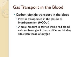
— Carbon dioxide transport in the blood
◦ Most is transported in the plasma as
bicarbonate ion (HCO3–)
◦ A small amount is carried inside red blood
cells on hemoglobin, but at different binding
sites than those of oxygen
53.
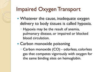
Impaired Oxygen Transport
—Whatever the cause, inadequate oxygen
delivery to body tissues is called hypoxia.
◦ Hypoxia may be the result of anemia,
pulmonary disease, or impaired or blocked
blood circulation.
— Carbon monoxide poisoning
◦ Carbon monoxide (CO) - odorless, colorless
gas that competes vigorously with oxygen for
the same binding sites on hemoglobin.
54.
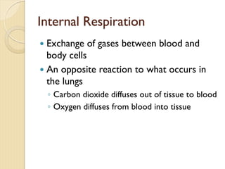
Internal Respiration
— Exchangeof gases between blood and
body cells
— An opposite reaction to what occurs in
the lungs
◦ Carbon dioxide diffuses out of tissue to blood
◦ Oxygen diffuses from blood into tissue
55.
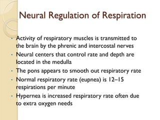
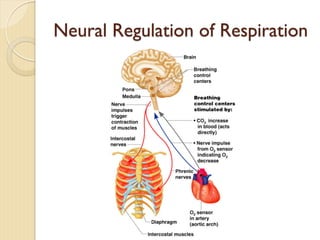
Neural Regulation ofRespiration
• Activity of respiratory muscles is transmitted to
the brain by the phrenic and intercostal nerves
• Neural centers that control rate and depth are
located in the medulla
• The pons appears to smooth out respiratory rate
• Normal respiratory rate (eupnea) is 12–15
respirations per minute
• Hypernea is increased respiratory rate often due
to extra oxygen needs
Factors Influencing RespiratoryRate
and Depth
— Physical factors
◦ Increased body temperature
◦ Exercise
◦ Talking
◦ Coughing
— Volition (conscious control)
— Emotional factors
58.
— Chemical factors
◦Carbon dioxide levels
– Level of carbon dioxide in the blood is the main
regulatory chemical for respiration
– Increased carbon dioxide increases respiration
– Changes in carbon dioxide act directly on the medulla
oblongata
Factors Influencing Respiratory Rate
and Depth
59.
— Chemical factors
◦Oxygen levels
– Changes in oxygen concentration in the blood are
detected by chemoreceptors in the aorta and carotid
artery
– Information is sent to the medulla oblongata
Factors Influencing Respiratory Rate
and Depth
60.
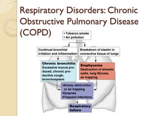
Respiratory Disorders: Chronic
ObstructivePulmonary Disease
(COPD)
— Exemplified by chronic bronchitis and
emphysema
— Major causes of death and disability in the
United States
61.
— Features ofthese diseases
◦ Patients almost always have a history of smoking
◦ Labored breathing (dyspnea) becomes
progressively more severe
◦ Coughing and frequent pulmonary infections are
common
Respiratory Disorders: Chronic
Obstructive Pulmonary Disease
(COPD)
62.
— Features ofthese diseases
◦ Most victimes retain carbon dioxide, are hypoxic
and have respiratory acidosis
◦ Those infected will ultimately develop respiratory
failure
Respiratory Disorders: Chronic
Obstructive Pulmonary Disease
(COPD)
63.
Emphysema
— Alveoli enlargeas adjacent chambers break through
— Chronic inflammation promotes lung fibrosis
— Airways collapse during expiration
— Patients use a large amount of energy to exhale
— Overinflation of the lungs leads to a permanently
expanded barrel chest
— Cyanosis appears late in the disease
64.
Chronic Bronchitis
— Mucosaof the lower respiratory passages
becomes severely inflamed
— Mucus production increases
— Pooled mucus impairs ventilation and gas
exchange
— Risk of lung infection increases
— Pneumonia is common
— Hypoxia and cyanosis occur early
Lung Cancer
— Accountsfor 1/3 of all cancer deaths in
the United States
— Increased incidence associated with
smoking
— Three common types
◦ Squamous cell carcinoma
◦ Adenocarcinoma
◦ Small cell carcinoma
67.
Sudden Infant Deathsyndrome
(SIDS)
— Apparently healthy infant stops breathing
and dies during sleep
— Some cases are thought to be a problem
of the neural respiratory control center
— One third of cases appear to be due to
heart rhythm abnormalities
68.
Asthma
— Chronic inflamedhypersensitive
bronchiole passages
— Response to irritants with dyspnea,
coughing, and wheezing
69.
Developmental Aspects ofthe
Respiratory System
— Lungs are filled with fluid in the fetus
— Lungs are not fully inflated with air until two
weeks after birth
— Surfactant that lowers alveolar surface
tension is not present until late in fetal
development and may not be present in
premature babies
70.
— Important birthdefects
◦ Cystic fibrosis – oversecretion of thick mucus
clogs the respiratory system
◦ Cleft palate
Developmental Aspects of the
Respiratory System
71.
Aging Effects
— Elasticityof lungs decreases
— Vital capacity decreases
— Blood oxygen levels decrease
— Stimulating effects of carbon dioxide
decreases
— More risks of respiratory tract infection
72.
Respiratory Rate ChangesThroughout
Life
— Newborns – 40 to 80 respirations per
minute
— Infants – 30 respirations per minute
— Age 5 – 25 respirations per minute
— Adults – 12 to 18 respirations per minute
— Rate often increases somewhat with old age


![RespiratoryVolumes and Capacities
• Normal breathing moves about 500 ml of air with
each breath (tidal volume [TV])
• Many factors that affect respiratory capacity
• A person’s size
• Sex
• Age
• Physical condition
• Residual volume of air – after exhalation, about
1200 ml of air remains in the lungs](https://image.slidesharecdn.com/respiratory1-250325143049-de50d360/85/Respiratory-System-Anatomy-and-Physiology-41-320.jpg)
![Gas Transport in the Blood
— Oxygen transport in the blood
◦ Inside red blood cells attached to hemoglobin
(oxyhemoglobin [HbO2])
◦ A small amount is carried dissolved in the
plasma](https://image.slidesharecdn.com/respiratory1-250325143049-de50d360/85/Respiratory-System-Anatomy-and-Physiology-51-320.jpg)


![Lesson6 [2 Oth Oct 2008]](https://cdn.slidesharecdn.com/ss_thumbnails/lesson62othoct2008-091022023449-phpapp01-thumbnail.jpg?width=640&height=640&fit=bounds)


![CTEV [ clubfoot] DR ARUN LAL ,DR MOHAMED ASHRAF travancore medical college k...](https://cdn.slidesharecdn.com/ss_thumbnails/ctevclubfootdrarunlaldrmohamedashraftravancoremedicalcollegekollamkeralaindia-260208063247-18fc466c-thumbnail.jpg?width=640&height=640&fit=bounds)

![ONFH[AVN HIP] -TRIPLE REGIME -A NOVAL SURGICAL CONCEPT .pptx](https://cdn.slidesharecdn.com/ss_thumbnails/onfhavnhip2026koaconcalicutdrgokuldevdrmashraf-260210064517-213ec005-thumbnail.jpg?width=640&height=640&fit=bounds)


![PERI-PROSTHETIC FRACTURE NAIL-PLATE CONSTRUCT [NPC].pptx](https://cdn.slidesharecdn.com/ss_thumbnails/drarunkumardrmohamedashrafperiprostheticfrasturenail-plateconstructnpc-260209164459-7e9d15a1-thumbnail.jpg?width=640&height=640&fit=bounds)