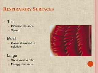
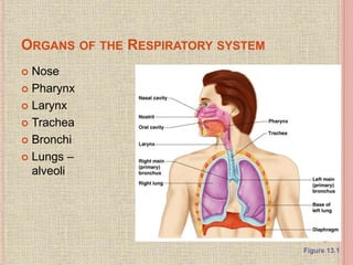
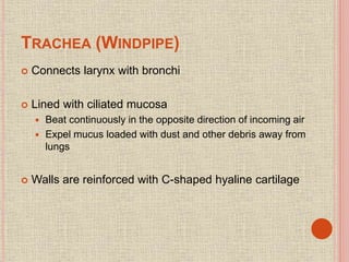
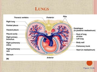
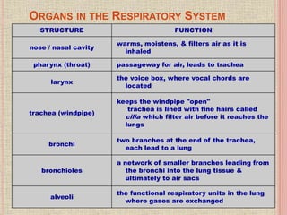
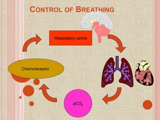
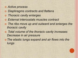
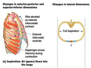
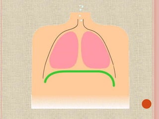
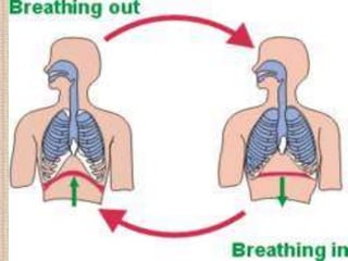
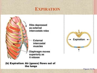
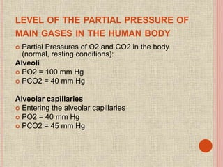
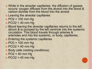
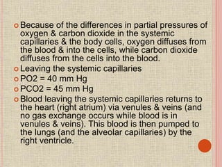
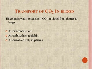
The document discusses the human respiratory system and gas exchange. It describes the major organs involved including the nose, pharynx, larynx, trachea, bronchi, and lungs. It explains the processes of pulmonary ventilation including inspiration and expiration. It discusses oxygen and carbon dioxide transport in the blood and partial pressures of gases in the body. It also covers common respiratory disorders like asthma, pneumonia, and emphysema.
lecture 5: it's good for as to take a breif about how does atmospheric air will pass to our lungs then to blood, for transportation and utilization of oxygen and excretion of carbon dioxide. Many issue are related when gas exchange is performed.
Respiration Process which involves taking in oxygen into the cells, using it for releasing energy by burning food and then eliminating the waste products like carbon dioxide and water from the body It is a catabolic process as the food is broken down into simpler form. In short, respiration is a biochemical activity taking place with in the protoplasm of the cell and results in the liberation of energy
2. Breathing and Respiration BREATHING 1. Mechanism by which organisms obtain oxygen from the air and release carbon dioxide 2. It is a physical process 3. It involves lungs of the organism RESPIRATION 1. It includes breathing and oxidation of food in the cells of the organism to release energy 2. It is a biochemical process 3. It involves the mitochondria in the cells where food is oxidized to release energy
This ppt is all about Respiratory System in Anatomy and Physiology and to familiarize the importance of the lungs and the parts of our respiratory system
"Discover the latest and most comprehensive PowerPoint presentation on the Respiratory System! Our presentation is perfect for students, educators, and healthcare professionals.
Features:
Detailed diagrams and animations to help you visualize the inner workings of the respiratory system
Accurate and up-to-date information to keep you informed on the latest research and discoveries
User-friendly layout that makes it easy to navigate and understand
Additional materials, such as quizzes and worksheets to supplement your learning
Don't miss out on this essential resource for your education or practice. Get your hands on the Respiratory System PowerPoint presentation today!"
Please note the above is an example, some characteristics of PPT or resource you are selling might be different and you need to adjust the content accordingly.
Respiratory system and includes gases exchanges, functions, factors, disorder...Manmeetkaur215
Respiratory system and gases exchanges. Function of organs, factors, disorders all are included in these slides with suitable diagrams and description.
The respiratory system consists of all the organs involved in breathing. These include the nose, pharynx, larynx, trachea, bronchi and lungs. The respiratory system does two very important things: it brings oxygen into our bodies, which we need for our cells to live and function properly; and it helps us get rid of carbon dioxide, which is a waste product of cellular function. The nose, pharynx, larynx, trachea and bronchi all work like a system of pipes through which the air is funnelled down into our lungs. There, in very small air sacs called alveoli, oxygen is brought into the bloodstream and carbon dioxide is pushed from the blood out into the air. When something goes wrong with part of the respiratory system, such as an infection like pneumonia, it makes it harder for us to get the oxygen we need and to get rid of the waste product carbon dioxide. Common respiratory symptoms include breathlessness, cough, and chest pain.
Respiratory System Shaikh Sameer Pharm D I year.pdfSami
Respiratory system anatomy
Lungs
Mechanism of respiration
Regulation of respiration
Transport of respiratory gases
Respiratory volumes and capacities
Some important disorders
DISSERTATION on NEW DRUG DISCOVERY AND DEVELOPMENT STAGES OF DRUG DISCOVERYNEHA GUPTA
The process of drug discovery and development is a complex and multi-step endeavor aimed at bringing new pharmaceutical drugs to market. It begins with identifying and validating a biological target, such as a protein, gene, or RNA, that is associated with a disease. This step involves understanding the target's role in the disease and confirming that modulating it can have therapeutic effects. The next stage, hit identification, employs high-throughput screening (HTS) and other methods to find compounds that interact with the target. Computational techniques may also be used to identify potential hits from large compound libraries.
Following hit identification, the hits are optimized to improve their efficacy, selectivity, and pharmacokinetic properties, resulting in lead compounds. These leads undergo further refinement to enhance their potency, reduce toxicity, and improve drug-like characteristics, creating drug candidates suitable for preclinical testing. In the preclinical development phase, drug candidates are tested in vitro (in cell cultures) and in vivo (in animal models) to evaluate their safety, efficacy, pharmacokinetics, and pharmacodynamics. Toxicology studies are conducted to assess potential risks.
Before clinical trials can begin, an Investigational New Drug (IND) application must be submitted to regulatory authorities. This application includes data from preclinical studies and plans for clinical trials. Clinical development involves human trials in three phases: Phase I tests the drug's safety and dosage in a small group of healthy volunteers, Phase II assesses the drug's efficacy and side effects in a larger group of patients with the target disease, and Phase III confirms the drug's efficacy and monitors adverse reactions in a large population, often compared to existing treatments.
After successful clinical trials, a New Drug Application (NDA) is submitted to regulatory authorities for approval, including all data from preclinical and clinical studies, as well as proposed labeling and manufacturing information. Regulatory authorities then review the NDA to ensure the drug is safe, effective, and of high quality, potentially requiring additional studies. Finally, after a drug is approved and marketed, it undergoes post-marketing surveillance, which includes continuous monitoring for long-term safety and effectiveness, pharmacovigilance, and reporting of any adverse effects.
Local Advanced Lung Cancer: Artificial Intelligence, Synergetics, Complex Sys...Oleg Kshivets
Overall life span (LS) was 1671.7±1721.6 days and cumulative 5YS reached 62.4%, 10 years – 50.4%, 20 years – 44.6%. 94 LCP lived more than 5 years without cancer (LS=2958.6±1723.6 days), 22 – more than 10 years (LS=5571±1841.8 days). 67 LCP died because of LC (LS=471.9±344 days). AT significantly improved 5YS (68% vs. 53.7%) (P=0.028 by log-rank test). Cox modeling displayed that 5YS of LCP significantly depended on: N0-N12, T3-4, blood cell circuit, cell ratio factors (ratio between cancer cells-CC and blood cells subpopulations), LC cell dynamics, recalcification time, heparin tolerance, prothrombin index, protein, AT, procedure type (P=0.000-0.031). Neural networks, genetic algorithm selection and bootstrap simulation revealed relationships between 5YS and N0-12 (rank=1), thrombocytes/CC (rank=2), segmented neutrophils/CC (3), eosinophils/CC (4), erythrocytes/CC (5), healthy cells/CC (6), lymphocytes/CC (7), stick neutrophils/CC (8), leucocytes/CC (9), monocytes/CC (10). Correct prediction of 5YS was 100% by neural networks computing (error=0.000; area under ROC curve=1.0).
- Video recording of this lecture in English language: https://youtu.be/kqbnxVAZs-0
- Video recording of this lecture in Arabic language: https://youtu.be/SINlygW1Mpc
- Link to download the book free: https://nephrotube.blogspot.com/p/nephrotube-nephrology-books.html
- Link to NephroTube website: www.NephroTube.com
- Link to NephroTube social media accounts: https://nephrotube.blogspot.com/p/join-nephrotube-on-social-media.html
Knee anatomy and clinical tests 2024.pdfvimalpl1234
This includes all relevant anatomy and clinical tests compiled from standard textbooks, Campbell,netter etc..It is comprehensive and best suited for orthopaedicians and orthopaedic residents.
Rasamanikya is a excellent preparation in the field of Rasashastra, it is used in various Kushtha Roga, Shwasa, Vicharchika, Bhagandara, Vatarakta, and Phiranga Roga. In this article Preparation& Comparative analytical profile for both Formulationon i.e Rasamanikya prepared by Kushmanda swarasa & Churnodhaka Shodita Haratala. The study aims to provide insights into the comparative efficacy and analytical aspects of these formulations for enhanced therapeutic outcomes.
Here is the updated list of Top Best Ayurvedic medicine for Gas and Indigestion and those are Gas-O-Go Syp for Dyspepsia | Lavizyme Syrup for Acidity | Yumzyme Hepatoprotective Capsules etc
micro teaching on communication m.sc nursing.pdfAnurag Sharma
Microteaching is a unique model of practice teaching. It is a viable instrument for the. desired change in the teaching behavior or the behavior potential which, in specified types of real. classroom situations, tends to facilitate the achievement of specified types of objectives.
Title: Sense of Taste
Presenter: Dr. Faiza, Assistant Professor of Physiology
Qualifications:
MBBS (Best Graduate, AIMC Lahore)
FCPS Physiology
ICMT, CHPE, DHPE (STMU)
MPH (GC University, Faisalabad)
MBA (Virtual University of Pakistan)
Learning Objectives:
Describe the structure and function of taste buds.
Describe the relationship between the taste threshold and taste index of common substances.
Explain the chemical basis and signal transduction of taste perception for each type of primary taste sensation.
Recognize different abnormalities of taste perception and their causes.
Key Topics:
Significance of Taste Sensation:
Differentiation between pleasant and harmful food
Influence on behavior
Selection of food based on metabolic needs
Receptors of Taste:
Taste buds on the tongue
Influence of sense of smell, texture of food, and pain stimulation (e.g., by pepper)
Primary and Secondary Taste Sensations:
Primary taste sensations: Sweet, Sour, Salty, Bitter, Umami
Chemical basis and signal transduction mechanisms for each taste
Taste Threshold and Index:
Taste threshold values for Sweet (sucrose), Salty (NaCl), Sour (HCl), and Bitter (Quinine)
Taste index relationship: Inversely proportional to taste threshold
Taste Blindness:
Inability to taste certain substances, particularly thiourea compounds
Example: Phenylthiocarbamide
Structure and Function of Taste Buds:
Composition: Epithelial cells, Sustentacular/Supporting cells, Taste cells, Basal cells
Features: Taste pores, Taste hairs/microvilli, and Taste nerve fibers
Location of Taste Buds:
Found in papillae of the tongue (Fungiform, Circumvallate, Foliate)
Also present on the palate, tonsillar pillars, epiglottis, and proximal esophagus
Mechanism of Taste Stimulation:
Interaction of taste substances with receptors on microvilli
Signal transduction pathways for Umami, Sweet, Bitter, Sour, and Salty tastes
Taste Sensitivity and Adaptation:
Decrease in sensitivity with age
Rapid adaptation of taste sensation
Role of Saliva in Taste:
Dissolution of tastants to reach receptors
Washing away the stimulus
Taste Preferences and Aversions:
Mechanisms behind taste preference and aversion
Influence of receptors and neural pathways
Impact of Sensory Nerve Damage:
Degeneration of taste buds if the sensory nerve fiber is cut
Abnormalities of Taste Detection:
Conditions: Ageusia, Hypogeusia, Dysgeusia (parageusia)
Causes: Nerve damage, neurological disorders, infections, poor oral hygiene, adverse drug effects, deficiencies, aging, tobacco use, altered neurotransmitter levels
Neurotransmitters and Taste Threshold:
Effects of serotonin (5-HT) and norepinephrine (NE) on taste sensitivity
Supertasters:
25% of the population with heightened sensitivity to taste, especially bitterness
Increased number of fungiform papillae
Title: Sense of Smell
Presenter: Dr. Faiza, Assistant Professor of Physiology
Qualifications:
MBBS (Best Graduate, AIMC Lahore)
FCPS Physiology
ICMT, CHPE, DHPE (STMU)
MPH (GC University, Faisalabad)
MBA (Virtual University of Pakistan)
Learning Objectives:
Describe the primary categories of smells and the concept of odor blindness.
Explain the structure and location of the olfactory membrane and mucosa, including the types and roles of cells involved in olfaction.
Describe the pathway and mechanisms of olfactory signal transmission from the olfactory receptors to the brain.
Illustrate the biochemical cascade triggered by odorant binding to olfactory receptors, including the role of G-proteins and second messengers in generating an action potential.
Identify different types of olfactory disorders such as anosmia, hyposmia, hyperosmia, and dysosmia, including their potential causes.
Key Topics:
Olfactory Genes:
3% of the human genome accounts for olfactory genes.
400 genes for odorant receptors.
Olfactory Membrane:
Located in the superior part of the nasal cavity.
Medially: Folds downward along the superior septum.
Laterally: Folds over the superior turbinate and upper surface of the middle turbinate.
Total surface area: 5-10 square centimeters.
Olfactory Mucosa:
Olfactory Cells: Bipolar nerve cells derived from the CNS (100 million), with 4-25 olfactory cilia per cell.
Sustentacular Cells: Produce mucus and maintain ionic and molecular environment.
Basal Cells: Replace worn-out olfactory cells with an average lifespan of 1-2 months.
Bowman’s Gland: Secretes mucus.
Stimulation of Olfactory Cells:
Odorant dissolves in mucus and attaches to receptors on olfactory cilia.
Involves a cascade effect through G-proteins and second messengers, leading to depolarization and action potential generation in the olfactory nerve.
Quality of a Good Odorant:
Small (3-20 Carbon atoms), volatile, water-soluble, and lipid-soluble.
Facilitated by odorant-binding proteins in mucus.
Membrane Potential and Action Potential:
Resting membrane potential: -55mV.
Action potential frequency in the olfactory nerve increases with odorant strength.
Adaptation Towards the Sense of Smell:
Rapid adaptation within the first second, with further slow adaptation.
Psychological adaptation greater than receptor adaptation, involving feedback inhibition from the central nervous system.
Primary Sensations of Smell:
Camphoraceous, Musky, Floral, Pepperminty, Ethereal, Pungent, Putrid.
Odor Detection Threshold:
Examples: Hydrogen sulfide (0.0005 ppm), Methyl-mercaptan (0.002 ppm).
Some toxic substances are odorless at lethal concentrations.
Characteristics of Smell:
Odor blindness for single substances due to lack of appropriate receptor protein.
Behavioral and emotional influences of smell.
Transmission of Olfactory Signals:
From olfactory cells to glomeruli in the olfactory bulb, involving lateral inhibition.
Primitive, less old, and new olfactory systems with different path
These lecture slides, by Dr Sidra Arshad, offer a quick overview of the physiological basis of a normal electrocardiogram.
Learning objectives:
1. Define an electrocardiogram (ECG) and electrocardiography
2. Describe how dipoles generated by the heart produce the waveforms of the ECG
3. Describe the components of a normal electrocardiogram of a typical bipolar lead (limb II)
4. Differentiate between intervals and segments
5. Enlist some common indications for obtaining an ECG
6. Describe the flow of current around the heart during the cardiac cycle
7. Discuss the placement and polarity of the leads of electrocardiograph
8. Describe the normal electrocardiograms recorded from the limb leads and explain the physiological basis of the different records that are obtained
9. Define mean electrical vector (axis) of the heart and give the normal range
10. Define the mean QRS vector
11. Describe the axes of leads (hexagonal reference system)
12. Comprehend the vectorial analysis of the normal ECG
13. Determine the mean electrical axis of the ventricular QRS and appreciate the mean axis deviation
14. Explain the concepts of current of injury, J point, and their significance
Study Resources:
1. Chapter 11, Guyton and Hall Textbook of Medical Physiology, 14th edition
2. Chapter 9, Human Physiology - From Cells to Systems, Lauralee Sherwood, 9th edition
3. Chapter 29, Ganong’s Review of Medical Physiology, 26th edition
4. Electrocardiogram, StatPearls - https://www.ncbi.nlm.nih.gov/books/NBK549803/
5. ECG in Medical Practice by ABM Abdullah, 4th edition
6. Chapter 3, Cardiology Explained, https://www.ncbi.nlm.nih.gov/books/NBK2214/
7. ECG Basics, http://www.nataliescasebook.com/tag/e-c-g-basics
Basavarajeeyam is an important text for ayurvedic physician belonging to andhra pradehs. It is a popular compendium in various parts of our country as well as in andhra pradesh. The content of the text was presented in sanskrit and telugu language (Bilingual). One of the most famous book in ayurvedic pharmaceutics and therapeutics. This book contains 25 chapters called as prakaranas. Many rasaoushadis were explained, pioneer of dhatu druti, nadi pareeksha, mutra pareeksha etc. Belongs to the period of 15-16 century. New diseases like upadamsha, phiranga rogas are explained.
1. GASEOUS EXCHANGE IN HUMAN
Zakia khatoon
Syyeda urooj
Nelofar Hanif
Diya Fatima
Kainat Abbas
2. INTRODUCTION
Oxygen is needed for aerobic respiration
CO₂ is produce as a result of aerobic respiration.
Exchange of these gases with the environment.
Need Respiratory Surface
3. RESPIRATORY SURFACES
• Thin
– Diffusion distance
– Speed
• Moist
– Gases dissolved in
solution
• Large
– SA to volume ratio
– Energy demands
4. ORGANS OF THE RESPIRATORY SYSTEM
Nose
Pharynx
Larynx
Trachea
Bronchi
Lungs –
alveoli
Figure 13.1
5. FUNCTION OF THE RESPIRATORY SYSTEM
Oversees gas exchanges between the blood and
external environment
Exchange of gasses takes place within the alveoli
Passageways to the lungs purify, warm, and
humidify the incoming air
6. THE NOSE
The only externally visible part of the respiratory
system
Air enters the nose through the external nares
(nostrils)
The interior of the nose consists of a nasal cavity
divided by a nasal septum
7. ANATOMY OF THE NASAL CAVITY
Lateral walls have projections called conchae
Increases surface area
Increases air turbulence within the nasal cavity
The nasal cavity is separated from the oral cavity by the
palate
Anterior hard palate (bone)
Posterior soft palate (muscle)
8. ANATOMY OF THE NASAL CAVITY
Olfactory receptors are located in the mucosa on
the superior surface
The rest of the cavity is lined with respiratory
mucosa
Moistens air
Traps incoming foreign particles
Paranasal sinuses
Cavities within bones surrounding the nasal cavity
9. PHARYNX (THROAT)
Muscular passage from nasal cavity to larynx
Three regions of the pharynx
Nasopharynx – superior region behind nasal cavity
Oropharynx – middle region behind mouth
Laryngopharynx – inferior region attached to larynx
The oropharynx and laryngopharynx are common
passageways for air and food
10. STRUCTURES OF THE PHARYNX
Auditory tubes enter the nasopharynx
Tonsils of the pharynx
Pharyngeal tonsil (adenoids) in the nasopharynx
Palatine tonsils in the oropharynx
Lingual tonsils at the base of the tongue
11. LARYNX (VOICE BOX)
Routes air and food into proper channels
Plays a role in speech
Made of eight rigid hyaline cartilages and a spoon-
shaped flap of elastic cartilage (epiglottis)
Vocal cords - vibrate with expelled air to create
sound (speech)
12. STRUCTURES OF THE LARYNX
Thyroid cartilage
Largest hyaline cartilage
Protrudes anteriorly (Adam’s apple)
Epiglottis
Superior opening of the larynx
Routes food to the larynx and air toward the trachea
Glottis
opening between vocal cords
13. TRACHEA (WINDPIPE)
Connects larynx with bronchi
Lined with ciliated mucosa
Beat continuously in the opposite direction of incoming air
Expel mucus loaded with dust and other debris away from
lungs
Walls are reinforced with C-shaped hyaline cartilage
14. PRIMARY BRONCHI
Formed by division of the trachea
Enters the lung at the hilus
(medial depression)
Right bronchus is wider, shorter,
and straighter than left
Bronchi subdivide into smaller
and smaller branches
15. BRONCHIOLES
Smallest branches
of the bronchi
All but the smallest
branches have
reinforcing cartilage
Terminal
bronchioles end in
alveoli (grape like
sacs).
Figure 13.5a
16. LUNGS
Ocupy most of the thoracic cavity
Apex is near the clavicle (superior
portion)
Each lung is divided into lobes by
fissures
Left lung – two lobes
Right lung – three lobes
18. ORGANS IN THE RESPIRATORY SYSTEM
STRUCTURE FUNCTION
nose / nasal cavity
warms, moistens, & filters air as it is
inhaled
pharynx (throat) passageway for air, leads to trachea
larynx
the voice box, where vocal chords are
located
trachea (windpipe)
keeps the windpipe "open"
trachea is lined with fine hairs called
cilia which filter air before it reaches the
lungs
bronchi
two branches at the end of the trachea,
each lead to a lung
bronchioles
a network of smaller branches leading from
the bronchi into the lung tissue &
ultimately to air sacs
alveoli
the functional respiratory units in the lung
where gases are exchanged
19. RESPIRATORY DISRUPTIONS
• Smoking
– Inhibit cilia movement causes ‘smoker’s cough’
• Thicken bronchioles and reduce elasticity
• Alveoli rupture
– Stopping allows cilia and alveoli damage to reverse
• Premature birth (37 weeks or less)
– Surfactant production incomplete
• Keeps alveoli from collapsing
– Each breath requires large effort
• Emphysema
– Bronchi swell, tearing alveoli
– Reduced SA for gas exchange
• Pneumonia
– Fluid in alveoli
• Bronchitis inflames of bronchioles
20. VENTILATION/BREATHING
The movement of air between the environment and
the lungs via inhalation and exhalation.
Mechanical ventilation: using artificial methods to
assist breathing
Medical ventilator: a mechanical ventilator, a
machine designed to move breathable air into and
out of the lungs.
22. MECHANISM OF BREATHING
(PULMONARY VENTILATION)
Mechanical process
Depends on volume changes in the thoracic cavity
Volume changes lead to pressure changes, which lead to
equalize pressure of flow of gases
2 phases
Inspiration – flow of air into lung
Expiration – air leaving lung
25. .
Active process
Diaphragms contracts and flattens
Thoracic cavity enlarges
External intercostals muscles contract
The ribs move up and outward and enlarges the
thoracic cavity
Total volume of the thoracic cavity increases
Decrease in air pressure
The elastic lungs expand and air flows into the
lungs
26.
27.
28.
29. EXPIRATION
Passive process dependent up on natural lung
elasticity
As muscles relax, air is pushed out of the lungs
Forced expiration can occur mostly by contracting
internal intercostal muscles to depress the rib cage
31. MEASURES OF PULMONARY VENTILATION
Respiratory volumes – values determined by using
a spirometer
Tidal Volume (TV) – amount of air inhaled or exhaled with
each breath under resting conditions
Inspiratory Reserve Volume (IRV) – amount of air that
can be inhaled during forced breathing in addition to
resting tidal volume
Expiratory Reserve Volume (ERV) – amount of air that
can be exhaled during forced breathing in addition to tidal
volume
Residual Volume (RV) – Amount of air remaining in the
lungs after a forced exhalation.
32. TRANSPORT OF GASES
The exchange of gases (O₂ & CO₂) between the
alveoli & the blood occurs by simple diffusion.
O₂ diffusing from the alveoli into the blood & CO₂
from the blood into the alveoli.
Diffusion requires a concentration gradient. So, the
concentration (or pressure) of O₂ in the alveoli must
be kept at a higher level than in the blood & the
concentration (or pressure) of CO₂ in the alveoli
must be kept at a lower lever than in the blood.
33. PARTIAL PRESSURE
The partial pressure exerted by each gas in a
mixture equals the total pressure times the
fractional composition of the gas in the mixture.
Total atmospheric pressure (at sea level) is about
760 mm Hg and, further, that air is about 21%
oxygen, then the partial pressure of oxygen in the
air is 0.21 times 760 mm Hg or 160 mm Hg.
34. LEVEL OF THE PARTIAL PRESSURE OF
MAIN GASES IN THE HUMAN BODY
Partial Pressures of O2 and CO2 in the body
(normal, resting conditions):
Alveoli
PO2 = 100 mm Hg
PCO2 = 40 mm Hg
Alveolar capillaries
Entering the alveolar capillaries
PO2 = 40 mm Hg
PCO2 = 45 mm Hg
35. While in the alveolar capillaries, the diffusion of gasses
occurs: oxygen diffuses from the alveoli into the blood &
carbon dioxide from the blood into the alveoli.
Leaving the alveolar capillaries
PO2 = 100 mm Hg
PCO2 = 40 mm Hg
Blood leaving the alveolar capillaries returns to the left
atrium & is pumped by the left ventricle into the systemic
circulation. This blood travels through arteries &
arterioles and into the systemic, or body, capillaries.
Entering the systemic capillaries
PO2 = 100 mm Hg
PCO2 = 40 mm Hg
Body cells (resting conditions)
PO2 = 40 mm Hg
PCO2 = 45 mm Hg
36.
37. Because of the differences in partial pressures of
oxygen & carbon dioxide in the systemic
capillaries & the body cells, oxygen diffuses from
the blood & into the cells, while carbon dioxide
diffuses from the cells into the blood.
Leaving the systemic capillaries
PO2 = 40 mm Hg
PCO2 = 45 mm Hg
Blood leaving the systemic capillaries returns to
the heart (right atrium) via venules & veins (and
no gas exchange occurs while blood is in
venules & veins). This blood is then pumped to
the lungs (and the alveolar capillaries) by the
right ventricle.
38. TRANSPORT OF CO₂ IN BLOOD
Three main ways to transport CO₂ in blood from tissues to
lungs
As bicarbonate ions
As carboxyhaemoglobin
As dissolved CO₂ in plasma
39.
40. AS BICARBONATE IONS
Most of the carbon dioxide approximately 70% is carried in
blood as bicarbonate ions.
CO₂ entering blood cells reacts with water and form carbonic
acid in the presence of carbonic anhydrase.
Carbonic acid is unstable and splits into H+ and HCO₃⁻
H+ combine with H₄bO₂ that dissociates O₂ from Hb and
diffuse into cells for aerobic respiration.
The bicarbonate ion then diffuses outside the RBC in the
plasma and combines with Sodium ions to from Sodium
bicarbonate (NaHCO3).
41. CONT..
Loss of bicarbonate ions from RBC causes positive
charge inside RBC which is balanced by diffusion of
chloride (Cl-) ion from plasma into the RBC.
This exchange of Cl- ion and HCO3- ion between
plasma and RBC is known as chloride shift.
This phenomenon of chloride shift maintains the
electrical neutrality of cell.
42. O₂ HEMOGLOBIN DISSOCIATION CURVE
The following four factors decrease the affinity, or
strength of attraction, of Hb for O 2 and result in a
shift of the O 2‐Hb dissociation curve to the right
1. Increase in temperature.
2. Increase in partial pressure of CO 2 (pCO 2). The
effect of CO2 on Oxygen dissociation curve is
known as Bohr effect.
3. Increase in acidity (decrease in pH).
43. It has been found that increase in concentration of
CO2 decreases the amount of oxyhaemoglobin
formation.
according to Bohr effect, for any particular partial
pressure of Oxygen, the affinity of Haemoglobin
toward Oxygen decreases and favors dissociation
of oxyhaemoglobin when the partial pressure of
carbondioxide increases.
It means, higher CO2 concentration causes the
dissociation of HbO2 releasing free O2.
Increase in PCO2 shifts the O2 dissociation curve
downwards. Higher PCO2 lowers the affinity of
hemoglobin for O2.
44.
45. AS CARBAMINOHAEMOGLOBIN (23%)
some CO2 combines with Haemoglobin to form
carbaminohaemoglobin in RBCs.
CO2 + NHbNH2————–HbNH.COOH
(carbaminohaemoglobin).
finally, CO2 are carried to lungs and expelled out by
expiration process of breathing
46. AS DISSOLVED CO₂ IN PLASMA
some CO₂ dissolved in the plasma to form carbonic acid
carbon dioxide mixed with water of blood plasma to
form carbonic acid.
CO₂+ H₂O——————H₂CO₃
47. CO₂ POISONING/
HYPERCAPNIA OR HYPERCARBIA
Causes: breathing volcanic gas.
rebreathing exhaled air (e.g., from breathing air in a
bag, sleeping in a sealed tent, sleeping with a blanket
over one's head)
scuba diving, hypoventilation, lung diseases.
breathing in confined spaces or areas with poor air
circulation, such as a sealed room, a tunnel, cargo.
Symptoms:
high blood pressure, flushed skin, headache and
twitching muscles.
At severe conditions panic, irregular heartbeat,
hallucinations, vomited and potentially
unconsciousness or even death.
48. RESPIRATORY DISORDERS
Pre-term birth can lead to infants with under-
developed lungs . Smoking and air pollution are two
common causes of respiratory problems.
Diorders include:
Obstructive conditions (e.g., emphysema,
bronchitis, asthma attacks)
Infectious, environmental: (e.g., pneumonia,
tuberculosis, asbestosis, particulate pollutants):
49. CONT…
Common Respiratory Disorders Include:
Swine flu, also called swine influenza, hog flu,
or pig flu, a respiratory disease of pigs that is
caused by an influenza virus.
Chronic Bronchitis: - Any irritant reaching the
bronchi and bronchioles will stimulate an increased
secretion of mucus. In chronic bronchitis the air
passages become clogged with mucus, and this
leads to a persistent cough.
50. CONT…
Emphysema: The delicate walls of the alveoli
break down, reducing the gas exchange area of the
lungs. The condition develops slowly and is seldom
a direct cause of death.
Asthma: - Periodic constriction of the bronchi and
bronchioles makes it more difficult to breathe.
Pneumonia :- An infection of the alveoli. It can be
caused by many kinds of both bacteria and viruses.
Tissue fluids accumulate in the alveoli reducing the
surface area exposed to air. If enough alveoli are
affected, the patient may need supplemental
oxygen.