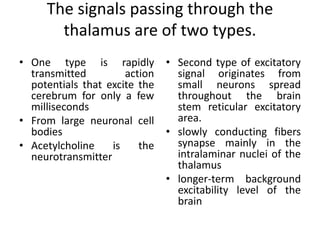
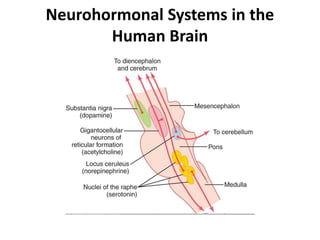
Objectives
• To provide a brief overview of the structure
and function of the reticular formation.
• And its role in excitation of cerebrum.
• The parts of the limbic system and its
functions
Activating-Driving Systems of the
Brain
• Without continuous transmission of nerve signals
from the lower brain into the cerebrum, the
cerebrum becomes useless.
Nerve signals in the brain stem activate the cerebral
part of the brain in two ways:
• (1) by directly stimulating a background level of
neuronal activity in wide areas of the brain and
• (2) by activating neurohormonal systems that
release specific facillitatory or inhibitory
hormone-like neurotransmitter substances into
selected areas of the brain.
The signals passing through the
thalamus are of two types.
• One type is rapidly
transmitted action
potentials that excite the
cerebrum for only a few
milliseconds
• From large neuronal cell
bodies
• Acetylcholine is the
neurotransmitter
• Second type of excitatory
signal originates from
small neurons spread
throughout the brain
stem reticular excitatory
area.
• slowly conducting fibers
synapse mainly in the
intralaminar nuclei of the
thalamus
• longer-term background
excitability level of the
brain
Excitation of the Excitatory Area by Peripheral
Sensory Signals
Excitability of brain stem & in turn cerebrum
depend on the number and type of sensory
signals from the periphery
Pain signals in particular increase activity in this
excitatory area and therefore strongly excite
the brain to attention.
Increased Activity of the Excitatory Area
Caused by Feedback Signals Returning from the
Cerebral Cortex
• Signals are sent from the cortex to the brain
stem excitatory area, which in turn sends still
more excitatory signals to the cortex.
• This is a general mechanism of positive
feedback that allows any beginning activity in
the cerebral cortex to support still more
activity, thus leading to an "awake" mind.
A Reticular Inhibitory Area Is Located
in the Lower Brain Stem
• This is the reticular inhibitory area, located
medially and ventrally in the medulla.
• This area can inhibit the reticular facilitory area of
the upper brain stem and thereby decrease
activity in the superior portions of the brain as
well.
• One of the mechanisms for this is to excite
serotonergic neurons; these in turn secrete the
inhibitory neurohormone serotonin at crucial
points in the brain.
In addition to these areas there are different
neurohormonal systems
Neurohormonal Systems in the
Human Brain
The locus ceruleus and the
norepinephrine system.
• The locus ceruleus is a small area located
bilaterally and posteriorly at the juncture
between the pons and mesencephalon.
• Nerve fibers from this area spread
throughout the brain, they secrete
norepinephrine.
• This system probably plays an important
role in causing dreaming, thus leading to a
type of sleep called rapid eye movement
sleep (REM sleep).
• The substantia nigra and the dopamine
system
They secrete dopamine. *
• The dopamine is believed to act as an inhibitory
transmitter in the basal ganglia, but in some
other areas of the brain it is possibly excitatory.
The raphe nuclei and the serotonin system.
In the midline of the pons and medulla are several thin
nuclei called the raphe nuclei.
Many of the neurons in these nuclei secrete serotonin.
They send fibers into the diencephalon and a few fibers
to the cerebral cortex; still other fibers descend to the
spinal cord.
The serotonin secreted at the cord fiber endings
suppress pain,
& in the diencephalon and cerebrum almost certainly
plays an essential inhibitory role to help cause normal
sleep.
The gigantocellular neurons of the reticular
excitatory area and the acetylcholine system.
• The fibers from these large cells divide
immediately into two branches, one passing
upward to the higher levels of the brain and the
other passing downward through the
reticulospinal tracts into the spinal cord.
• The neurohormone secreted at their terminals is
acetylcholine.
• In most places, the acetylcholine functions as an
excitatory neurotransmitter. Activation of these
acetylcholine neurons leads to an acutely awake
and excited nervous system.
Other Neurotransmitters and Neurohormonal
Substances Secreted in the Brain
enkephalins, gamma-aminobutyric acid,
glutamate, vasopressin, adrenocorticotropic
hormone, α-melanocyte stimulating hormone
(α-MSH), neuropeptide-Y (NPY), epinephrine,
histamine, endorphins, angiotensin II, and
neurotensin
The Limbic System and the
Hypothalamus
Limbic system
• The word "limbic" means "border.“*
• The term limbic system has been expanded to
mean the entire neuronal circuitry that controls
emotional behavior and motivational drives.
• Hypothalamus is a major part of the limbic
system with its related structures.
• In addition to their roles in behavioral control,
these areas control many internal conditions
of the body, such as body temperature,
osmolality of the body fluids, and the drives to
eat and drink and to control body weight.
These internal functions are collectively called
vegetative functions of the brain, and their
control is closely related to behavior.
• Anatomically, the limbic structures include the
subcallosal, the cingulate, and the
parahippocampal gyri, the hippocampal
formation, the amygdaloid nucleus, the
mammillary bodies, and the anterior thalamic
nucleus, the septal nuclei and portions of
basal ganglia.
Functions of Limbic system
Aggression. Stimulation of
certain areas of the amygdala
electrical stimulation of the amygdala and
hypothalamus
surgical removal of the limbic system can
result in an absence of fear
hypothalamus contains a feeding center
hypothalamus contains a satiety center
• Sexual drive can be stimulated from several
areas of the hypothalamus, especially the
most anterior and most posterior portions of
the hypothalamus.
Major reward centers are located along the
course of the medial forebrain bundle, especially
in the lateral and ventromedial nuclei of the
hypothalamus.
potent areas for punishment have been found in the central gray
area surrounding the aqueduct of Sylvius in the mesencephalon
and extending upward into the periventricular zones of the
hypothalamus and thalamus.
Less potent punishment areas are found in some locations in
the amygdala and hippocampus.

The limbic system and the hypothalamus

  • 1.
    Objectives • To providea brief overview of the structure and function of the reticular formation. • And its role in excitation of cerebrum. • The parts of the limbic system and its functions
  • 2.
    Activating-Driving Systems ofthe Brain • Without continuous transmission of nerve signals from the lower brain into the cerebrum, the cerebrum becomes useless. Nerve signals in the brain stem activate the cerebral part of the brain in two ways: • (1) by directly stimulating a background level of neuronal activity in wide areas of the brain and • (2) by activating neurohormonal systems that release specific facillitatory or inhibitory hormone-like neurotransmitter substances into selected areas of the brain.
  • 4.
    The signals passingthrough the thalamus are of two types. • One type is rapidly transmitted action potentials that excite the cerebrum for only a few milliseconds • From large neuronal cell bodies • Acetylcholine is the neurotransmitter • Second type of excitatory signal originates from small neurons spread throughout the brain stem reticular excitatory area. • slowly conducting fibers synapse mainly in the intralaminar nuclei of the thalamus • longer-term background excitability level of the brain
  • 5.
    Excitation of theExcitatory Area by Peripheral Sensory Signals Excitability of brain stem & in turn cerebrum depend on the number and type of sensory signals from the periphery Pain signals in particular increase activity in this excitatory area and therefore strongly excite the brain to attention.
  • 6.
    Increased Activity ofthe Excitatory Area Caused by Feedback Signals Returning from the Cerebral Cortex • Signals are sent from the cortex to the brain stem excitatory area, which in turn sends still more excitatory signals to the cortex. • This is a general mechanism of positive feedback that allows any beginning activity in the cerebral cortex to support still more activity, thus leading to an "awake" mind.
  • 7.
    A Reticular InhibitoryArea Is Located in the Lower Brain Stem • This is the reticular inhibitory area, located medially and ventrally in the medulla. • This area can inhibit the reticular facilitory area of the upper brain stem and thereby decrease activity in the superior portions of the brain as well. • One of the mechanisms for this is to excite serotonergic neurons; these in turn secrete the inhibitory neurohormone serotonin at crucial points in the brain.
  • 8.
    In addition tothese areas there are different neurohormonal systems
  • 9.
    Neurohormonal Systems inthe Human Brain
  • 10.
    The locus ceruleusand the norepinephrine system. • The locus ceruleus is a small area located bilaterally and posteriorly at the juncture between the pons and mesencephalon. • Nerve fibers from this area spread throughout the brain, they secrete norepinephrine. • This system probably plays an important role in causing dreaming, thus leading to a type of sleep called rapid eye movement sleep (REM sleep).
  • 11.
    • The substantianigra and the dopamine system They secrete dopamine. * • The dopamine is believed to act as an inhibitory transmitter in the basal ganglia, but in some other areas of the brain it is possibly excitatory.
  • 12.
    The raphe nucleiand the serotonin system. In the midline of the pons and medulla are several thin nuclei called the raphe nuclei. Many of the neurons in these nuclei secrete serotonin. They send fibers into the diencephalon and a few fibers to the cerebral cortex; still other fibers descend to the spinal cord. The serotonin secreted at the cord fiber endings suppress pain, & in the diencephalon and cerebrum almost certainly plays an essential inhibitory role to help cause normal sleep.
  • 13.
    The gigantocellular neuronsof the reticular excitatory area and the acetylcholine system. • The fibers from these large cells divide immediately into two branches, one passing upward to the higher levels of the brain and the other passing downward through the reticulospinal tracts into the spinal cord. • The neurohormone secreted at their terminals is acetylcholine. • In most places, the acetylcholine functions as an excitatory neurotransmitter. Activation of these acetylcholine neurons leads to an acutely awake and excited nervous system.
  • 14.
    Other Neurotransmitters andNeurohormonal Substances Secreted in the Brain enkephalins, gamma-aminobutyric acid, glutamate, vasopressin, adrenocorticotropic hormone, α-melanocyte stimulating hormone (α-MSH), neuropeptide-Y (NPY), epinephrine, histamine, endorphins, angiotensin II, and neurotensin
  • 15.
    The Limbic Systemand the Hypothalamus
  • 16.
    Limbic system • Theword "limbic" means "border.“* • The term limbic system has been expanded to mean the entire neuronal circuitry that controls emotional behavior and motivational drives.
  • 17.
    • Hypothalamus isa major part of the limbic system with its related structures. • In addition to their roles in behavioral control, these areas control many internal conditions of the body, such as body temperature, osmolality of the body fluids, and the drives to eat and drink and to control body weight. These internal functions are collectively called vegetative functions of the brain, and their control is closely related to behavior.
  • 18.
    • Anatomically, thelimbic structures include the subcallosal, the cingulate, and the parahippocampal gyri, the hippocampal formation, the amygdaloid nucleus, the mammillary bodies, and the anterior thalamic nucleus, the septal nuclei and portions of basal ganglia.
  • 23.
  • 24.
  • 25.
    electrical stimulation ofthe amygdala and hypothalamus surgical removal of the limbic system can result in an absence of fear
  • 26.
  • 27.
  • 28.
    • Sexual drivecan be stimulated from several areas of the hypothalamus, especially the most anterior and most posterior portions of the hypothalamus.
  • 29.
    Major reward centersare located along the course of the medial forebrain bundle, especially in the lateral and ventromedial nuclei of the hypothalamus.
  • 30.
    potent areas forpunishment have been found in the central gray area surrounding the aqueduct of Sylvius in the mesencephalon and extending upward into the periventricular zones of the hypothalamus and thalamus. Less potent punishment areas are found in some locations in the amygdala and hippocampus.

Editor's Notes

  • #14 The norepinephrine generally excites the brain to increased activity. However, it has inhibitory effects in a few brain areas because of inhibitory receptors at certain neuronal synapses.
  • #15 *Other neurons located in adjacent regions also secrete dopamine, but they send their endings into more ventral areas of the brain, especially to the hypothalamus and the limbic system.
  • #20 *Originally, the term "limbic" was used to describe the border structures around the basal regions of the cerebrum in the border zone between the cerebral cortex and the hypothalamus.
  • #27 *importance of reticular formation regarding limbic system